Friday, January 29, 2021

Lupine Publishers | Paediatric Dental Procedures Under General Anaestheisa-A Safe Surgery

 Lupine Publishers | Journal of Pediatric Dentistry


Abstract

This article provides an overall review of the indications and safety of paediatric dental procedures performed under general anaesthesia. This pharmacological management of children with anxiety, systemic health considerations and special needs in a controlled hospital setting facilitates the provision of safe and effective treatment with successful outcomes.

Introduction

The relationship between an infant, child or an adolescent patient and a dentist is very important because this interaction influences the life of a child outside a dental surgery. Dental treatment requires an understanding of a child at their level of development since they differ in their personality, cognitive ability, motor skills, language milestones, fears and experience of life and pain. The three basic temperaments which may influence a child are classified as easy, difficult and slow to warm up. A child with an easy temperament is flexible. Children with a difficult temperament respond best to confidence whereas children who are slow to warm up require patience and sensitivity. A variety of approaches and techniques may be therefore be employed by the dental team to inform and communicate with a child for the purpose of structuring their behavior with a child [1,2]. A safe and effective pain control in children and adolescents employs over lapping categories of behavioral techniques, local anaesthesia (LA), conscious sedation and general anaesthesia (GA). Selection of the most optimum management in a child is influenced by the cognitive ability, medical status and the complexity of the dental procedure [3,4].

Pharmacological Behaviour Management in Children

Children perceive pain according to their stage of cognitive development. Management of pain and anxiety in a child is an integral component of paediatric dentistry. Hence the behaviour management skills for this purpose include communication, empathy, active listening, coaching, tolerance and flexibility. Recent trends indicate an increasing use of general anaesthesia for pharmacological pain and anxiety management in paediatric dentistry. The indications for dental treatment under GA include restoration of carious teeth, endodontic treatment in caries or trauma, prophylactic scaling to prevent periodontal pathology, exposure of ectopic teeth, removal of supernumeraries, extraction or removal of permanent molars, clearance of unrestorable asymptomatic teeth and extractions for balancing /or compensating [5,6]. General anaesthesia should be administered within a hospital setting [7] with trained personnel immediately available to assist the anaesthetist with the resuscitation of a collapsed patient, support and maintain a collapsed patient pending recovery or for supervised transfer to a critical care facility or to a separate hospital [8,9].

Indications for General Anaesthesia In Paediatric Dentistry

The need for dental treatment of children under general anaesthesia represents the final choice for solution. A minor risk of complication from the anaesthetic always persists hence a decision should strike a balance between the risk and benefit. Indications for general anaesthesia in paediatric dentistry include contraindication to the use of local anaesthesia in an acute orofacial infection, preceding failure of local anaesthesia or sedation; inability of a child to cope with the proposed treatment owing to disability, language barrier, immaturity or psychological disorder; extensive treatment; facial swelling; facial trauma; severe cellulitis; abscesses and caries in multiple quadrants, orthodontic extraction of permanent premolars; physical, emotional or learning impairment or a combination of two or more of these and patient/career preference where other techniques have been already tried. Care should be taken when systemic medical conditions co-exist, airway has anatomical or functional abnormality and congenital syndromes like epidermolysis bullosa or mucopolysaccharidoses are present. A pre-procedure visit assesses for satisfactory completion of work and consent. Post procedure home care instructions should include dietary advice, analgesics, fluorides and follow up visits. The elective anaesthesia should be delayed for a period of 2-3 weeks in a child presenting with an upper respiratory tract infection on the day of planned surgery [10,11].

Preanaesthetic Assessment for General Anaesthesia

The preanaesthetic assessment should take place in a separate hospital visit. It should include dental, medical, and preliminary anaesthetic assessments prior to the day of the elective surgery. The history should include information about different factors like behavioral issues (autism, extreme anxiety, developmental delay, needle phobia), preexisting syndromes (cleft, velocardiofacial syndrome, Down’s Syndrome), cardiac disease (congenital defects, heart murmurs), respiratory disease (asthma, cystic fibrosis), airway problems (micrognathia, cleft, prior tracheostomy, known history of intubation difficulty, sleep apnoea, croup, cleft palate); neurological disease (epilepsy, cerebral palsy, previous brain injury), endocrine and metabolic disorder (genetic metabolic disorder, diabetes), haematological disorders (haemoglobinopathies, thrombocytopenia, haemophilia), neuromuscular disorder (muscular dystrophy), allergies (latex), medication (anticoagulants, insulin). Any change to the medication necessitates a consultation with the specialist [12,13].

The airway assessment allows careful intraoperative airway management, limited mouth opening is evaluated in facial swelling due to dentofacial infection or trauma. This may be precluded in urgent clinical cases or in geographical or social limitations. Information pertaining to fasting, pain management and home arrangements after discharge is imparted. Duration of fasting for solids and milk is 6 hours, for breast milk is 4 hours and for clear fluid is 2 hours. Children with ASA 1 or 2 are amenable to a day stay theatre whereas a severe systemic disease requires an overnight stay for maintaining the airway, ensuring food is tolerated, pain management and hemostasis. The potential risks for general anaesthesia and the proposed treatment plan should be discussed prior to obtaining an informed consent [12,13].

Consent

A consent form is a prerequisite in a child less than 14 years of age. The form should be signed by a parent or guardian in the presence of a dentist as a third-party witness. A child between 14-16 years of age can provide a responsible informed consent however the parent or guardian should also consent with the dentist witnessing the signature (Gillick competent). The consent for treatment in a child 16 years and over must be from their own accord [14,15].

Operating Theatre Environment

Strategies should be employed to reduce the fear and anxiety of a child and to help cope with the environment in a theatre. These include minimizing the pre procedure waiting time, letting children wear their own clothes for comfort and allowing parents to stay with during the induction phase. Intraveinous cannulation maybe facilitated by a preoperative application of topical local anaesthetic creams (Ametop, EMLA, LMX4). Premedication with oral analgesics like paracetamol 15mg/kg, ibuprofen or both augment pain management when administered almost one hour prior to general anaesthesia. Special needs paediatric dental patients require specific preoperative arrangements such as sedative premedication. It is therefore safe to administer 0.2 -0.5 mg/kg Midazolam via oral, buccal or intranasal routes. Very uncooperative children may require 2-3mg/kg intramuscular ketamine in autism or developmental delay. Antibiotics for infective endocarditis are not prescribed in children undergoing dental surgery unless specifically advised by a cardiologist [16].

When administering general anaesthesia clinical observation of the patient is augmented by core standards of monitoring which assess the physiological state of patient, depth of anaesthesia and function of the anaesthetic equipment. These standards of monitoring should be uniform irrespective of the mode, location or duration of general anaesthesia. Airway is shared by the anaesthetist and the dentist amicably. Nasotracheal intubation with a preformed nasal RAE tube (Ring, Adair and Elwyn) provides a good access to either side of the mouth for the dentist and secures the airway for the anaesthetist in more extensive dental surgery procedures such as wisdom tooth extraction. Tracheal intubation via the oral route may be indicated when nasal intubation is contraindicated in trauma to adenoidal tissue in younger children. A throat pack reduced to the size of one third (ribbon gauze m30 cm moistened with saline) should be used during the procedure and removed at the conclusion of the procedure. A laryngeal mask airway device maybe used for longer procedures such as surgical extraction of teeth which are impacted, traumatized teeth and when the ventillation is spontaneous or controlled. A flexible LMA is more difficult to insert in children sometimes but the reinforced tubing allows better access to the teeth. Spontaneous ventilation is maintained via inhalation anaesthesia with sevoflurane in oxygen combined with air or nitrous oxide. The airway is maintained by performing a jaw thrust which may be improved by the pulling the mandible forward during the extraction of mandibular teeth. A pharyngeal pack prevents mouth-breathing and protects the airway from soiling. A Ferguson mouth-gag or McKesson mouthprop maintains mouth-opening during the extractions. Simple extractions can be performed under a face mask only technique. However, if a problem is anticipated with the airway then the anaesthetic technique should allow maintenance of spontaneous ventilation. Antisialagogue agents (atropine, glycopyrrolate) directed at induction may control excessive secretions. The eyes should be protected with taping and padding [16,17].

After induction of the anaesthesia, tracheal intubation may be aided by a neuromuscular blocking agent or a short-acting opioid agent like remifentanil or alfentanil. This is followed by controlled ventilation using a volatile agent or a target-controlled i.e. infusion of propofol for maintenance of general anaesthesia.

A spontaneous breathing technique for tracheal intubation may be used alternatively by sevoflurane in oxygen with or without nitrous oxide which is the preferred with limited mouth-opening. A difficult tracheal intubation will require fiberoptic intubation [18]. Analgesia is administered when the child patient is asleep. Local anaesthesia may cause distress due to numbness of the area around the mouth. NSAIDS like ibuprofen maybe prescribed 10mg/ kg every 6 hourly or Paracetamol 15mg/kg every 4 hourly may be used. Intravenous opioids may increase the chance of postoperative vomiting. Long and complex dental procedures may require an intraoperative administration of opioid analgesics like fentanyl, morphine or both. The use of remifentanil by intraoperative infusion may lead to haemodynamics stability in complicated surgical extractions or in total i.e. anaesthesia. If, however a drug is administered in a patient with spontaneous breathing there is an allied risk of apnoea. Anti-emetic agents, such as ondansetron, dexamethasone or both are indicated in some patients and always considered when administering opioid analgesics. Additional antiinflammatory effects of dexamethasone reduce post procedure swelling in some dental surgeries [17,18].

Postoperative management entails removal of the throat pack and suction of the oropharynx under direct vision. The residual neuromuscular block is reversed, anaesthetic agent is discontinued and 100% oxygen is administered. The patient is placed in a left lateral position with the removal of a tracheal tube or LMA in a spontaneously breathing or awake or deeply anaesthetized patient. A spontaneous breathing allows the respiratory and laryngeal reflexes to return thereby reducing the laryngeal aspiration of blood and secretions. A deeply anaesthetized state avoids complications like coughing and reduces the risk of laryngospasm. Regardless of the duration of treatment, the standards for recovery and discharge after general anaesthesia for dental surgery should be similar to any other procedure performed under general anaesthesia. In the period immediately after general anaesthesia for dental treatment, the child should be managed in an appropriately equipped postanaesthetic care unit by a designated member of staff who has received training in paediatric resuscitation. Supplemental oxygen should be administered until emergence from anaesthesia [17,18].

Emergence indicates the recovery phase of a child during which the child is awake and in a stable condition accompanied by the parents. The child may feel an unpleasant taste in the mouth, feels like the mouth is different due to missing teeth or presence of new crowns however the post-operative pain is not very severe. The dissolvable stitches dissolve in a week but non-dissolving stitches need to be removed. A small amount of bleeding from the socket may be managed with a clean gauze or handkerchief. It is moistened with warm water, rolled, placed over the socket and bit firmly on for at least 10 minutes. If this fails to achieve hemostasis after about 30 minutes, then professional help is advisable. Facial swelling disappears within a few days and is helped by wrapping something cool (frozen peas) in a towel and resting it on the swollen area for a few minutes. The diet should be soft and smooth diet to avoid discomfort and bleeding. Mouth rinses should be avoided on the day of extractions as they may induce bleeding however should be commenced two days after the operation alongside tooth brushing. Sometimes very young children may not be able to rinse adequately [17,18]. The medical notes should document information about postoperative instructions, consultation, type of procedure, performer of the procedure and contact details should be provided in case a complication arises. The responsibility of a discharge process is shared between the dentist, anaesthetist and the recovery nursing staff [18,19]. General anaesthesia may have to be repeated if the treatment fails, the adoption of preventive counselling fails or when children with medical or behavioral conditions require it as the most practical method of dental care provision [20].

Clinical Effectiveness

A primary tooth restored under GA is expected to exfoliate naturally. The most predictable and durable restorations for all types of carious lesions except the small ones are the Preformed metal (stainless steel) crowns. Pulp therapy in the primary teeth is performed with caution under GA considering the clinical failure rate of the medicament. A tooth extraction is contraindicated under special circumstances like haemangioma/lymphangioma in supporting tissues.

Complications Related to General Anaesthesia For Dental Treatment

Minor complications secondary to general anaesthesia for paediatric dentistry include postoperative nausea, headache, retching and vomiting in the presence of blood swallowed. Trauma might be sustained by the soft tissues or teeth adjacent to the operative site. Tracheal intubation or throat pack-based irritation may cause cough and sore throat postoperatively. Major complications include complete respiratory obstruction resulting from inhalation of foreign materials, airway obstruction from positioning of the throat pack or mouth-gag/prop, presence of blood or debris from nasal mask in a sitting position, injury to the neck from intraoperative positioning or dislocation of TMJ. It is rare to use halothane in general anaesthesia for paediatric dentistry since cardiac arrhythmias may occur intraoperatively and lead to a cardiac arrest owing to high levels of endogenous catecholamines, stimulation of the trigeminal nerve and epinephrine containing local anaesthetic agents.

Conclusion

Pain and anxiety management is a very important aspect of paediatric dentistry and includes the use of various pharmacological techniques such as local anaesthesia, sedation and general anaesthesia in that order. It requires a comprehensive preoperative assessment of a child to determine the cognitive ability, diagnose a condition and plan the most optimum method of management for the treatment plan proposed. It is important to have a wellequipped operating theatre and an experienced anaesthetic team in conjunction with the specialist dentist to ensure a thorough investigation, diagnosis, treatment outline, informed consent, pre procedure anaesthetic evaluation, timely and controlled induction of a general anaesthetic, peri procedure coordination between the anaesthetist and the dentist, optimum provision of dental treatment, post procedure patient evaluation, recovery with the parents, provision of post procedure instructions with regular follow ups. This pharmacological management will manage cases which are not amenable to chair side nonpharmacological management and ensure a positive developmental experience for a child.

Read More Lupine Publishers Pediatric Dentistry Journal Articles:
https://lupine-publishers-pediatric-dentistry.blogspot.com/

Friday, January 22, 2021

Lupine Publishers | Subluxation Injury in Primary Teeth: A Case Report

 Lupine Publishers | Journal of Pediatric Dentistry


Abstract

Subluxation, also called central dislocation, is often the result of an axial impact in the apical direction, moving the tooth within the alveolar bone. This is perhaps one of the most traumatic injuries, since the displacement of a tooth within its socket implies extensive and acute involvement of the periodontal ligament, bone damage and rupture of the neurovascular bundle. The close relationship between the apexes of the primary teeth and the developing permanent successor in turn can lead to multiple complications, which are greater when the permanent tooth is affected in the early stages of development.

Keywords: Subluxation; Dental Trauma; Primary Teeth

Introduction

Trauma to the oral hard and soft tissues is commonly seen in children. Among all facial injuries, dental injuries are the most common. As much as 18% of all injuries in children up to 6 years of age are seen in the oral region [1]. Injuries to the primary dentition are common, occurring with a significantly higher annual incidence than in the permanent dentition [2]. One third of all children in the primary dentition stage suffer from traumatic injuries to the mouth. This is possibly related to poor motor coordination and is sometimes due to the child’s inability to evaluate risks [3]. Resilience of the alveolar bone in young children causes dental luxations of the intrusive type to be more common than crown fractures [4-6]. Traumatic injuries to the primary dentition are often overlooked by parents mainly because less attention is given to the primary dentition and to the child’s inability to cope with the situation.

Case Report

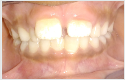
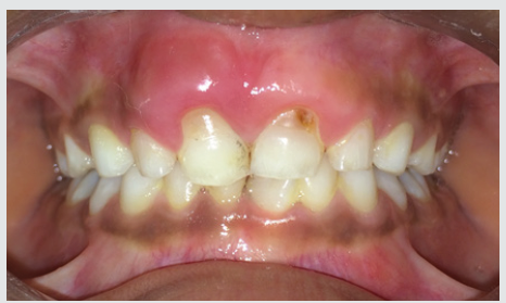
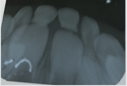
A male patient of 6 years old reported to the dental hospital along with his parents. The chief complaint of the patient was pain in upper anterior teeth. His height was 4 ft. 8 inch and weighed 32 kg. No relevant medical history was recorded. Family medical history was also not significant. It was his first dental visit. No other oral habits were present. On further questioning patient gave history of brushing by self once a day with adult size toothbrush using fluoridated toothpaste. He was well built, and his gait was normal with straight posture and mesomorphic body type. His facial profile was convex with competent lips.eth traumatized. On functional examination patient had nasal breathing and adult type of swallow pattern. He had 24 teeth present in his oral cavity. The parents informed that the child fell from a bicycle and got his teeth traumatized. On intra oral examination of the maxillary arch, the central incisors were found to be tender and mobile (Figure 1). Radiograph was recorded and it showed more than 50% root resorption of primary central incisors in the maxillary arch (Figure 2). Also, the soft tissue around 51 and 61 was swollen due to the injury. Hence, the diagnosis of subluxation was made.

Figure 1:

Lupinepublishers-openaccess-pediatric-dentistry-journal

Figure 2:

Lupinepublishers-openaccess-pediatric-dentistry-journal

Treatment Regimens

The overall principle of treatment is not to take any risk of damage to the permanent successor, which usually implies a very conservative approach. Use of topical anesthetics, local anesthesia, and sedation should be considered. Analgesics may improve the quality of care when pain is anticipated. The administration of a single dose of analgesic 1 h before injection and the use of a topical anesthetic will reduce the discomfort of a local anesthetic [7]. Adequate oral hygiene and a soft diet should be prescribed. Management of an intruded primary incisor depends on the following variables:

a) Direction of intrusion.

b) Degree of intrusion.

c) Presence of alveolar bone fracture.

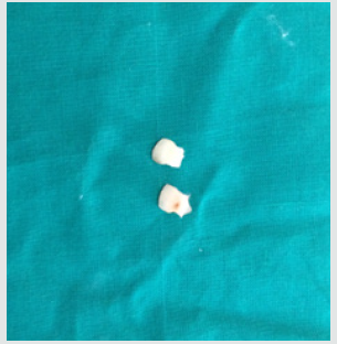
Figure 3:

Lupinepublishers-openaccess-pediatric-dentistry-journal

For the present case we did the extraction of 51 and 61 as already there was resorption of them (Figure 3). Extraction was done using local anesthesia. The soft tissues were cleansed and irrigated with betadine solution. The patient was recalled every 3 months for regular follow up until the permanent incisors were erupted in normal condition (Figure 4).

Figure 4:

Lupinepublishers-openaccess-pediatric-dentistry-journal

Discussion

Injury to the child patient is a traumatic experience on a physical as well as emotional and psychological level. In view of the patient’s tender age and the likelihood that the dental visit will be the patient’s first, managing the child’s traumatic injuries is a demanding task. The close relationship between the apex of the injured primary tooth and the underlying permanent tooth germ must be kept in mind. In the present case, we extracted the tooth and gave post extraction instructions. Antibiotic therapy was prescribed to prevent the spread of inflammation to the permanent tooth germ, as advocated by Kenny and Yacobi , Andreasen and Andreasen [3], Wilson [8], and Andreasen et al. [9]. After extraction, the irregularity of the resorbed root surface was evident, postextraction intra-orally. It was due to external inflammatory root resorption, confirming the radiographic diagnosis [10-15].

Conclusion

Special care should be taken during the evaluation and followup of traumatized immature teeth. The long-term prognosis of immature teeth is dependent on continued root formation. In cases of subluxation of primary teeth, it is essential to diagnose the direction of the displaced tooth to rule out injury to the successor and thus prevent the occurrence of sequelae in the permanent dentition. Regular follow up should be mandatory to observe the successors [15-18].

Read More Lupine Publishers Pediatric Dentistry Journal Articles:
https://lupine-publishers-pediatric-dentistry.blogspot.com/

Saturday, January 16, 2021

Lupine Publishers | Pediatric Oral Systemic Health: From Fetus to Adolescence

 Lupine Publishers | Journal of Pediatric Dentistry


Abstract

The oral systemic health link has now become well recognized and numerous publications have discussed the importance of the oral microbiome in the overall health of any individual. This is likely even more important in children and this influences their future health as they mature. What is particularly interesting is that the microbiome of a child is developed prior to birth and is related to the maternal oral microbiome. Early intervention to the mother prior to the child’s birth, with proper sleep, exercise and dietary adjustments such as the limitation of added sugars and use of coconut oil, for example, will influence the developing child’s microbiome. Direct influence with the polyols xylitol and erythritol to reduce pathogens, and probiotics to boost levels of commensals would also be required. Due to the extent of oral disease, and its tremendous associated costs, urgent action is required by all health professions.

Introduction

Every species depends on adaptation to survive. Humans have managed to survive and flourish while directly influencing the environment of the entire planet, encompassing all other forms of life. While there may be philosophical disagreements as to the inadvertent harm to our natural surroundings, the human species, homo sapiens, has been the only survivor of the hominins [1]. The biology of this success is intertwined in the coevolution of homo sapiens and the associated holobiome [2,3]. Chronic illnesses and debilitations appear to be increasing, requiring reflection into the evolutionary process, and the perturbations that have recently occurred creating this environment of now-declining health [4]. Current research would point to the “Hygiene hypothesis”, overuse of anti-microbials, dietary shifts and the resultant decrease in human microbiome diversity [5,6]. The old model of looking for an increase in pathogens is flawed. Indeed, the fault lies with the decrease in commensals that not only compete directly with the pathogens, but also modulate the immune response of the host [7]. To improve the health of children, we must first improve the microbiome of the mother. The maternal microbiome sets the stage for the child’s microbiome [8,9].

Pre-natal intervention has been studied with positive results reported by the supplementation of the mother with probiotics or polyols [10,11]. Published studies using xylitol that involve the nursing mother and child have demonstrated the decrease in the maternal transmission of mutans streptococci [12]. Certainly, intervention may be desired even earlier, preferably before pregnancy because it is also reported that antecedent use of antibiotics by the mother will influence the maternal microbiome [13]. The placental microbiome is most closely related to the maternal oral microbiome [14]. The presence of commensal bacteria in the placenta and developing fetus is essential to fetal immunological maturation [15]. The oral health of the expectant mother should then be considered primarily important to the oral systemic health of the fetus and later, the child. In addition, the placental microbiome appears to be developed quite early in the pregnancy, by maternal imprinting [14]. This maternal imprinting involves the transportation of viable commensals via circulating monocytes, properly creating a fetal microbiome to program the developing child [16]. Animal studies have demonstrated the transmission of maternal breast commensals into the amniotic fluid [17]. All this depends upon the mother actually having a healthy microbiome [18]. The maternal microbiome can be influenced in numerous ways including diet, exercise and probiotic supplementation [19-22]. Limiting added dietary sugar and the regular addition of polyols can help decrease the prevalence of pathogens before they are passed on to the child [12,23-25]. In the case of Early Childhood Caries, the reduction of maternal Candida albicans will reduce the biofilm formation by Streptococcus mutans potentially reducing the incidence of dental caries [26,27]. Some Lactobacilli, all probiotics such as Lactobacilli rhamnosus, have been demonstrated to inhibit Candida albicans [28-30]. Other supplements, such as N-acetyl cysteine, also have a reducing effect on Candida albicans levels [31-32]. Coconut oil in the form of Medium Chain Triglycerides (MCT) supplements has also been used to reduce levels of Candida albicans and is reportedly as effective as ketoconazole [33-35]. But it has also been reported that coconut oil has more beneficial components than just MCT, giving pause as to why whole coconut oil isn’t utilized more [36]. Regardless, MCT may increase exercise endurance and encourage weight loss [37]. In addition, another natural product, propolis has also been demonstrated to inhibit Candida albicans and other oral pathogens [38-40]. If the expectant mother increases exposure to coconut oil or N-AC, the inhibitory effects may be beneficial in preventing the onset of ECC or, possibly Candida albicans systemic disease if prematurely born [41].

Vitamin K2 has also been reported as being very beneficial as an anti-caries agent and for activation of proper bone and dentin formation in concert with vitamins A and D [42]. This research isn’t new, but recently furthered explored and reported [43]. Insufficient levels of Vitamin D have been linked to S-ECC in a number of studies [44-47]. With increased publications of the beneficial properties of these supplements, it is surprising that the dental profession has not enthusiastically adopted a more fully energetic policy on their role in preventing ECC, especially considering the lack of important micronutrients in the typical American fast food diet [48]. The method of birth has been greatly researched demonstrating that C-section results in an increase in childhood allergies and asthma [49]. The research implicates the lack of exposure to the bacteria of the birth canal and anus as being causative with the neonate’s microbiome lacking maternal commensals [50]. After birth, either by vaginal delivery or C-section, breast feeding provides the infant with Human Milk Oligosaccharides which are much more than just food for bacteria as originally proposed for the child [51]. The HMOs also are antiadhesive antimicrobials that serve as soluble decoy receptors, preventing pathogens attachment to the infant’s mucosal surfaces and thereby lowering the risk for viral, bacterial and protozoan parasite infections [52,53].

HMO’s also reportedly modulate epithelial and immune cell responses, reducing excessive mucosal leukocyte infiltration and activation, lowering the risk for necrotizing enterocolitis and providing the infant with sialic acid, a potentially essential nutrient for brain development and cognition [54,55]. Formula does not have the same protective properties that breastmilk does and sadly, many pediatric dentists criticize breast feeding as being cariogenic, even though published research links the associated dental caries to additional carbohydrate intake and night feeding [56,57]. The benefits of breast feeding have been well documented, and the need to adjust the preventive dentistry protocol to accommodate breast feeding should be evident [58]. Although the World Health Organization recommends two years, mothers probably should breast feed their infants for a least a year, the time interval reported to be the found in early hominins, Australopithecus africanus [59]. Another benefit from breast feeding, besides developing the microbiome and immune modulation, could be regulation of metals, especially zinc and copper, protecting the neurological development of the infant [60-62].

Streptococcus mutans has long been considered the key pathogen for the development of dental caries, the most prevalent chronic disease of humans [63-65]. Efforts to reduce the levels of Streptococcus mutans in infants and children with xylitol and preventing dental caries have been successful, raising the question as to why this is not standard dental practice. 66-67 However, other bacterial and fungal organisms have now been closely identified with the development of dental caries [68]. Scardovia wiggsiae is a Bacillus bacterium found extensively associated with Severe- Early Childhood Caries [69]. Scardovia wiggsiae and Slackia exigua have been reported to be involved in the early caries development [70]. Candida albicans, a fungal organism, helps with the biofilm production by increasing the extracellular polysaccharide matrix which protects Streptococcus mutans from anti-microbials and commensals such as Streptococcus oralis [71]. Lactobacilli inhibit the colonization of Candida albicans, hence decreasing the polysaccharide matrix, exposing the Streptococcus mutans to the bactericins or hydrogen peroxide of its natural competitors, other Streptococcus species [72]. Streptococcus oralis produces hydrogen peroxide that inhibits the anaerobic Streptococcus mutans growth [73,74]. Indeed, Probiora probiotic, a commercially available probiotic product, contains Streptococcus oralis, uberis and rattus, and claims to inhibit several key dental pathogens [75- 77]. Probiotics have been reported to be an important adjunct in preventive dental care [78-80].

Erythritol and xylitol are polyols that have been extensively researched and demonstrated to have notable anti-cariogenic and anti-periodontal disease properties [81,82]. Polyols (particularly the non-hexitol alditols or sugar alcohols erythritol and xylitol) have been found effective in inhibiting the transition to and maturation of biofilms from planktonic cells [83]. Xylitol clearly inhibited the formation of mixed species biofilms, which included Porphyromonas gingivalis in vitro [84]. Erythritol suppressed the maturation of gingivitis biofilms and contributed to a healthier oral ecosystem [85]. Porphyromonas gingivalis takes advantage of early colonizers (Streptococci and Candida) to provide attachment and protection within the biofilm matrix. Polyols can reduce extracellular polysaccharide production and interfere with biofilm matrix elaboration, thereby reducing adherence and biofilm development [86-88]. Streptococci and Candida utilize common dietary sugars sucrose and D-glucose for preferred energy sources, as well as for polysaccharide production. Higher glucose concentrations stimulate Candida growth. Compared with common D-sugars, xylitol induced the lowest adhesion and biofilm formation on either Streptococcus mutans or Candida albicans [89]. In addition, xylitol has been demonstrated to decrease the levels of cariogenic bacteria while having little effect on beneficial bacteria [90]. The discovery of bacteriophages specific for certain strains of Streptococcus mutans also show great promise in the management of pediatric oral health [91]. With the costs of dental disease rapidly escalating, now (2010) estimated at 442 billion US dollars, all effective measures to prevent oral disease should be urgently started in the pediatric population [92].

The Airway evaluation of the infant/toddler is of paramount importance during the first Age One examination [31]. Airway issues in children have been linked to future obesity, diabetes and behavioral issues [94,95]. Mouth breathing increases the oral microbiome pathogenic potential, as the incoming air will reduce the protective nature of the saliva [96]. Studies have demonstrated the correlation between oral disease and airway pathology [97,98]. Sleep Disturbed Breathing in children has been extensively reviewed in the literature, describing an ever-increasing pathologic chain of events [99,100]. Amongst the deleterious effects of mouth breathing are lower and mid-facial adaptations, orthodontic malocclusions, potential speech issues, esthetic concerns, sleep disturbed bruxism, and future temporomandibular joint dysfunctions [101-104]. The key to the future health of children is effective preventive care. What becomes a serious morbidity in adulthood started in childhood. Now more than ever, pediatric health care providers need to emphasize the connection between the oral health of children and their systemic health, with all the future ramifications now clearly reported in the scientific literature. The importance of the oral microbiome, its role as a “gateway” microbiome, and the systemic connection need to be more fully explained to patients, parents and all health care professionals.

Interestingly, the oral health care of the child starts before birth, requiring the participation of all involved in pre-natal care. It is now obvious that what is most important may be the microbiome, and how it is affected by the environment, diet, sleep, exercise, antibiotics, polyols and probiotics. The microbiome then modulates the immune system, allergies, resistance to pathogens, autoimmune responses, and ultimately patient health and longevity. At last, there seems to be great interest in the importance of pediatric and general oral health due to the crisis that poor oral health is bringing upon us [105]. We should be concerned that research studies from several countries have all reported neurotoxicity effects from relatively low levels of fluoride in children [106- 111]. Our over reliance on fluoride to create fluorapatite to inhibit decay seems inadequate at best. Perhaps this means that the time has come to treat a bacterial disease, as a bacterial disease. After all, dental caries and periodontal disease, and to a great extent downstream comorbidity including atherosclerosis, diabetes, strokes, inflammatory Alzheimer’s, diabetes, and many systemic illnesses, can be traced back to a “dysbiosis” started in infancy.

Read More Lupine Publishers Pediatric Dentistry Journal Articles:
https://lupine-publishers-pediatric-dentistry.blogspot.com/
Friday, January 8, 2021

Lupine Publishers | Pediatric Dentistry Condition- A Mini Review

 Lupine Publishers | Journal of Pediatric Dentistry


Abstract

Tooth decay is the most common chronic childhood disease and the World Health Organization has identified it as a worldwide problem with 60-90% prevalence among school-age children. Dental caries is a topical contagious infectious disease that affects people of all ages and in any area of the world. Oral hygiene is a part of public health and oral and dental diseases affect different aspects of quality of life. DMFT is one of the indicators that the World Health Organization has introduced to determine the severity and prevalence of caries. One of the goals of the World Health Organization is to keep the DMFT index of students at less than 2. Different factors are effective in occurrence of dental and interdental caries that we discuss about them in this mini review.

Keywords: Decay Missing Filled Index, Dental Caries, Oral Hygiene, Tooth Decay

Abbreviations: WHO: World Health Organization; DMFT: decay-missing-filled index; BSS: Basic Screening Survey

Introduction

Tooth decay is the most common chronic childhood disease and the World Health Organization [WHO] has identified it as a worldwide problem with 60-90% prevalence among school-age children [1]. According to statistics in European countries, 6.1% of children aged 6-12 have at least one decayed or missed tooth, and due to the prevalence of tooth decay in all social classes, this disease can impose heavy costs on society [2]. Also, according to the statistics in Iran, decay-missing-filled index [DMFT] was 0.2% among 6 to 9-year-old children and 0.9% to 1.5% among 12-yearold children. Also, DMFT was 1.7% in 3 to 6-year old children and 3.3 to 4.8% in 9-year old children [3]. Four important factors: host, germs of the oral environment, food and time, has a role in tooth decay, without each of which, tooth decay will not occur [4]. Therefore, oral hygiene is very important in preventing it. Prevalence of dental caries in 6 to 12-year old children is one of the most important health problems. This can directly and indirectly impair the health of children and teenagers, and this problem is common among low-income groups and groups that do not comply with oral hygiene standards, such as not using toothbrushes and floss, dental caries is much more severe and acute [5].

Dental Caries

Dental caries is a topical contagious infectious disease that affects people of all ages and in any area of the world. Oral hygiene is a part of public health and oral and dental diseases affect different aspects of quality of life [6,7]. DMFT is one of the indicators that the World Health Organization has introduced to determine the severity and prevalence of caries. Nowadays, general dental health programs are usually only concerned with determining the prevalence of dental caries. Therefore, to measure the prevalence of dental caries and to determine oral health status in the society, especially in teenagers, appropriate indicators have been introduced by reputable authorities such as the Association of State and Territorial Dental Directors [2011]. This indicator is called Basic Screening Survey. The main purpose of using this indicator is to provide a framework for obtaining cheap and easy oral health information. On the basis of this indicator, people are classified into two parts, and it is ultimately determined whether or not they have caries [8,9].

Dental caries in Iranian students

One of the goals of the World Health Organization is to keep the DMFT index of students at less than 2. There are many studies on the calculation of DMFT in Iran, including Hamisi et al. which studied 323 students in Qazvin and showed that the prevalence of non-caries was 14.3% and their DMFT was 12.1%. [10]. Also, in a study on 12-year-old children in Tehran and Isfahan, DMFT was 11.2, which was the highest rate of caries [11]. In another study, the rate of DMFT in children in Isfahan was reported 3.41, and the conclusion of this study was that it showed a high proportion of DMFT in decay [12]. A study in Sirjan [a city in Kerman province] showed that the prevalence of non-caries state in 12-year-old students was 34.1%, which means that about 60% of 12-yearold students in this city due to various reasons, particularly not using toothbrush and floss, had dental and interdental decay [13]. Also, in the U.S Department of Health and Human Services in New Hampshire, a large evaluation of oral health status among public school students was done using the BSS index and it showed that approximately 2.3% of students had dental caries [14].

Are There Any Differences Between Girls or Boys in Caries Status?

The results of some studies show that there is no significant relationship between the prevalence of caries in male and female students. Among these studies, we can mention Nabipour et al., Who did not report a significant difference in caries status between male and female students [15]. However, in some studies, such as the study by Boroumand et al., Caries in 3-6-year-old boys was less than that of girls [16]. However, in some studies, such as the study by Boroumand et al., Caries in 3-6-year-old boys was less than that of girls [16]. Also, Loyola-Pontigo et al. and Rosado-Casanova et al. reported a higher incidence of caries in girls than boys [17,18].

Some Other Effective Factors of Children’s Caries Status

Another factor affecting dental and interdental caries is the level of parents’ education. In a study by Campus et.al, there was a significant relationship between parents’ education and lower incidence of caries in children [19]. Ismail and Sohn also stated in their study that children whose parents had a college education had significantly lower dental caries than children whose parents had lower educational level [20]. Also, the differences in socioeconomic status of families can lead to a different status of caries in children, as Primosch in a study in this regard observed a changing status of caries in families with different structures and concluded that this difference may be the result of the different socio-economic status of families, which may affect children’s dietary habits as well as hygiene [21].

Acknowledgement

The authors thank Farzanegan Hazrat Zeinab High School of Rey city and student research center of Basirat especially Dr. Salmani for their supports.

Read More Lupine Publishers Pediatric Dentistry Journal Articles:
https://lupine-publishers-pediatric-dentistry.blogspot.com/
Lupine Publishers: Lupine Publishers | Mouth Guards: Guardians of the...

Lupine Publishers: Lupine Publishers | Mouth Guards: Guardians of the...:    Lupine Publishers | Journal of Pediatric Dentistry Abstract Children and teens are more prone to injuries while playing, with most of the...

Wednesday, January 6, 2021

Monday, January 4, 2021

Lupine Publishers: Lupine Publishers | Abundance, Generation Determin...

Lupine Publishers: Lupine Publishers | Abundance, Generation Determin...:   Lupine Publishers | Current Investigations in Agriculture and Current Research Abstract The present work was...

Lupine Publishers: Happy New Year!!!!

Lupine Publishers: Happy New Year!!!!:   Wishing you a Happy New Year with the hope that you will have many blessings in the year to come. This upcoming year will give us the opp...

Friday, January 1, 2021

Lupine Publishers | Mouth Guards: Guardians of the Dontium

 Lupine Publishers | Journal of Pediatric Dentistry


Abstract

Children and teens are more prone to injuries while playing, with most of the impact taken by the face especially the jaws and the teeth. The most significant factor in preventing sports-related or recreational orofacial injuries is wearing basic protective devices such as properly fitting helmets, face masks, or mouth guards. A mouth guard, custom-fitted by your dentist and worn every time you play or train, will protect against dental injury. Mouth guards are available in 3 different variants which are stock, self-adapting, custom-made mouth guards. The benefits far exceed the expenditure when considering the fees and discomfort associated with a traumatic dental injury

Keywords: Mouth Guard; Orofacial Structures; Teeth; Custom; Sports; Injuries

Introduction

Physical activity forms a very important part of life. There have been constant reports of sedentary lifestyle being associated with disease processes and over the time a lot of importance has been emphasized on physical activities be it the gym or the sports. Children and teens are more prone to injuries while playing, with most of the impact taken by the face especially the jaws and the teeth. Hence, it becomes very important to wear protective gear as these injuries might not only cause temporary impairment but also cause hindrance to the growth and development of the facial structures and also may lead to an unaesthetic appearance of the child at the later stage. The injuries occurring in sports can range from a simple ball-hit to a serious impairment of the head, face or the mouth. It is very important to wear protective gear when you are actively participating in any recreational outdoor events and also in some instances in the indoor activities with the risk of injuries. The most significant factor in preventing sports-related or recreational orofacial injuries is wearing basic protective devices such as properly fitting helmets, face masks, or mouth guards. The use of the mouth guard forms the basic minimal requirement for protecting your mouth, which should form an essential piece of the athletic equipment that the athlete should use as the standard gear equipment from a very early age.

A mouth guard, custom-fitted by your dentist and worn every time you play or train, will protect against dental injury. Well-fitted mouth guards prevent violent contact between the maxillary and mandibular dentition, which can result in soft tissue lacerations, tooth avulsions, tooth or bone fractures, endodontic injuries, and concussions [1]. This being said it has been found that injury to teeth are 60 times more likely when the athlete is not wearing the mouth guard than when he/she is wearing it. Statistically, sporting activities contribute to nearly one-third of all dental injuries [2-4]. Mouth guards help buffer an impact or blow that otherwise could cause broken teeth, jaw injuries or cuts to the lip, tongue or face. Mouth guards also may reduce the rate and severity of concussions Consequently, the dentist plays an important role in informing patients, athletes and their parents, and coaches of the importance of prevention, diagnosis, and treatment of orofacial injuries in sports and recreational activities [1].

A mouth guard should be able to fulfill the following basic requirements:

a) Encompass all maxillary teeth extending up to the distal surfaces of the second molars in class I and class II patients [4-7].

b) Encompass all mandibular teeth extending up to the distal surfaces of the second molar on class III patients [4-7].

c) Mouth guard may be abridged to cover until the distal surfaces of the first molars, in case it is known to trigger the gag reflex of the patient [4].

d) The labial flange should range to within 2mm of the sulcus [5].

e) The palatal flange should range around 2mm above the gingival margin [5].

f) The margins of the labial flange should be rounded.

g) The margins of the palatal edge should be tapered [5].

h) Be easy to clean [8,9].

i) Not impede with breathing or speech activity [4].

j) Be fabricated from a material approved by the U.S. Food and Drug Administration that can reduce the impact force to teeth, surrounding soft tissues, and bone [4,9].

k) Be comfortable and retentive and fit properly [9].

Mouth guards are available in different variants

A. Stock

B. Self-adapting

C. Custom-made

A. Stock

These are readily available over the counter in different sizes, made from polyurethane, a copolymer of vinyl acetate, or ethylene. As they are produced in bulk and are of standard sizes, they remain inexpensive, however they offer a low level of protection with little retention and is not so easily accepted by the athlete. The need to hold the mouth guard in place by clenching his or her teeth together is another disadvantage.

B. Self-Adapting

Also known as the “boil-and-bite” type mouth guard. It is readily available over the counter and made from ethylene-vinyl acetate (EVA). Herein, it is heated in hot water and then placed in the mouth to be adapted to the teeth by biting down. It is relatively inexpensive and can be replaced frequently in athletes with a mixed dentition or by individuals who are experiencing rapid growth. It has the property of re-adaptability. However, it is often bulky and does not retain its shape over time.

C. Custom-Made

The custom-made mouth guard is fabricated in a dental laboratory on a cast taken from an impression made by a dentist. The custom-made mouth guard offers the best fit and the most protection of any of the protective devices [4,5,9]. It is usually made of a thermoplastic material that is heated and adapted to the cast under pressure or with a vacuum form machine. Due to the laborious work involved in the fabrication of this type of appliance, it is expensive but retentive. The American College of Prosthodontists (ACP) recommends the use of custom mouth guards for all contact sports and for any recreational activities that may hypothetically end in orofacial injuries. Accurate maxillary and mandibular alginate impressions in centric occlusion registration recorded by a qualified dentist at approximately 5-mm opening anteriorly shall be used for fabrication of the custom trays as recommended by the ACP.4,5 The standard thickness is 4-mm, however 5- or 6-mm thickness is recommended as it will be able to protect the athlete better in case of extreme sports [4].

Mouth guards can be either single-layered or multi-layered. Currently, the most commonly used materials in the construction of custom mouth guards are EVA copolymer, soft acrylic resin, polyvinyl chloride, polyvinyl acetate-polyethylene (pEVA), and elastomers [1,8,9]. Many different designs of multi-layered materials are available. The most frequently used is a double layer made of similar materials. Dual laminated mouth guards possess an outer hard shell of styrolbutadiene co-polymerisate, and a soft inner layer of ethylene copolymer and vinyl acetate. This design of a more rigid outer material with an inner softer material will reduce the impact force transferred to the teeth due to the shock-absorbing capability of the softer layer [4,5]. The critical areas in terms of energy absorption and transmitted forces are the incisal edges of the anterior teeth and the attached (marginal) gingiva. Therefore, an optimal thickness of the device is achieved by the application of vacuum forming pressure-lamination technique of two layers of a thermoplastic sheet (EVA copolymer) and if necessary, by placing two layers of protective air-cells against the critical areas. An acrylic-resin-based elastomer may be processed over the thermoplastic sheet to improve protection for the athlete [4]. Custom-made mouth guards have proved to be the most effective means of prevention of injuries to the orofacial structures [1,4,5]. They are superior in quality, comfort, retention, and prevention of injuries when compared to stock or self-adapting devices. Although custom-made mouth guards are the most expensive type of protective oral device, they are the most highly recommended.

Conclusion

The benefits far exceed the expenditure when considering the fees and discomfort associated with a traumatic dental injury. Further, it becomes the duty of the dentist to create awareness among the athletes and make it a habit for them to wear mouth guard as a part of their equipment for sports.

Read More Lupine Publishers Pediatric Dentistry Journal Articles:
https://lupine-publishers-pediatric-dentistry.blogspot.com/
980 nm Diode Laser: A Good Choice for the Treatment of Pyogenic Granuloma

Abstract Pyogenic granuloma is a benign non/neo plastic mococutanous lesion . It is a reactional response to constant minor trauma and ca...