Showing posts with label Open Access Publishers. Show all posts

Tuesday, February 11, 2020

Lupine Publishers | Varied clinical and Oral Presentation of Beckwith – Wiedemann Syndrome - Report of a Case from Saudi Arabia

Lupine Publishers | Journal of Pediatric Dentistry Impact Factor


Abstract

Beckwith – Wiedemann syndrome is congenital, genetic and epigenetic pathologies with low prevalence and diverse clinical presentations. It is characterized by triad of omphalocele, macroglossia and gigantism. This syndrome has been widely studied with a current emphasis on improvement of prenatal diagnostic techniques and a multidisciplinary approach towards treatment. We report a case of BWS from Saudi Arabia, with unique presentations and misleading history which delayed diagnosis, due to cultural and religion constraints.
Keywords:Congenital; Epigenetic; Genetic; Prenatal

Introduction

Genetic and epigenetic changes or a human genomic imprinting disorder is characterized by phenotypic variability which might shows its occurrence either as sporadic or inherited. The pathology presents wide range of effect on psychological and social wellbeing of patients and families. One such congenital, multigenic, multisystem human genomic imprinting disorder with complex molecular etiology and variable complex phenotype is Beckwith – Wiedemann Syndrome (BWS). Beckwith-Wiedemann Syndrome is most common overgrowth syndrome described by Beckwith in 1963 and Wiedemann in 1964 with similar findings. It is rare congenital deformity with low prevalence but at same time have high prevalence within genetic abnormalities of overgrowth [1]. The presentation of triad features of omphalocele (exomphalos), macroglossia and gigantism was described earlier as EMG syndrome which now is referred as Beckwith – Widemann Syndrome. The incidence of BWS reported is approximately 1:13700 births and the major cause is thought till date is genetic and epigenetic defects within the chromosome 11p15.5 regions [2].
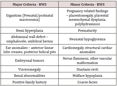
BWS presents wide array of clinical manifestations such as congenital abdominal wall defects as hernia (exomphalos), gigantism, macroglossia, nevus flammeus, ear pits/hearing loss, midface hypoplasia, cardiac anomalies, hemihypertrophy, genitourinary anomalies and musculoskeletal abnormalities. To standardize the diagnostic criteria various attempts have been made to classify the major and minor criteria. Elliot et al described the diagnosis of BWS with the presence of either three major features (abdominal wall defect, macroglossia, gigantism) or two major and three minor features (ear pits, nevus flammeus, hemi hyperplasia, nephromegaly, neonatal hypoglycemia) [3]. In spite of diverse clinical presentations of BWS, most of the cases do not show characteristic features at birth but develop later in life. Also, children with BWS have significantly increased risk of cancer during early childhood which need strict follow up and monitoring. Here, we present a case of BWS with unique dental and medical presentation and its differential diagnosis with literature review.

Case Report

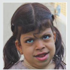
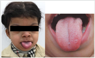
A 5-year-old female patient, accompanied by her mother, presented to the dental unit with complaint of decay tooth in upper front region of mouth. Extra oral examination revealed dysmorphic features, coarse facies and developmental problems (Figure 1). Intra oral examination of hard tissue showed high arched palate, decayed teeth in relation to 51, 52, 55, 61, 62, 74, 75, 84,85. Oral soft tissue examination revealed macroglossia, enlargement of fungiform papillae and mild loss of filiform papillae (Figure 2). Speech and feeding difficulty were noticed due to macroglossia. History revealed she is the youngest 7th child born out of consanguineous marriage in 30th week by cesarian section. She has a chronic history of constipation for 9 months of age. She passes hard stool once in every 8 to 10 days, by spending long time in washroom. It is associated with decrease in appetite and abdominal pain. She was given Movicol (half the adult dose) twice a day for constipation without any medical prescription. She was also tried with lactulose, glycerin suppository and mineral oil. Under medical supervision fleet enema and contrast enema were performed to relieve constipation and to rule out Hirschsprung disease.
Figure 1: Photograph showing dysmorphic features and hypertelorism.
Lupinepublishers-openaccess-pediatric-dentistry-journal
Figure 2: Macroglossia with enlarged fungiform papillae and loss of filiform papillae.
Lupinepublishers-openaccess-pediatric-dentistry-journal
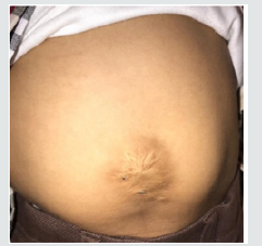
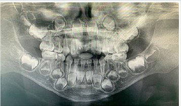
Other medical findings noticed omphalocele, ear pits, large child at 90th centiles, large rounded eyes with hypertelorism, abdominal soft lax, enlargement of kidney, distention of left renal pelvis with significantly distended urinary bladder, abnormal anatomy of the colon located in left abdomen and partial colonic non – rotation with no evidence of obstruction (Figure 3). Based on the clinical and past medical history a diagnosis of Beckwith – Wiedemann Syndrome (BWS) was made. Series of laboratory investigation were reviewed which presented negative urine examination, alpha – fetoprotein, karyotype, microarray and methylation analysis for BMS. Patient was advised for gene analysis and targeting testing for parents. The gene analysis of CDKN1C gene showed heterozygous alteration consistent with BWS but targeting gene tests were refused by parents. Panoramic radiograph was advised considering the patient chief complaint, which revealed multiple developing permanent tooth buds, protrusion of anterior teeth, open bite and increase in mandibular dimension (Figure 4). Under preventive measures the patient was treated for the decayed teeth and is under follow up from past 6 months.
Figure 3: Photograph showing abdominal wall defect with surgical scar.
Lupinepublishers-openaccess-pediatric-dentistry-journal
Figure 4: Panaromic radiograph showing multiple developing permanent tooth buds, open bite and increased mandibular dimension.
Lupinepublishers-openaccess-pediatric-dentistry-journal

Discussion

Diagnostic criteria for BWS is still a matter of research due to its varied clinical presentations and overlapping features with other various conditions. The presence of major and minor findings is generally helpful in establishing the clinical diagnosis (Table 1). The oral findings as mentioned in the literature and observed in our case has been tabulated in Table 2 [4,5]. The incidence of BWS is difficult to assess in Saudi Arabia, as most of the cases goes undiagnosed and unnoticed. Also attributed to its diverse clinical presentation and difficulty in diagnosing. In the present case, features of macroglossia, macrosomia, omphalocele, abdominal wall defect (treated immediately after birth and surgical scar observed clinically), Renal involvement, ear crease, high arched palate, open bite and increased mandibular dimension, leads to the diagnosis of BWS. Various molecular mechanisms and alterations have been involved in BWS such as abnormal methylation of H19DMR, loss of imprinting of IGF2, chromosomal rearrangements, loss of imprinting of LIT1, uniparental disomy of 11p15 and CDKN1C mutations [2]. The full gene analysis of CDKN1C gene profile were suggestive of BWS in our case and the alteration is thought to be located in the allele inherited from the mother. Parental testing was advised which was refused by the parents. There are various endocrine and overgrowth syndromes that was considered in the differential diagnosis. These included Simpson-Golabi-Behmel syndrome (mutation in X-linked gene, GPC3), Perlman syndrome (Increased risk of neonatal mortality), Costello syndrome (missense mutation in HRAS), Sotos syndrome (Mutation in NSD1) and Mucopolysaccharidosis type IV (lysosomal storage disorder) [6]. Oral findings like macroglossia of BWS needs differentiation from other lesions like lymphangioma, idiopathic muscular hypertrophy, hemangioma, rabdomyomas, amyloidosis, cretinism and acromegaly.
Table 1: Presenting major and minor features of BWS.
lupinepublishers-openaccess-pediatric-dentistry-journal
Table 2: Oral findings of BWS.
lupinepublishers-openaccess-pediatric-dentistry-journal
The overall risk of BWS for tumor development/malignancies is estimated to range from 4 – 21%. The tumors reported with BWS are mainly embryonal tumors such as Wilms tumor, hepatoblastoma, rabdomyosarcoma, adrenocortical carcinoma and neuroblastoma [7]. The prenatal diagnosis with current technology is increasing representing an important tool to determine some features of BWS before birth. In our case, parents were highly orthodox and refuse to share the detailed prenatal and ultrasonic reports. Few misguided information’s were given by mother which was later clarified with the reports from the subsequent medical hospitals. Patient’s parents were advised for periodic follow up with genetic counselling and the possibility of surgical interventions in the medical units, but they refused to follow and changed the hospitals every time. Hence, an effort was put forward to retrieve the information’s related to the patient while giving her the primary treatment for which she reported to our dental unit. This suggest the need of awareness required in the country like Saudi Arabia, where most of the cases goes unreported/unnoticed or parent’ consent not given or the cultural and religion barriers that prevent reporting such cases. Though the patient was treated with dental fillings, the follow up of the patients is been restricted by the family members.

Conclusion


Beckwith – Wiedemann Syndrome patients usually grow and do well despite being at increased risk of childhood cancer. Hence, strict follow up, awareness of parents and cancer screening is mandatory. Families, physicians and dentists should determine screening schedule including abdominal ultrasound in every three months, blood test to measure alpha-fetoprotein in every six weeks, dental check-up in every six months and other symptomatic treatment schedule as and when required.

Read More Lupine Publishers Pediatric Dentistry Journal Articles : https://lupine-publishers-pediatric-dentistry.blogspot.com/
Read More Lupine Publishers Articles : https://lupinepublishers.blogspot.com/

Wednesday, October 30, 2019

Lupine Publishers | Isolation and Characterization of Candida Species from Dental Caries in Deciduous Teeth

Lupine Publishers | Journal of Pediatric Dentistry


Abstract

The present study showed the presence of Candida tropicalis as a mayor fungus isolated of dental caries in deciduous teeth.
Keywords: Dental Caries, C Tropicalis

Introduction

Candida species is the most frequent fungus found in the oral cavity [1]. This microorganism provokes a pathology known as candidiasis in many forms [2], however, this yeast can be found in dental decay lesions, gingival and periodontal disease [3]. Candida albicans is the most frequent species of microorganism in all these lesions [4], however, other Candida species as Candida tropicalis, C glabrata, C. Krusei, C. guillermondii are less present in oral cavity [5]. Dental caries, is the most frequent lesions over world and its etiology is eminently microbial, being the Streptococcus mutans who produce the teeth demineralization and destruction [6]. The main of this study is to isolate and characterize the Candida species from dental caries in deciduous teeth.

Materials and Methods

Fifty children, female and male, from pediatric dentistry of Universidad Andina del Cusco, between 4 and 6 years old with dental caries are selected. Before remove and rehabilitate the dental caries, with a dental spoon excavator it was collected a caries sample and stored in 0.9% NaCl [7]. After that, the samples were sonicated and 100 ul aliquot was placed in CHROM Agar Candida medium (CHRO Magar, Paris, France) and were incubated for 48 hours at 37°C [7]. It followed the CHRO Magar Candida manual instructions to determine the presence of Candida species.

Results

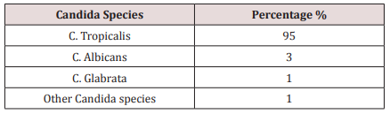
The Candida species most present in the dental caries in deciduous teeth were the C. tropicalis. Other species of Candida are found in less percentages (Table 1).
Table 1.
lupinepublishers-openaccess-journal-pediatric-dentistry

Discussion

Candida species is the most fungus found in oral cavity being the C Albicans the most pathological yeast of the Candida species [4]. This microorganism was found in many oral lesions as candidiasis, dental caries, gingival and periodontal disease [2,3]. Other Candida non albicans are founded in less frequency. However, C. tropicalis and C. glabrata has been described as emerging pathogens in recent years [8]. In the present study, C. tropicalis was presented in the most cases of dental caries in deciduous teeth, being this data corroborated with other studies who the main pathogen is the C. tropicalis. Most studies, in fact, found that the C. albicans as the mayor pathogen isolated from dental caries [6,9,10]. This difference of data can be explained by the geographical location of patients where Candida species can be found in amounts depending on the geographical area. In this study, other Candida species, can be found in less amounts. Despite limitations, the data obtained in the present study demonstrated the high rate of C. Tropicalis in dental caries in deciduous teeth, however, has not been determined which factor is involved in the pathogenesis of dental caries produced by C. tropicalis. It is also important study the oral microbiome in dental caries to dilucidated the role of Candida species, mainly C. tropicalis, in the development of dental caries in deciduous teeth.

Conclusion

Candida tropicalis is the most fungi founded in dental caries lesion in deciduous teeth in child between 4 and 6 years old.

For more Lupine Publishers Open Access Journals Please visit our website:
http://lupinepublishers.us/
For more Open Access Journal on Pediatric Dentistry articles Please Click Here:
https://lupinepublishers.com/pediatric-dentistry-journal/

To Know More About Open Access Publishers Please Click on Lupine Publishers

Follow on Linkedin : https://www.linkedin.com/company/lupinepublishers
Follow on Twitter   :  https://twitter.com/lupine_online

Saturday, October 26, 2019

Lupine Publishers | Color Changes of Pediatric Dental Bridges

Lupine Publishers | Journal of Pediatric Dentistry

Abstract

Dental technology that depended on the standardized lost-wax casting technology has been greatly improved with the introduction of dental CAD ⁄ CAM systems. The aim of the present study was to compare between the color changes of CAD/CAM acrylic and manually performed acrylic bridges used for pediatric patients. Forty study casts of children aged 2 - 4 years old of both genders, with prematurely lost one of the maxillary central incisors and the adjacent lateral incisor was carious and considered to be abutment tooth were involved in this study for construction of cantilever bridges. For each cast, two bridges were constructed; 1st one is CAD/CAM acrylic bridge and the other one is manually performed acrylic bridge. After immersing the bridges in saturated chocolate solution for different time intervals, color changes of the bridges were measured using 3Shape scanner system.
Keywords: Color; Changes; CAD/CAM; Bridges; Pediatric; Patient

Introduction

Trauma and/or dental caries is the common causes those result in premature loss of teeth in children. Cosmetic/aesthetic restoration of such condition considers to be challenging in the pediatric dental field. In case of premature tooth loss in anterior incisal segment there will result in arch space loss and teeth’s inclination that causing a collapse of the anterior teeth and midline shifting [1], as well as may lead to parafunctional habits [2]. Mahmoud (2009) found that anterior tooth loss had effect on patient’s quality of life and gave negative effects on him/his [3]. Al Rawi (2017) found that placement of cantilever acrylic bridges for restoring the aesthetic dental appearance of preschool children resulted in positive successes both to the child and parents [4]. Extrinsic discoloration of teeth and oral prostheses is stains caused by foods or beverages. In pediatric patients such stain mostly occurred due to colored foods such as beets or chocolate as well as berries and candies [5, 6]. This study considered to be the first step of our series studies deal with determining different physical and mechanical properties of the prostheses used for pediatric patients we planned to carry out (in vitro and in vivo studies). Starting with the present study that aimed to compare between the color changes of CAD/CAM and manually performed acrylic bridges used for pediatric patients. After immersing the bridges in chocolate solution for different time intervals, color changes of the bridges were measured using 3Shape scanner system.

Material and Methods

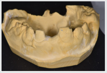
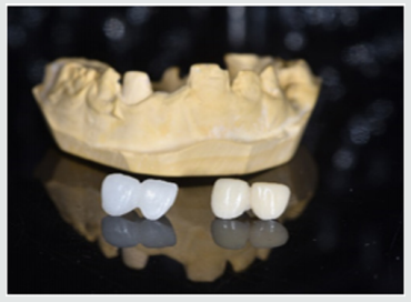
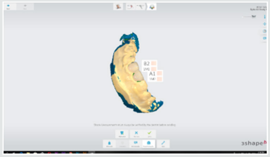
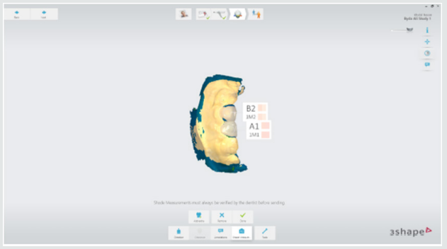
This study starting with collection of forty study casts of children aged 2-4 years old of both gender, with prematurely lost one of the maxillary central incisors and the adjacent lateral incisor was carious and considered to be abutment tooth were involved in this study for construction of cantilever bridges (Figure 1). For each cast, two bridges were constructed; 1st one is CAD/CAM acrylic bridge and the other one is manually performed acrylic bridge (Figure 2). Construction CAD/CAM bridge: The cast was 3D scanned by special scanner (710 3D) (smart optics Sensortechnik GmbH, Germany). The design of the bridge was carried out using Exocad Program (smart optics Sensortechnik GmbH, Germany). Acrylic block (Poly-methyl methacrylate) of classic shade A1 (Ivoclar vivadent, Switzerland) was used for fabrication of the bridge using CAD/ CAM machine (Charly dental, ZI Fonlabour, France). The bridge was finished and polished very well [4]. Construction of manually acrylic bridge: Wax pattern was fabricated on cast then followed the technique of typical wax loss; the heat-cure acrylic (Ivoclar vivadent, AG, FL-9494 Schaan/Liechtenstein) of classic shade A1 was used for bridge fabrication. Finally, surface finishing and polishing was done [7]. Saturated chocolate solution was prepared using 15g chocolate powder (MacChocolate TM, Malaysia) with 100ml distilled water. Baseline color readings for acrylic bridges were taken then immersed in chocolate solution for different time intervals (one week and two weeks) and maintained in incubator of 37 °C, Fresh chocolate solution was prepared every day. Before color measurements after one week and two weeks’ time intervals, the bridges were rinsed with distilled water for 30 seconds, cleaned with a soft bristle toothbrush and then dried with tissue paper [8]. Color measurement was carried out in the facial surfaces at the center third of the abutment and the center third of the pontic part of each bridge as shown in Figure 3. Color measurements of the bridges were measured using 3Shape scanner system (3 Shape A/S, Holmens Kanal 7.1060 Copenhagen K Denmark) and according to the software program of the system, Classic shade (Ivoclar vivadent, Switzerland) was depended.
Figure 1: One of the study casts involved in this study
lupinepublishers-openaccess-journal-pediatric-dentistry
Figure 2: One of the study casts involved in this study
lupinepublishers-openaccess-journal-pediatric-dentistry
Figure 3: Demonstrated the color shade measurement of the abutment and pontic portions of the acrylic bridge.
lupinepublishers-openaccess-journal-pediatric-dentistry

Results

Table 1 demonstrated the color shade of all samples at the baseline and after one-week and two weeks-time intervals. The results of the present study revealed that for all samples, the color measurement demonstrated that in CAD/CAM group even with using A1 shade acrylic block but at the baseline measurement the abutment revealed A0 shade while the pontic revealed A1 shade. Meanwhile, in manual group the abutment measured to be A1 shade and the pontic gave B1 shade. The results demonstrated that for all samples there were no changes in the color shade of CAD/CAM and manually fabricated acrylic bridges after one-week time interval, meanwhile, there were significantly color changes of all abutment and pontic portions of all samples of both bridge types after two weeks-time interval immersed in chocolate solution (Figure 4 & 5).
Figure 4: color shade measurement of CAD/CAM acrylic bridge after two weeks.
lupinepublishers-openaccess-journal-pediatric-dentistry
Figure 5: Color shade measurement of manual acrylic bridge after two weeks.
lupinepublishers-openaccess-journal-pediatric-dentistry
Table 1: Demonstrated the color shade of all samples at the baseline and after one-week and two weeks-time intervals.
lupinepublishers-openaccess-journal-pediatric-dentistry

Discussion

Restorations in the oral cavity are exposed to several factors that make them vulnerable to color changes, such as temperature, humidity, food and beverages. In the oral environment, restorative materials are also subjected to numerous other liquids, to temperature and load stress, and to tooth brushing. The success of restorations depends not only on mechanical and physical properties, but also on the esthetic appearance [9]. The color measurement in this study demonstrated that in CAD/CAM group even with using A1 shade acrylic block but at the baseline measurement the abutment revealed A0 shade while the pontic revealed A1 shade. These occurred because the thickness of the abutment was only about 0.5mm lead to that the color measured of the abutment was lighter than the pontic portion. Meanwhile, in manual group the abutment measured to be A1 shade and the pontic gave B1 shade. These results agreed with other studies those found the thickness of the material significantly affected the color shade of the prostheses [10,11].
The results demonstrated that the color changes demonstrated only after two weeks-time intervals immersed in chocolate solution. Even the color shades recorded in the CAD/CAM group considered to be lighter than in manual fabricated group, the discoloration from chocolate solution was probably due to adsorption of color colorant of chocolate solution at the surface of the prostheses.
The CAD/CAM bridges fabricated from blocks of pre-polymerized acrylic resin those had a hydrophobic surface that repels water [12]. As well as, perfect polishing surfaces of the bridges involved in this study revealed the limited discoloration that occurred agreed with other research [13]. As the duration of immersion increased, the color change values of both types of prostheses were recorded by 3Shape scanner system. Thus, the time is considered to be important factor in the staining of the dental prostheses and these results agreed with others [14,15]. Fabrication of dental prostheses with the help of CAD/ CAM technology is related to the advantages of high-density polymers based on highly cross linked polymethylmethacrylate [16]. Those advantages include; good esthetic, low water solubility and absorption, sufficient strength, low toxicity, easy repair with simple fabrication technique [17]. The using of hot cure acrylic for fabrication of dental prostheses even of some advantages but the main disadvantages include porosity with the presence of residual monomer which is a potential allergen, increased finishing time, brittle and uneven thickness [18]. A limitation of this study is that it was an in vitro study and need to be collected with in vivo study to measure the degree of color changes of the prostheses with presenting the effect of saliva and oral hygiene measures. Further clinical and in vitro studies are necessary to evaluate the susceptibility of CAD/CAM and manually acrylic bridges to discoloration by other beverages and nutrients.

Conclusion

Color considered as the most important factors for aesthetic appearance of dental restorations. In addition to the optimal chemo mechanical properties of acrylic resins, their availability in different color-shades has increased their application in fixed and removable prostheses. Acrylic resins can have acquired discoloration over time because of the process of adsorption and liquid molecules adhere to resin materials which was decreased their effect with using of CAD/CAM technology over conventional methods of acrylic resin prostheses fabrication.

For more Lupine Publishers Open Access Journals Please visit our website:
http://lupinepublishers.us/
For more Open Access Journal on Pediatric Dentistry articles Please Click Here:
https://lupinepublishers.com/pediatric-dentistry-journal/

To Know More About Open Access Publishers Please Click on Lupine Publishers

Follow on Linkedin : https://www.linkedin.com/company/lupinepublishers
Follow on Twitter   :  https://twitter.com/lupine_online

Lupine Publishers | Management of Perinatal and Infant Oral Health

Lupine Publishers | Journal of Pediatric Dentistry

Abstract

Mothers in the perinatal stage and infants should be identified and evaluated for the risk of dental caries. Early childhood caries can lead to detrimental consequences in the primary dentition. This entails that oral health care advice regarding oral hygiene, diet, fluoride and dental management be provided to minimize the risk factors and optimize the protective factors to improve the longterm oral health outcomes for both the mother and her infant.
Keywords: Perinatal Oral Health; Infant Oral Health; Early Childhood Caries

Introduction

The perinatal period is vital for the holistic well-being of pregnant women. It is defined as a time span which commences when the 20th to 28th week of gestation is completed and ends at 1 to 4 weeks subsequent to birth of a child [1]. Early childhood caries (ECC) and severe form of ECC (s-ECC) start as soon as teeth start to erupt, develop on every surface of a primary tooth, have a rapid progression with a long-term detrimental impact on the primary dentition [2]. The long term sequelae of ECC include a greater risk of new carious lesions in both the primary and permanent dentitions, [3,4] high cost of treatment, [5] hospital stay and emergency room visits [6,7] loss of school time, [8] diminished cognitive ability [9] and a poor oral health-related quality of life [10]. Hence the oral health of both the mother and the future child are instrumental in preventing and arresting the disease process to manage early childhood caries during this phase [11].

Epidemiology

This chronic, infectious disease affects the general population however it is 32 times more likely to occur in infants from low socioeconomic status, with high sugar diet and whose mothers have a low education level [12-14] It affects 1-17% children in developed and 70% children in under-developed countries [15]. Epidemiologic evidence shows that the highest prevalence of ECC is reported from Africa and South-East Asia [16]. The prevalence of ECC among Indian children between 8–48 months is 44% [17]. A study from Sri Lanka reports an incidence of 23% ECC among 1-2-year old’s [18]. North American prevalence of ECC ranges from 11-72% and over 28 % children have caries by the time they reach kindergarten [19,20]. Pakistan has a variation in prevalence of ECC ranging 27.9% - 51% [21,22].

Anticipatory Guidance According to Caries Risk

New mothers and infants are seen by the medical health care professionals earlier and more often than dentists. It is therefore important that they understand the dynamic multifactorial etiology and risk factors for ECC prevention counselling in pregnant women/caregivers and encouraging a dental home visit at age 1 [23]. In some instances, pregnant women may defer dental care, experience unwillingness of dentists to provide oral care [24-27] or may be unaware of the implications of poor oral health for their pregnancy [28,29]. Hence early identification of mothers with poor oral health/high caries risk and timely delivery of educational information and prevention for themselves and their unborn child can help reduce the incidence of ECC, prevent the need for dental rehabilitation and improve their oral health [30-32]. Caries-risk assessment for infants allows the determination of relative risk for dental disease to prevent disease by identifying and minimizing risk factors (plaque accumulation, diet, lack of topical/systemic fluoride, high frequency of sugar containing medicines) and optimizing protective factors (oral hygiene practices, fluoride and fissure sealants) when the primary dentition starts to erupt [33]. The current trend shows more emphasis on prevention and arrest of the disease processes to manage ECC. This is attributed to the costly and high-risk restorative treatment for ECC since it often entails the use of sedation and/or general anesthesia and a high recurrence rate [34,35]. The chronic disease management approach encompasses engagement of parents to facilitate preventive measures and temporary restorations of the lesion to defer advanced restorative care [36]. An active surveillance methodology entails monitoring caries progression in children and setting up prevention programs for managing incipient carious lesions [37]. An Interim therapeutic restorations (ITR) is a form of temporary tooth restoration in young children until compliance improves and conventional cavity preparation and restoration is possible [38].

Oral Health Care Advice to Pregnant or Lactating Mothers

Physicians, dentists, and nurses impart educational advice for mothers during the perinatal period. The preventive advice should include timely brushing with fluoridated toothpastes and use of sugar free gums. The dietary advice should address the quality and quantity of nutritional food along with food cravings that may raise the caries risk. Dental procedures which are considered safe during all trimesters of pregnancy include oral assessment, prophylaxis, local anaesthetic, regular treatment and radiographs with shielding (optimal in second trimester). If, however there is discomfort the elective treatment may be deferred. Breast feeding of infants should be tailored with food over a year or longer but should not be ad libitum. It provides nutritional, developmental and psychological health advantages with a significant decrease in the risk for acute and chronic diseases. It may also transfer maternal medication to infants under 6 months hence use cautiously. It provides awareness of health consequences of tobacco use and exposure to secondhand smoke in children [39-43].

Oral Health Care Advice for Infants

An infant should be taken for an initial evaluation to a dental home by the age of one by the pediatricians and the general practitioners. This attains the medical and dental history of both the child and parents, allows oral assessment with a demonstration on age appropriate gum and tooth cleaning, brushing the teeth twice a day with an optimum level of fluoridated toothpaste (smear or rice sized for children under 3), dietary advice (avoid sugar by bottle, sippy cup, sugar between meals, 4-6 ounces of 100% fruit juice per day for 4-6 year old children, systemically administered fluoride (if the drinking water is unfluoridated) and professional fluoride application if caries risk is high, injury prevention advice for facial trauma (objects, cords, pacifiers, car seats, electric cords), advice on teething with excessive salivation areas of intermittent discomfort (oral analgesics, chilled teething rings, over the counter teething gels), management of atypical frenum attachments (frenectomy or frenuloplasty to facilitate breast feeding) and counselling regarding non-nutritive habits such as digit or pacifier sucking, abnormal tongue thrust or bruxism (wean before skeletal dysplasia or malocclusion) [33,44,45].

Conclusion

It is very important to design and implement caries assessment in order to identify the caries risk for infants and expectant mothers/lactating mothers. This will allow effective education on oral health via motivational interviewing techniques to help improve oral behaviour and timely implementation of caries preventive measures to help change the trajectory of oral health of a mother and her infant.

For more Lupine Publishers Open Access Journals Please visit our website:
http://lupinepublishers.us/
For more Open Access Journal on Pediatric Dentistry articles Please Click Here:
https://lupinepublishers.com/pediatric-dentistry-journal/
To Know More About Open Access Publishers Please Click on Lupine Publishers

Saturday, September 28, 2019

Lupine Publishers | Early Interventions to Promote Pediatric Oral Health

Lupine Publishers | Journal of Pediatric Dentistry

Introduction

It is well established that dental caries is the most prevalent and preventable chronic disease among children [1]. Preschool years are a critical period of development; poor oral health can create lifelong consequences for one’s overall health outcomes. If oral health needs are not addressed earlier, it may negatively impact a child’s ability to eat, sleep, learn or socialize, further damaging the child’s psychological and social dimensions of well-being [1,2]. Another negative outcome is the need for dental surgery as it accounts for 31% of all surgeries among children under the age of 6 [3]. Caries in childhood is a predictor for adult oral health; this may affect other health conditions such as diabetes or cardiovascular disease over time [2]. Caries is a multifactorial chronic disease influenced by biological, lifestyle, and behavior factors [4]. Risk factors for early childhood caries include: bacteria transmission from mother to infant [5]; the social determinants of health [2]; parental knowledge [6], attitudes and behaviors towards oral health (e.g., diet, pacifier use, and daily tooth brushing); prolonged bottlefeeding practices [7], and cultural beliefs around primary or “milk teeth” [8,9]. Oral health is connected to socioeconomic status; those with higher income are more likely to access a dentist and have dental insurance coverage [10]. It is important to identify effective interventions targeting preschool children in order to collaborate with the Family Health Division, other Regional departments, and community partners to meet the emerging oral health needs of our community.

Future Directions

The recommendations made decades ago to promote early childhood oral health by establishing a dental home before the first birthday, and providing education and preventive interventions, are crucial components of effective care [11]. Now, as then, dental professionals play a pivotal role by assessing and monitoring the individualized risk of each pediatric patient and applying the latest evidence-based approaches to disease prevention and treatment [12]. Effective care requires a constant review the literature, ongoing assessment of the rapidly evolving understanding of the oral microbiome and its effect on caries progression and implementing management protocols as early as possible. Beyond the clinic, however, it is equally important for dental teams to provide parents/caregivers with the knowledge and skills to make appropriate dietary and lifestyle choices for their children, while ensuring proper oral hygiene and regular dental visits. It is only through these combined efforts that oral disease can be prevented.

Key Takeaways

a) Establishing a dental home before a child’s first birthday ensures a safe place for comprehensive care and allows clinicians to develop recommendations specific to that patient’s individualized risk.
b) Assessing caries risk and implementing preventive strategies are critical elements of pediatric care, particularly for preschoolers and children with special
c) health care needs.
d) Parents and caregivers must be given the means to mitigate the child’s caries risk through effective self-care and healthy lifestyle choices, and by working with dental teams to verify whether the risk management regimen has been effective.
e) It is only through the combined efforts of dental professionals and parents/caregivers that oral disease can be prevented.

For more Lupine Publishers Open Access Journals Please visit our website:
For more Open Access Journal on Pediatric Dentistry articles Please Click Here:

To Know More About Open Access Publishers Please Click on Lupine Publishers
Follow on Twitter   :  https://twitter.com/lupine_online

980 nm Diode Laser: A Good Choice for the Treatment of Pyogenic Granuloma

Abstract Pyogenic granuloma is a benign non/neo plastic mococutanous lesion . It is a reactional response to constant minor trauma and ca...