Showing posts with label Dental and Oral Health Journals Impact Factor. Show all posts

Saturday, May 15, 2021

Lupine Publishers | An Assessment of Emergency Care Following Tooth Avulsion Among A Selected Population of Nigerian School Children

 Lupine Publishers | Journal of Pediatric Dentistry


Abstract

Aim: To assess the knowledge of school children on the first aid administered after traumatic tooth avulsion.

Methods: A structured questionnaire was used to obtain information from children of six primary and six secondary schools in Port Harcourt, Nigeria. Information elicited included respondents’ socio-demographics, exposure to dental trauma and knowledge on emergency care for tooth avulsion. Data collected was analysed using SPSS Version 22 and level of significance was set at p <0.05.

Results:There were 411pupils; 194 males and 217 females with a mean age of 12.5 (±2.6) years. One hundred and eighty-three (44.5%) had previous dental injuries. Three hundred and twenty-three (78.6%) would seek treatment in dental clinic after tooth avulsion. Only 16 (3.9%) would replace the tooth in its socket, 160 (38.9%) would take it to the dental clinic and 192 (46.7%) will throw the tooth on a roof top. Only 164 (39.9%) would seek immediate treatment. Twenty-two (5.4%) will use milk as storage/ transport media. There were statistically significant differences between
a) The males and females in their time for seeking treatment (p=0.01).
b) The public and private school pupils on where to go for treatment (p=0.013) and transport media use (p=0.00).
c) The primary and secondary school pupils on replantation of avulsed tooth (p=0.03) and transport media use (p=0.00).

Discussion: Although majority knew who a dentist is, their knowledge of emergency care when avulsion occurs is low; they require oral health education to bridge the gap in their knowledge.

Keywords:Tooth avulsion; traumatic dental injuries; emergency care; Nigerian school children; tooth replantation; transport media

Introduction

The school is an educational institution for learning both within [1] and outside [2] the classrooms under the guidance of teachers [3]. However, recreation and sport activities [2-4] in schools when unsupervised may predispose school children to traumatic dental injuries (TDI). Such injuries occur more among males than females and more commonly in urban children than rural children [5-8]. Sixty percent of TDI have been reported to occur during sporting activities among school aged children [9,10]. Of these, tooth avulsion; the complete displacement of the tooth out of the alveolar socket, is the most severe type of TDI and it occurs in both the primary and permanent dentitions [11]. In the permanent dentition, it occurs commonly among children aged between 6-12 years, [5,8,11,12] during the period in the tooth eruption cycle when root formation is incomplete and the periodontal ligament surrounding erupting teeth are loosely structured and provide very minimal resistance to an extrusive force. Tooth avulsion has an incidence of between 0.5 to 16% [8-14] among children and an incidence of 20.8% was reported among Nigerian adolescents [15].
Tooth loss, as a result of trauma, in addition to being distressing has both functional and psychosocial consequences in the permanent dentition [9,11,16]. First aid carried out minimizes the negative emotional/social consequences and ensuing cost of treatment that may result from premature tooth loss. Timely intervention at the site of the accident and immediate presentation to a dental clinic for professional care would result in a favourable outcome [7,10,16]. Replantation is the treatment option for avulsed permanent teeth and the treatment outcome is dependent on prompt and appropriate intervention at the site of the accident [3,17]. Other factors include short extra oral time, suitable transport media, immaturity of the root apex and patients’ general health [16,18,19]. The first aid carried out at the time of injury includes replacing the tooth within the alveolar socket and immediate referral to the dental clinic [8,13] for replantation procedure. However, if the tooth cannot be replaced into its original position, a suitable storage and transport medium would help maintain the viability of the periodontal ligament cells while seeking prompt professional attention in the dental clinic. The transport and storage media include Hanks Balanced Salt Solution (HBSS), Eagle’s medium, Normal saline, Via span, propolis, milk and coconut water [13,18,19]. The most recommended medium based on maintenance of PDL cell viability, availability, low cost and long shelf life is milk as reported in the reviews by Adnan et al. [18] and Udoye et al. [19]. Parents, teachers and students, especially, are almost always present where these injuries occur and need to be knowledgeable on what to do immediately it occurs. The knowledge of parents, [17,20] school teachers [3,9,10,20-23] and students [24,25] has been assessed in several studies both within and outside Nigeria. Most of these showed a poor awareness and low knowledge of first aid measures following tooth avulsion. Though the knowledge of students has been conducted in a study in Nigeria, it was done over a decade ago in another geopolitical region [24]. The aim of this study was to assess the level of knowledge of first aid administered following tooth avulsion among school children in the South-South geopolitical region in Nigeria.

Materials and Methods

Ethical clearance was obtained from the Research and Ethics Committee of the University of Port Harcourt Teaching Hospital. Consent was sought and obtained from the State Universal Basic Education Board and the proprietors of the private schools. Consent and assent were sought from the head teachers and pupils after being assured of confidentiality, respectively. Children aged 8 to 15 years were selected from six (three public and three private) primary and six (three public and three private) secondary schools using a multistage sampling technique. A structured questionnaire [24] was used to obtain information from each of the children. The information elicited were the socio-demographics such as age as at the last birthday and sex, other information were experience of exposure to dental trauma, response to past traumatic injuries, knowledge of the emergency treatment with particular focus on tooth avulsion and knowledge on the process of replantation. The information collected was entered into data spreadsheet and analyzed using the IBM Statistical Package for Social Sciences (SPSS) software, Version 22.0 (SPSS Inc., Chicago, IL, USA). Descriptive summary statistics was obtained for demographic variables and knowledge of emergency care. Pearson’s chi square was used to assess the differences in knowledge between groups and statistical significance was set at p<0.05.

Results

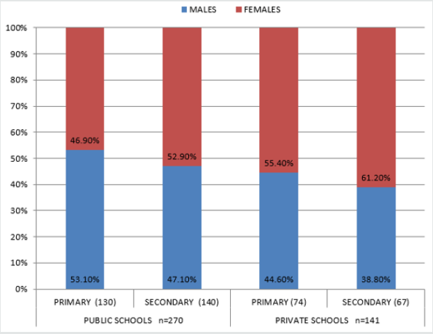
Figure 1: The distribution of the school pupils according to the type of schools, level of education and gender.
Lupinepublishers-openaccess-pediatric-dentistry-journal

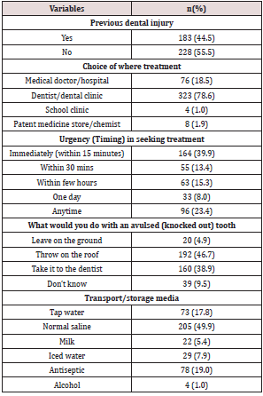
Four hundred and eleven students comprising 194 males and 217 females with a mean age of 12.5 (+2.6) participated in this study. Two hundred and four (49.6%) primary and 207 (50.4%) secondary school pupils participated in the study; 270 (65.7%) attended public schools while 141 (34.3%) attended private schools. Details in Figure 1. One hundred and eighty-three (44.5%) had history of previous dental injuries. Their responses to the questions on their knowledge of avulsion revealed that 323 (78.6%) would go to a dentist if they had tooth avulsion, 76 (18.5%) would see a medical doctor and 4 (1%) would go to the school clinic. Only 16 (3.9%) would replace the tooth in its socket. Although 164 (39.9%) would seek immediate treatment, 160 (38.9%) would take the tooth to the dentist. Two hundred and five (49.9%) would use normal saline as transport medium and only 22 (5.4%) would use milk. When avulsion occurs, 192 (46.7%) would throw it on roof top, while 160 (38.9%) would take the tooth to the dental clinic. Details in Table 1.

Table 1: The knowledge of the school pupils on traumatic dental emergencies (tooth avulsion).
Lupinepublishers-openaccess-pediatric-dentistry-journal

Gender of the subjects

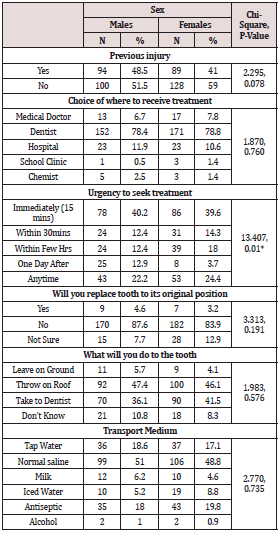
When the gender was considered equal proportion of males and females would seek treatment from a dental clinic (78%) and immediate treatment within 15 minutes (40%). Though 40% of males and females would seek treatment within 15 minutes of the injury, there were statistically significant differences between the males and females (p=0.01) in their timing for seeking treatment (Table 2).

Table 2: The relationship between the Gender and knowledge to the process of tooth replantation.
Lupinepublishers-openaccess-pediatric-dentistry-journal

*p<0.05 is statistically significant

Private and public schools

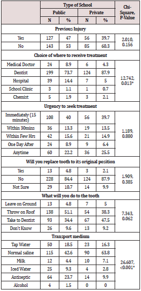
Following avulsion, 4.8%public and 2.1%private school pupils would attempt replacing the tooth within the tooth socket at the site of injury. Three hundred and twenty-three (78.6%) would go to a dental clinic for treatment, however only 164 (39.9%) would seek dental treatment immediately after the injury. More (51.1%) public school pupils compared to 38%private pupils had cultural beliefs on throwing an avulsed tooth on roof tops (p=0.06). Twelve (4.4%) and 10 (7.1%) public and private school pupils, respectively would use milk as a transport medium while 63.8% private as against 42.6% public school pupils preferred normal saline as transport medium. There were statistically significant differences between the public and private school pupils on where to go for treatment (p=0.013) and transport media (p=0.00) following avulsion (Table 3).

Table 3: The relationship between the pupil’s school type and the knowledge of the process of tooth replantation.
Lupinepublishers-openaccess-pediatric-dentistry-journal

*p<0.05 is statistically significant

Level of education

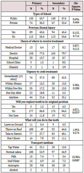
Table 4 shows that 52.9% of primary school pupils and 44.9% secondary school pupils preferred normal saline while 7.4% of primary school pupils preferred milk compared to 3.4% secondary school pupils. There were statistically significant differences between the primary and secondary school pupils on replacing the avulsed tooth back to the sockets (p=0.03), transport media (p=0.00).

Table 4: The association between the type of school, knowledge of emergency dental care and the level of education of the pupils.
Lupinepublishers-openaccess-pediatric-dentistry-journal

*p<0.05 is statistically significant

Discussion

Tooth avulsion is known to commonly occur among children in the mixed dentition phase hence the sample for this study was taken from among the school children. Though school children have teachers in the school premises, sometimes other pupils are the ones present when tooth avulsion occurs. The prognosis of treated avulsed tooth is dependent on prompt treatment which relies greatly on what is done at the site of the accident. Appropriate transport and storage media within the recommended period of storage will help maintain viability of the periodontal ligament cells, thus a favourable treatment outcome [18,19]. In this study 44.5% of the children had experienced dental trauma compared to 6-12.8% reported prevalence of TDI among the school aged children [15]. A good proportion (78.6%) of the participants would choose to receive treatment in a dental clinic. This demonstrates good awareness of whom a dentist is and understanding of the role a dentist plays in the healthcare. Such knowledge may have been fostered by series of outreaches and awareness programmes previously done in most of the schools [22]. However, there was a statistically significant difference in the level of knowledge between the private and public schools (p=0.013), the private school participants seem to know better. This finding is contrary to that reported in South Western Nigeria where the children preferred going to see a medical doctor thereby showing less awareness of the dentists’ role [24].

It was observed that 53.3% would seek professional care within 30 minutes after tooth injury, 23% may not seek care based on their choice of seeking care at “anytime”. This is a concern for a condition that requires urgent care and an off shoot of poor awareness of the importance of prompt treatment following dental trauma. The first aid measures at the site of injury include replacing the tooth in its original position [3,13] or placing in a suitable transport medium [13,18,19]. Only 16 (3.9%) would replace the tooth in its original position in the socket and there was statistically significant difference between the primary and secondary school pupils (p=0.03). This value is less than the 17.8% reported in the south west Nigeria [24]. When the gender and type of schools were considered there were no statistically significant differences between the males and females (p=0.19) and schools (p=0.39). There must be an understanding of tooth anatomy and most importantly there should be a formal teaching on what to do when tooth avulsion takes place. Though the most suitable transport medium listed was milk, [18,19] a good majority preferred normal saline, probably because normal saline appears more medicinal than milk. In this study it was observed that (46.7%) school children had a strong cultural belief on what should be done to a tooth that has been avulsed. This was shown by their response that they will throw the avulsed tooth on roof tops. This belief was commoner among pupils in public schools. The finding buttresses what was reported in a similar study in another geographical location within Nigeria with a different culture [24]. This belief stems from the myth that when a lizard sees an exfoliated tooth, that the succedaneous tooth would not erupt, so to prevent this from happening, the exfoliated teeth were thrown on roof tops away from the lizards [25]. The practice of throwing on roof tops has also been observed in some Asian countries like India, China, Japan, Korea, and Vietnam, although for different reasons [26]. The implication of this, is that the avulsed permanent tooth that would have had a chance of survival from replantation procedure in the dental clinic were being thrown away.

Conclusion

Traumatic dental injuries though a common occurrence amongst these school children (44.5%), their knowledge on first aid measures administered when tooth avulsion occurs before professional intervention is low. Although the children were aware of whom to see when they have dental injuries, the children still hold on to cultural beliefs which could be detrimental to the maintenance of the integrity of the dental arch hence oral health. These gaps in knowledge on first aid following tooth avulsion should be addressed by improving oral health education on traumatic dental injuries with emphasis on emergency care of tooth avulsion in schools. The curriculum on health education in schools should include oral care during dental emergencies.

Conflict of Interest

None of the authors have any conflicts of interest that should be disclosed.

Read More Lupine Publishers Pediatric Dentistry Journal Articles:
https://lupine-publishers-pediatric-dentistry.blogspot.com/

Tuesday, February 11, 2020

Lupine Publishers | Varied clinical and Oral Presentation of Beckwith – Wiedemann Syndrome - Report of a Case from Saudi Arabia

Lupine Publishers | Journal of Pediatric Dentistry Impact Factor


Abstract

Beckwith – Wiedemann syndrome is congenital, genetic and epigenetic pathologies with low prevalence and diverse clinical presentations. It is characterized by triad of omphalocele, macroglossia and gigantism. This syndrome has been widely studied with a current emphasis on improvement of prenatal diagnostic techniques and a multidisciplinary approach towards treatment. We report a case of BWS from Saudi Arabia, with unique presentations and misleading history which delayed diagnosis, due to cultural and religion constraints.
Keywords:Congenital; Epigenetic; Genetic; Prenatal

Introduction

Genetic and epigenetic changes or a human genomic imprinting disorder is characterized by phenotypic variability which might shows its occurrence either as sporadic or inherited. The pathology presents wide range of effect on psychological and social wellbeing of patients and families. One such congenital, multigenic, multisystem human genomic imprinting disorder with complex molecular etiology and variable complex phenotype is Beckwith – Wiedemann Syndrome (BWS). Beckwith-Wiedemann Syndrome is most common overgrowth syndrome described by Beckwith in 1963 and Wiedemann in 1964 with similar findings. It is rare congenital deformity with low prevalence but at same time have high prevalence within genetic abnormalities of overgrowth [1]. The presentation of triad features of omphalocele (exomphalos), macroglossia and gigantism was described earlier as EMG syndrome which now is referred as Beckwith – Widemann Syndrome. The incidence of BWS reported is approximately 1:13700 births and the major cause is thought till date is genetic and epigenetic defects within the chromosome 11p15.5 regions [2].
BWS presents wide array of clinical manifestations such as congenital abdominal wall defects as hernia (exomphalos), gigantism, macroglossia, nevus flammeus, ear pits/hearing loss, midface hypoplasia, cardiac anomalies, hemihypertrophy, genitourinary anomalies and musculoskeletal abnormalities. To standardize the diagnostic criteria various attempts have been made to classify the major and minor criteria. Elliot et al described the diagnosis of BWS with the presence of either three major features (abdominal wall defect, macroglossia, gigantism) or two major and three minor features (ear pits, nevus flammeus, hemi hyperplasia, nephromegaly, neonatal hypoglycemia) [3]. In spite of diverse clinical presentations of BWS, most of the cases do not show characteristic features at birth but develop later in life. Also, children with BWS have significantly increased risk of cancer during early childhood which need strict follow up and monitoring. Here, we present a case of BWS with unique dental and medical presentation and its differential diagnosis with literature review.

Case Report

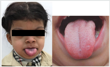
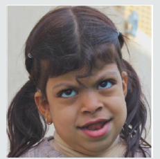
A 5-year-old female patient, accompanied by her mother, presented to the dental unit with complaint of decay tooth in upper front region of mouth. Extra oral examination revealed dysmorphic features, coarse facies and developmental problems (Figure 1). Intra oral examination of hard tissue showed high arched palate, decayed teeth in relation to 51, 52, 55, 61, 62, 74, 75, 84,85. Oral soft tissue examination revealed macroglossia, enlargement of fungiform papillae and mild loss of filiform papillae (Figure 2). Speech and feeding difficulty were noticed due to macroglossia. History revealed she is the youngest 7th child born out of consanguineous marriage in 30th week by cesarian section. She has a chronic history of constipation for 9 months of age. She passes hard stool once in every 8 to 10 days, by spending long time in washroom. It is associated with decrease in appetite and abdominal pain. She was given Movicol (half the adult dose) twice a day for constipation without any medical prescription. She was also tried with lactulose, glycerin suppository and mineral oil. Under medical supervision fleet enema and contrast enema were performed to relieve constipation and to rule out Hirschsprung disease.
Figure 1: Photograph showing dysmorphic features and hypertelorism.
Lupinepublishers-openaccess-pediatric-dentistry-journal
Figure 2: Macroglossia with enlarged fungiform papillae and loss of filiform papillae.
Lupinepublishers-openaccess-pediatric-dentistry-journal
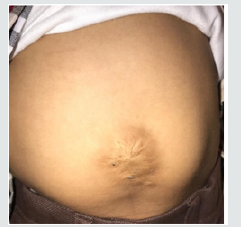
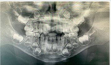
Other medical findings noticed omphalocele, ear pits, large child at 90th centiles, large rounded eyes with hypertelorism, abdominal soft lax, enlargement of kidney, distention of left renal pelvis with significantly distended urinary bladder, abnormal anatomy of the colon located in left abdomen and partial colonic non – rotation with no evidence of obstruction (Figure 3). Based on the clinical and past medical history a diagnosis of Beckwith – Wiedemann Syndrome (BWS) was made. Series of laboratory investigation were reviewed which presented negative urine examination, alpha – fetoprotein, karyotype, microarray and methylation analysis for BMS. Patient was advised for gene analysis and targeting testing for parents. The gene analysis of CDKN1C gene showed heterozygous alteration consistent with BWS but targeting gene tests were refused by parents. Panoramic radiograph was advised considering the patient chief complaint, which revealed multiple developing permanent tooth buds, protrusion of anterior teeth, open bite and increase in mandibular dimension (Figure 4). Under preventive measures the patient was treated for the decayed teeth and is under follow up from past 6 months.
Figure 3: Photograph showing abdominal wall defect with surgical scar.
Lupinepublishers-openaccess-pediatric-dentistry-journal
Figure 4: Panaromic radiograph showing multiple developing permanent tooth buds, open bite and increased mandibular dimension.
Lupinepublishers-openaccess-pediatric-dentistry-journal

Discussion

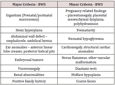
Diagnostic criteria for BWS is still a matter of research due to its varied clinical presentations and overlapping features with other various conditions. The presence of major and minor findings is generally helpful in establishing the clinical diagnosis (Table 1). The oral findings as mentioned in the literature and observed in our case has been tabulated in Table 2 [4,5]. The incidence of BWS is difficult to assess in Saudi Arabia, as most of the cases goes undiagnosed and unnoticed. Also attributed to its diverse clinical presentation and difficulty in diagnosing. In the present case, features of macroglossia, macrosomia, omphalocele, abdominal wall defect (treated immediately after birth and surgical scar observed clinically), Renal involvement, ear crease, high arched palate, open bite and increased mandibular dimension, leads to the diagnosis of BWS. Various molecular mechanisms and alterations have been involved in BWS such as abnormal methylation of H19DMR, loss of imprinting of IGF2, chromosomal rearrangements, loss of imprinting of LIT1, uniparental disomy of 11p15 and CDKN1C mutations [2]. The full gene analysis of CDKN1C gene profile were suggestive of BWS in our case and the alteration is thought to be located in the allele inherited from the mother. Parental testing was advised which was refused by the parents. There are various endocrine and overgrowth syndromes that was considered in the differential diagnosis. These included Simpson-Golabi-Behmel syndrome (mutation in X-linked gene, GPC3), Perlman syndrome (Increased risk of neonatal mortality), Costello syndrome (missense mutation in HRAS), Sotos syndrome (Mutation in NSD1) and Mucopolysaccharidosis type IV (lysosomal storage disorder) [6]. Oral findings like macroglossia of BWS needs differentiation from other lesions like lymphangioma, idiopathic muscular hypertrophy, hemangioma, rabdomyomas, amyloidosis, cretinism and acromegaly.
Table 1: Presenting major and minor features of BWS.
lupinepublishers-openaccess-pediatric-dentistry-journal
Table 2: Oral findings of BWS.
lupinepublishers-openaccess-pediatric-dentistry-journal
The overall risk of BWS for tumor development/malignancies is estimated to range from 4 – 21%. The tumors reported with BWS are mainly embryonal tumors such as Wilms tumor, hepatoblastoma, rabdomyosarcoma, adrenocortical carcinoma and neuroblastoma [7]. The prenatal diagnosis with current technology is increasing representing an important tool to determine some features of BWS before birth. In our case, parents were highly orthodox and refuse to share the detailed prenatal and ultrasonic reports. Few misguided information’s were given by mother which was later clarified with the reports from the subsequent medical hospitals. Patient’s parents were advised for periodic follow up with genetic counselling and the possibility of surgical interventions in the medical units, but they refused to follow and changed the hospitals every time. Hence, an effort was put forward to retrieve the information’s related to the patient while giving her the primary treatment for which she reported to our dental unit. This suggest the need of awareness required in the country like Saudi Arabia, where most of the cases goes unreported/unnoticed or parent’ consent not given or the cultural and religion barriers that prevent reporting such cases. Though the patient was treated with dental fillings, the follow up of the patients is been restricted by the family members.

Conclusion


Beckwith – Wiedemann Syndrome patients usually grow and do well despite being at increased risk of childhood cancer. Hence, strict follow up, awareness of parents and cancer screening is mandatory. Families, physicians and dentists should determine screening schedule including abdominal ultrasound in every three months, blood test to measure alpha-fetoprotein in every six weeks, dental check-up in every six months and other symptomatic treatment schedule as and when required.

Read More Lupine Publishers Pediatric Dentistry Journal Articles : https://lupine-publishers-pediatric-dentistry.blogspot.com/
Read More Lupine Publishers Articles : https://lupinepublishers.blogspot.com/

Friday, December 13, 2019

Lupine Publishers | Mouth Guards: Guardians of the Dontium

Lupine Publishers| Journal of Pediatric Dentistry with High Impact Factor

Abstract

Children and teens are more prone to injuries while playing, with most of the impact taken by the face especially the jaws and the teeth. The most significant factor in preventing sports-related or recreational orofacial injuries is wearing basic protective devices such as properly fitting helmets, face masks, or mouth guards. A mouth guard, custom-fitted by your dentist and worn every time you play or train, will protect against dental injury. Mouth guards are available in 3 different variants which are stock, self-adapting, custom-made mouth guards. The benefits far exceed the expenditure when considering the fees and discomfort associated with a traumatic dental injury
Keywords: Mouth Guard; Orofacial Structures; Teeth; Custom; Sports; Injuries

Introduction

Physical activity forms a very important part of life. There have been constant reports of sedentary lifestyle being associated with disease processes and over the time a lot of importance has been emphasized on physical activities be it the gym or the sports. Children and teens are more prone to injuries while playing, with most of the impact taken by the face especially the jaws and the teeth. Hence, it becomes very important to wear protective gear as these injuries might not only cause temporary impairment but also cause hindrance to the growth and development of the facial structures and also may lead to an unaesthetic appearance of the child at the later stage. The injuries occurring in sports can range from a simple ball-hit to a serious impairment of the head, face or the mouth. It is very important to wear protective gear when you are actively participating in any recreational outdoor events and also in some instances in the indoor activities with the risk of injuries. The most significant factor in preventing sports-related or recreational orofacial injuries is wearing basic protective devices such as properly fitting helmets, face masks, or mouth guards. The use of the mouth guard forms the basic minimal requirement for protecting your mouth, which should form an essential piece of the athletic equipment that the athlete should use as the standard gear equipment from a very early age.
A mouth guard, custom-fitted by your dentist and worn every time you play or train, will protect against dental injury. Well-fitted mouth guards prevent violent contact between the maxillary and mandibular dentition, which can result in soft tissue lacerations, tooth avulsions, tooth or bone fractures, endodontic injuries, and concussions [1]. This being said it has been found that injury to teeth are 60 times more likely when the athlete is not wearing the mouth guard than when he/she is wearing it. Statistically, sporting activities contribute to nearly one-third of all dental injuries [2-4]. Mouth guards help buffer an impact or blow that otherwise could cause broken teeth, jaw injuries or cuts to the lip, tongue or face. Mouth guards also may reduce the rate and severity of concussions Consequently, the dentist plays an important role in informing patients, athletes and their parents, and coaches of the importance of prevention, diagnosis, and treatment of orofacial injuries in sports and recreational activities [1].
A mouth guard should be able to fulfill the following basic requirements:
a) Encompass all maxillary teeth extending up to the distal surfaces of the second molars in class I and class II patients [4-7].
b) Encompass all mandibular teeth extending up to the distal surfaces of the second molar on class III patients [4-7].
c) Mouth guard may be abridged to cover until the distal surfaces of the first molars, in case it is known to trigger the gag reflex of the patient [4].
d) The labial flange should range to within 2mm of the sulcus [5].
e) The palatal flange should range around 2mm above the gingival margin [5].
f) The margins of the labial flange should be rounded.
g) The margins of the palatal edge should be tapered [5].
h) Be easy to clean [8,9].
i) Not impede with breathing or speech activity [4].
j) Be fabricated from a material approved by the U.S. Food and Drug Administration that can reduce the impact force to teeth, surrounding soft tissues, and bone [4,9].
k) Be comfortable and retentive and fit properly [9].
Mouth guards are available in different variants
A. Stock
B. Self-adapting
C. Custom-made
A. Stock
These are readily available over the counter in different sizes, made from polyurethane, a copolymer of vinyl acetate, or ethylene. As they are produced in bulk and are of standard sizes, they remain inexpensive, however they offer a low level of protection with little retention and is not so easily accepted by the athlete. The need to hold the mouth guard in place by clenching his or her teeth together is another disadvantage.
B. Self-Adapting
Also known as the “boil-and-bite” type mouth guard. It is readily available over the counter and made from ethylene-vinyl acetate (EVA). Herein, it is heated in hot water and then placed in the mouth to be adapted to the teeth by biting down. It is relatively inexpensive and can be replaced frequently in athletes with a mixed dentition or by individuals who are experiencing rapid growth. It has the property of re-adaptability. However, it is often bulky and does not retain its shape over time.
C. Custom-Made
The custom-made mouth guard is fabricated in a dental laboratory on a cast taken from an impression made by a dentist. The custom-made mouth guard offers the best fit and the most protection of any of the protective devices [4,5,9]. It is usually made of a thermoplastic material that is heated and adapted to the cast under pressure or with a vacuum form machine. Due to the laborious work involved in the fabrication of this type of appliance, it is expensive but retentive. The American College of Prosthodontists (ACP) recommends the use of custom mouth guards for all contact sports and for any recreational activities that may hypothetically end in orofacial injuries. Accurate maxillary and mandibular alginate impressions in centric occlusion registration recorded by a qualified dentist at approximately 5-mm opening anteriorly shall be used for fabrication of the custom trays as recommended by the ACP.4,5 The standard thickness is 4-mm, however 5- or 6-mm thickness is recommended as it will be able to protect the athlete better in case of extreme sports [4].
Mouth guards can be either single-layered or multi-layered. Currently, the most commonly used materials in the construction of custom mouth guards are EVA copolymer, soft acrylic resin, polyvinyl chloride, polyvinyl acetate-polyethylene (pEVA), and elastomers [1,8,9]. Many different designs of multi-layered materials are available. The most frequently used is a double layer made of similar materials. Dual laminated mouth guards possess an outer hard shell of styrolbutadiene co-polymerisate, and a soft inner layer of ethylene copolymer and vinyl acetate. This design of a more rigid outer material with an inner softer material will reduce the impact force transferred to the teeth due to the shock-absorbing capability of the softer layer [4,5]. The critical areas in terms of energy absorption and transmitted forces are the incisal edges of the anterior teeth and the attached (marginal) gingiva. Therefore, an optimal thickness of the device is achieved by the application of vacuum forming pressure-lamination technique of two layers of a thermoplastic sheet (EVA copolymer) and if necessary, by placing two layers of protective air-cells against the critical areas. An acrylic-resin-based elastomer may be processed over the thermoplastic sheet to improve protection for the athlete [4]. Custom-made mouth guards have proved to be the most effective means of prevention of injuries to the orofacial structures [1,4,5]. They are superior in quality, comfort, retention, and prevention of injuries when compared to stock or self-adapting devices. Although custom-made mouth guards are the most expensive type of protective oral device, they are the most highly recommended.

Conclusion

The benefits far exceed the expenditure when considering the fees and discomfort associated with a traumatic dental injury. Further, it becomes the duty of the dentist to create awareness among the athletes and make it a habit for them to wear mouth guard as a part of their equipment for sports.

For more Lupine Publishers Open Access Journals Please visit our website:
http://lupinepublishers.us/
For more Open Access Journal on Pediatric Dentistry articles Please Click Here: https://lupinepublishers.com/pediatric-dentistry-journal/
To Know More About Open Access Publishers Please Click on Lupine Publishers

Friday, December 6, 2019

Lupine Publishers | Comparison of the Efficacy of Plaque Removal of Listerine Smart Rinse Kids and Vi– One Junior Fluoridated Mouthwash in Children Aged 6 To 10 Years

Lupine Publishers | Journal of Pediatric Dentistry with High Impact Factor


Abstract

Objectives: In this study, a comparative study was done on the effects of Vi-One and Listerine fluoridated mouthwashes on the reduction of dental plaque in pediatric patients between 7-11 years of age in dental clinics of Sepideh and apple in Shiraz, Iran. Listerine Smart Rinse Kids is a product of the United States and Vi-One Junior mouth wash is the domestic production of the country at Rozhin Corporation. This research was conducted by Mohammad Karimi and Hassan Dehghan in 2018-2019.
Material and Methods: In this study, 100 individuals were selected and divided into two groups of 50. During the study, no other method of controlling the plaque was used. In this method, the first group first used Vi-One mouthwash for 10 days and after two weeks of rest and minimizing dental plaque, they used Listerine for 10 days. While the second group used Listerine first, then they applied Vi- One in the same way. The results of this review were then evaluated.
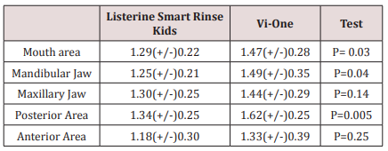
Results: The mean of plaque index in total mouth and in the posterior teeth area with the use of Listerine Smart Rinse Kids was lower than that of Vi-One Junior mouth rinse. In another words, Listerine had a better effect on plaque removal than the Vi-One mouthwash in the posterior mandibular region.
Conclusion: The results show that although Listerine mouthwash had a better effect on dental plaque removal, none of the two mouthwashes had a significant difference in effects on maxillary and mandibular jaws.
Keywords: Dental Plaque; Vi- One, Listerine, Fluoridated Mouthwash; Periodontal Diseases; Plaque Index

Introduction

Currently, dental caries and gingivitis are common oral and dental diseases in this country. One element that can prevent tooth decay is Fluoride. In the oral health program of the country, fluoride mouth wash 2% was used to prevent dental caries in elementary school students all over the country [1]. In the other hand, dental plaque is an important factor in the formation of dental caries and periodontal diseases. Leo and his colleagues have identified dental plaque as the main cause of gingivitis [2]. With the use of mouthwashes, one can control the dental plaque, chemically [3,4]. In fact, mechanical plaque removal is one of the most common and effective methods for preventing caries and inflammation of the gum [5]. Fluoride mouthwash usage is contraindicated in children younger than six years of age due to the risk of swallowing and causing systemic toxicity and fluorosis [6-8]. Symptoms of acute oral fluoride toxicity in children include severe nausea, vomiting, hyper salivation, abdominal pain, and diarrhea [9]. In severe or fatal cases, these symptoms can be followed by convulsions, cardiac arrhythmias, and coma [10- 12]. Laboratory and animal data have shown that prevention the accumulation of plaque and consequently, reduction in dental plaque can be achieved when fluorides is applied topically which inhibits the bacterial multiplication [13]. The fluoride from mouth rinse is retained in dental plaque and saliva to help prevent dental caries [14]. In one review, the average caries reduction in nonfluoridated communities attributable to fluoride mouth rinse was 31% [15]. Another study in Sweden reported that the use of fluoride mouthwash along with brushing has a significant effect in decreasing of dental caries [16]. Listerine Smart Rinse Kids has been used for the purpose of this study. This product is an alcoholfree mouthwash. The ingredients include Sodium fluoride 0.02% (0.01% w/v fluoride ion), Water, Sorbitol, flavor, phosphoric acid, Sucralose, Cetylpyridinium chloride, disodium phosphate, sodium saccharin, menthol, blue 1 and green 3 [17]. One study reported that use of this mouthwash can strengthen teeth 99% better than brushing alone [18]. Another source indicated that it gives 12- hour cavity protection [19]. Vi-one Junior Mouthwash is specially designed for children. This mouth rinse contains Sodium fluoride 0.05%, Cetylpyridinium chloride 0.05% and Disodium phosphate agents. The respective flavors contain sugar-free and harmless sweetener. This brand also is an alcohol-free product [20]. The purpose of this study was to compare the efficacy of two types of mouthwashes, one the domestic mouthwash (Vi-One Junior) and the other, the brand name Listerine Smart Rinse kids fluoridated mouthwash in the removal of the dental plaque.

Material and Methods

This study was a cross-over clinical trial. The eligibilities for entering in our study were as follow:
a) Children having at least 20 teeth with no large restorative area.
b) No history of periodontal Diseases.
c) Not having any Prosthodontic or Orthodontic appliances.
The condition for withdrawal from the study, if there was any sign of reactions to any of these mouth rinses. There was no obligation to have any food regimen.
The study population consisted of 100 patients who were in a 50-member group. Before taking oral mouthwash, plaque index was minimized, and all subjects underwent tooth scaling at the beginning and, if necessary, teeth polishing were done before taking mouthwash. Oral hygiene was assessed via a plaque index. First, in both groups, the Silness-Löe plaque index was recorded. It is an Index for evaluating the thickness of the plaque in the gingival region, which measures the thickness of plaque on all surfaces (M, B, D, and L) [21].
Coding for the plaque index was carried out according to the criteria [22]:
a) Code 0: No plaque
b) Code 1: A film of plaque is adhering to the free gingival margin and adjacent area of the tooth. The plaque may be seen in situ only after application of disclosing solution or by using the probe on the tooth surface.
c) Code 2: Moderate accumulation of soft deposits can be seen with the naked eye within the gingival pocket, the tooth, or gingival margin.
d) Code 3: Abundance of soft matter can be seen within the gingival pocket and/or on the tooth, and gingival margin.
In this index, each tooth is divided into four surface area but in our purposes in the present study, we modified the surfaces area from 4 to 6; thus, we have three surfaces in the buccal area (Mesiobuccal, Midbuccal, and Distobuccal) and three surfaces in the lingual area (Mesiolingual, Midlingual and Distolingual). The first group used Vi-One & Listerine mouthwash (kids mouthwash), for 10 days in the following way. Needless to say, this process was supervised by parents at home. The kids have to gargle 5 cc ’s of Vi-One mouthwash 2 times per day for 30 seconds, and during this period of time, no other plaque control methods and tooth brushing should be used. At the end of the period of 10 days, the plaque index was recorded again. Then, the subjects were given a week to rest and stop using the mouthwash while they had permission to start brushing like before. Again, the plaque index was minimized for patients with polishing the teeth, and they used Listerine mouthwash for 10 days. In the same way, 5 cc ‘s of the mouthwash twice daily was used for 30 seconds, and at the end of the one-week period, the plaque was recorded. For the second group, in the first 10 days, mouthwash. Listerine was prescribed and in the second 10 days, the Vi-One mouthwash was applied. All procedures were performed according to the above pattern.

Results

Paired T-test was used for statistical analysis of the findings. The findings showed when Listerine Smart Rinse Kids was used; the mean of plaque index in all area of the mouth (especially in the mandibular jaw and the posterior region) was significantly less than the time Vi-One was applied. However, there was no significant efficacy difference between the use of both types of mouthwash in the upper jaw and the anterior region.
There was no significant difference between the mean plaque index in Vi-One mouthwash between upper and lower jaw, and there was no significant difference between the maxillary and lower jaw in the case of Listerine Smart Rinse Kids either. The presence of this indicator in both types of mouthwash in the anterior region was significantly less than the posterior region. The mean and standard deviation of the plaque index in both groups, as well as in different regions of the mouth, are listed in Table 1.
Table 1: The amount of dental plaque in terms of area and type of Mouthwash.
lupinepublishers-openaccess-journal-pediatric-dentistry

Discussion

In general, the anti-plaque properties of mouthwashes are completed through bactericidal and bacteriostatic effects, separation of microorganisms from dental surfaces, loosening of joints to these surfaces or lowering of the surface tension of the tooth [2,21,23]. Some mouthwashes can be useful for preventing tooth decay or periodontitis [6,24]. Furthermore, mouthwashes are recommended for children and adolescents with orthodontic appliances or adults who need deep cleansing (such as curettage) [6]. These types of mouthwashes were generally used before and after surgery (especially Chlorhexidine) and have a very positive effect on the treatment of gum and ulcerative inflammation [6]. Use of this type of mouthwash should not last longer than 2 to 3 weeks due to some side effects such as staining the teeth and soft tissue staining, increased calculus deposition, unpleasant taste, burning sensation, and mucosal irritation [6]. It’s time to use this mouthwash after brushing and before bedtime, and it’s best not to eat anything after half an hour after use. Fluoride-containing mouthwashes are another type of mouth rinse that has a fairly large use. These mouthwashes have a significant effect on teeth strengthening. Fluoride in the mouthwash cause bonding with enamel and dentin, and with bonding with calcium and phosphorus, they form Fluorapatite, which is more resistant to caries than Hydroxyapatite. Fluorides also accelerate the mineralization, repair the decayed teeth surfaces, and help to increase the reverse processing of damaged tooth surface area [25,26]. Fluoride also reduces the effect of oral bacteria on teeth. It is done by interfering with the function and formation of the microorganisms. The best fluoride mouthwash protects the teeth against the acids which are produced by dental plaques. “Neglecting the oral hygiene of children leads to the accumulation of plaque and as a consequent the formation of dental calculus which will have a devastating effect on the both child’s gums and teeth” [27].
In one study, the statics showed an alcohol-free mouthwash containing a combination of 0.075% CPC and 0.05% Na F produces statistically significant reductions in dental plaque and gingivitis after three and six months compared to baseline [28]. In another research, Jessica E. Koopman, et al argued that the oral microbial community displayed remarkable resilience towards the disturbances it was presented with. The effects of the fluoride mouthwash on the microbial composition were trivial [29]. On the other side, in another study, the research showed that all four fluoride mouth rinses were effective in decreasing the plaque levels of S. Mutans [30]. In this study, we investigated the effect of two mouthwashes of Listerine Smart Rinse Kids and Vi-One in which Vi-One mouthwash in the posterior region was less efficient than the Listerine mouthwash, and the interesting point that most kids mentioned the taste of Listerine was more acceptable. Given that the contents of sodium fluoride were equal in both mouthwashes, due to the fact that Listerine mouthwash was more acceptable than the mouthwash, it could be related to the other materials present in this product which can be a part of the manufacturer’s secrets. This difference in taste can be a factor in the effect of improving Listerine’s efficacy in the posterior regions.

Conclusion

Listerine Smart Rinse Kids had a better effect on plaque removal than the Vi-One mouthwash in the posterior mandibular region. Both types of mouthwash had a better effect on the anterior region than the posterior region, but none of the two mouth rinses had a different effect on the maxillary and lower jaw. Although many popular types of mouthwash may help to control dental plaque and gingivitis, they should only be used as an adjunct to other oral hygiene measures such as brushing and flossing. Fluoride mouthwashes should be encouraged in children above the age of 6 with a high risk of caries.

For more Lupine Publishers Open Access Journals Please visit our website:
http://lupinepublishers.us/
For more Open Access Journal on Pediatric Dentistry articles Please Click Here:
https://lupinepublishers.com/pediatric-dentistry-journal/

To Know More About Open Access Publishers Please Click on Lupine Publishers


Follow on Linkedin : https://www.linkedin.com/company/lupinepublishers

Follow on Twitter   :  https://twitter.com/lupine_online

Friday, November 29, 2019

Lupine Publishers | Acute Primary Herpetic Gingivostomatitis In A Child: Strategies for Pain Suppression and to Improve Oral Intake

Lupine Publishers | Journal of Pediatric Dentistry

Abstract

This case report describes the management strategies and the evolution of the acute herpetic gingivostomatitis condition in a 3-year-old female child with a focus on suppressing pain and to improve oral intake with approaches to medicine and dentistry.
Keywords: Herpect Stomatitis; Drug Therapy; Child

Introduction

Herpetic gingivostomatitis is a condition that most often results from initial gingiva (gums) and oral mucosa infection with herpes simplex virus type 1 (HSV-1). While herpetic gingivostomatitis is the most common cause of gingivostomatitis in children before the age of 5, it can also occur in adults. The condition is characterized by a prodrome of fever followed by an eruption of painful, ulcerative lesions of the gingiva and mucosa, and often, yellow, perioral, vesicular lesions. HSV-1 is usually spread from direct contact or via droplets of oral secretions or lesions from an asymptomatic or symptomatic individual. Once a patient is infected with the herpes simplex virus, the infection can recur in the form of herpes labialis with intermittent re-activation occurring throughout life [1]. The pathogenesis of herpetic gingivostomatitis involves replication of the herpes simplex virus, cell lysis, and eventual destruction of mucosal tissue. Exposure to HSV-1 at abraded surfaces allows the virus to enter and rapidly replicate in epidermal and dermal cells. This results in the clinical manifestation of perioral blisters, erosions of the lips and mucosa, and eventual hemorrhagic crusting. Sufficient viral inoculation and replication allow the virus to enter sensory and autonomic ganglia, where it travels intraaxonally to the ganglionic nerve bodies. HSV-1 most commonly infects the trigeminal ganglia, where the virus remains latent until reactivation most commonly in the form of herpes labialis [2]. While most children with primary gingivostomatitis will be asymptomatic, some will experience considerable pain and discomfort and are at risk of dehydration. There are no large, well designed studies to clearly determine appropriate therapy for all children [3]. Professionals who treat children in this age group must be able to diagnose and treat common oral manifestations when necessary and should refer the child to a pediatrician for effective treatment if the presence of any systemic alteration is suspected [4]. Herpetic infections commonly affect the dental profession’s anatomical area of responsibility and the diagnosis and management of such infections fall in the purview of oral healthcare providers. To administer competent care to patients with herpetic infections, clinicians must understand the disease, its treatment, the impact the disease or its treatment may have on the patient and the extent to which the presence of a herpetic infection may impact on caregivers in the clinical process [5]. The purpose of this case report was to describe the treatment recommended for a child diagnosed with acute herpetic gingivostomatitis associated with tonsillitis and the ways to suppress pain and to improve oral intake from the perspective of medicine and dentistry.

Case Report

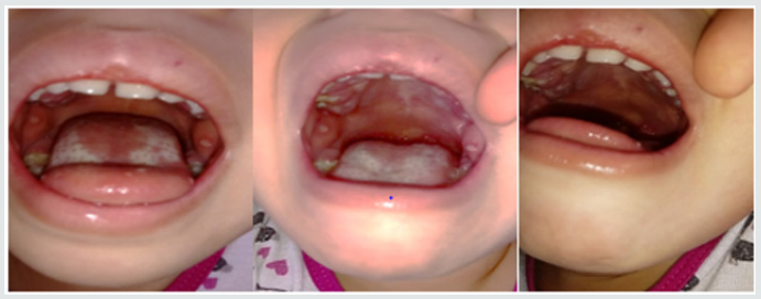
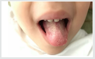
Parents of a 3-year-old and female child sought pediatrician due to inflammation in the throat of their daughter, with fever and irritability for two days, then their child feels pain in the mouth, and the drooling starts with the appearance of diffuse lesions in the oral mucosa, complaining of pain and having difficulty feeding. There was the prescription of antibiotics (amoxicillin and clavulanate potassium for oral suspension), anti-inflammatory and antipyretic. Intraoral cleaning with gauze and saline was recommended and the request for a new consultation, to eliminate the possibility of fungal contamination. The diagnosis of acute and viral primary herpetic gingivostomatitis was established (Figure 1). On intraoral examination, gingiva appeared fiery red in color and multiple vesicles were present on the attached mucosa. Multiple vesicles and ulcers were seen along the lateral border and anterior surface of the tongue. Both sided buccal mucosa revealed multiple vesicles. Her parents also complained about his bad breath during this period due to poor oral hygiene. Submandibular lymphatic glands of the kid were enlarged [6]. The pediatric dentistry was consulted because the child persisted with much pain, unable to sleep or eat (Figure 2). There was then the option of laser applications, with faster healing of ulcers and greater pain relief. There was substantial improvement in food, oral hygiene and sleep. The patient will perform control examinations, with simultaneous evaluation by pediatrician and pediatric dentistry.
Figure 1: Child oral examination two days under antibiotic prescription.
lupinepublishers-openaccess-journal-pediatric-dentistry
Figure 2: Aspect of the child’s tongue on the fourth day of drug treatment.
lupinepublishers-openaccess-journal-pediatric-dentistry

For more Lupine Publishers Open Access Journals Please visit our website:
For more Open Access Journal on Pediatric Dentistry articles Please Click Here:

To Know More About Open Access Publishers Please Click on Lupine Publishers

Friday, November 15, 2019

Lupine Publishers | Do Highly Aggressive Bacteria Cause Dental Caries in Some Children?

Lupine Publishers | Journal of Dental and oral health journals impact factor


Short Communication

Perhaps the question that many pediatric dentists may have is whether a group of bacteria play an important role in the development of dental caries in children. The results of the new research that was carried out at Umea University in Sweden, can answer this question. Researchers at Umea University discovered a new issue in relation to cariogenic different types of Streptococcus Mutans bacteria. They also investigated the adhesion performance of bacteria on children teeth with common caries and increased risk of dental caries. The results of the study, published in the EBio Medicine Journal, could lead to the development of a better way to identify high-risk patients and treat their caries. Dental caries is one of the diseases of the lifestyle, often due to poor oral and dental health and nutritional habits, which results in decreasing the level of acidic pH in the mouth [1-3]. The more likely we will see the development of dental caries if the teeth are exposed to a low salivary pH in a long period of time [4]. Low PH levels have a harmful effect on enamel; increases the growth of acid-producing bacteria, such as Streptococcus Mutans [5-8]. In this five-year study, the saliva of a large number of children was analyzed, and dental health care of these children was supervised. The researchers proved that high-risk children have more invasive types of cariogenic bacteria, and the adherence of these bacteria makes them more aggressive and more susceptible to survival. However, the results of this study showed that in some high-risk children at high risk of rot, there are certain types of highly invasive bacteria, S Mutans, which can cause caries irrespective of lifestyle. These invasive strains have unique sticky proteins called SpaP and Cnm, which increase the ability of the bacteria to survive in the antibacterial saliva of the mouth. One out of five Swedish children has such a dangerous strain and is at high risk for dental caries. These children do not respond to traditional caries prevention or treatment, and their lifestyle variables cannot predict the risk of caries [8]. Chronic dental caries and loose teeth are also risk factors for systemic diseases, such as cardiovascular disease [9,10]. Overall, 70% of tooth loss is due to tooth decay [11,12]. This article explains how up to half of the highrisk children are threatened by highly invasive types of S. Mutans.
These species can also increase the risk of cardiovascular disease and other systemic diseases in the future. Highly aggressive types also differ in terms of adhesion performance. Through biochemical studies, researchers discovered the association between the binding of SpaP and Cnm proteins and their adherence to saliva and DMBT1 protein in saliva [8]. They also showed that higher binding ability has led to an increase in dental caries over a five-year study period. In the end, to conclude this article, Dr. Stromberg believes other high-risk children have a genetic defect in their salivary receptors for bacteria, and the damaged genes may include the same genes that are involved in autoimmune diseases. But it is still important to emphasize that caries is affected by oral and dental health habits in many low to moderate risk people [8]. On the other hand, from new information on the identified types of bacteria, and their manner to start tooth decay, it can be used to improve dental care. Furthermore, the presence of these bacteria can be applied as biological markers for the early detection of high-risk patients. Moreover, their adhesion performance also can be considered as new targets for treatments [8].

For more Lupine Publishers Open Access Journals Please visit our website:
http://lupinepublishers.us/
For more Open Access Journal on Pediatric Dentistry articles Please Click Here:
https://lupinepublishers.com/pediatric-dentistry-journal/

To Know More About Open Access Publishers Please Click on Lupine Publishers


Follow on Linkedin : https://www.linkedin.com/company/lupinepublishers
Follow on Twitter   :  https://twitter.com/lupine_online

Saturday, October 26, 2019

Lupine Publishers | Color Changes of Pediatric Dental Bridges

Lupine Publishers | Journal of Pediatric Dentistry

Abstract

Dental technology that depended on the standardized lost-wax casting technology has been greatly improved with the introduction of dental CAD ⁄ CAM systems. The aim of the present study was to compare between the color changes of CAD/CAM acrylic and manually performed acrylic bridges used for pediatric patients. Forty study casts of children aged 2 - 4 years old of both genders, with prematurely lost one of the maxillary central incisors and the adjacent lateral incisor was carious and considered to be abutment tooth were involved in this study for construction of cantilever bridges. For each cast, two bridges were constructed; 1st one is CAD/CAM acrylic bridge and the other one is manually performed acrylic bridge. After immersing the bridges in saturated chocolate solution for different time intervals, color changes of the bridges were measured using 3Shape scanner system.
Keywords: Color; Changes; CAD/CAM; Bridges; Pediatric; Patient

Introduction

Trauma and/or dental caries is the common causes those result in premature loss of teeth in children. Cosmetic/aesthetic restoration of such condition considers to be challenging in the pediatric dental field. In case of premature tooth loss in anterior incisal segment there will result in arch space loss and teeth’s inclination that causing a collapse of the anterior teeth and midline shifting [1], as well as may lead to parafunctional habits [2]. Mahmoud (2009) found that anterior tooth loss had effect on patient’s quality of life and gave negative effects on him/his [3]. Al Rawi (2017) found that placement of cantilever acrylic bridges for restoring the aesthetic dental appearance of preschool children resulted in positive successes both to the child and parents [4]. Extrinsic discoloration of teeth and oral prostheses is stains caused by foods or beverages. In pediatric patients such stain mostly occurred due to colored foods such as beets or chocolate as well as berries and candies [5, 6]. This study considered to be the first step of our series studies deal with determining different physical and mechanical properties of the prostheses used for pediatric patients we planned to carry out (in vitro and in vivo studies). Starting with the present study that aimed to compare between the color changes of CAD/CAM and manually performed acrylic bridges used for pediatric patients. After immersing the bridges in chocolate solution for different time intervals, color changes of the bridges were measured using 3Shape scanner system.

Material and Methods

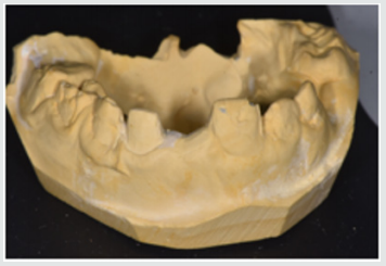
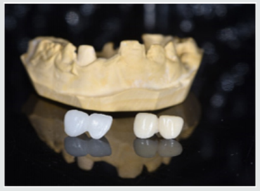
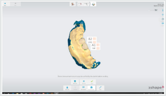
This study starting with collection of forty study casts of children aged 2-4 years old of both gender, with prematurely lost one of the maxillary central incisors and the adjacent lateral incisor was carious and considered to be abutment tooth were involved in this study for construction of cantilever bridges (Figure 1). For each cast, two bridges were constructed; 1st one is CAD/CAM acrylic bridge and the other one is manually performed acrylic bridge (Figure 2). Construction CAD/CAM bridge: The cast was 3D scanned by special scanner (710 3D) (smart optics Sensortechnik GmbH, Germany). The design of the bridge was carried out using Exocad Program (smart optics Sensortechnik GmbH, Germany). Acrylic block (Poly-methyl methacrylate) of classic shade A1 (Ivoclar vivadent, Switzerland) was used for fabrication of the bridge using CAD/ CAM machine (Charly dental, ZI Fonlabour, France). The bridge was finished and polished very well [4]. Construction of manually acrylic bridge: Wax pattern was fabricated on cast then followed the technique of typical wax loss; the heat-cure acrylic (Ivoclar vivadent, AG, FL-9494 Schaan/Liechtenstein) of classic shade A1 was used for bridge fabrication. Finally, surface finishing and polishing was done [7]. Saturated chocolate solution was prepared using 15g chocolate powder (MacChocolate TM, Malaysia) with 100ml distilled water. Baseline color readings for acrylic bridges were taken then immersed in chocolate solution for different time intervals (one week and two weeks) and maintained in incubator of 37 °C, Fresh chocolate solution was prepared every day. Before color measurements after one week and two weeks’ time intervals, the bridges were rinsed with distilled water for 30 seconds, cleaned with a soft bristle toothbrush and then dried with tissue paper [8]. Color measurement was carried out in the facial surfaces at the center third of the abutment and the center third of the pontic part of each bridge as shown in Figure 3. Color measurements of the bridges were measured using 3Shape scanner system (3 Shape A/S, Holmens Kanal 7.1060 Copenhagen K Denmark) and according to the software program of the system, Classic shade (Ivoclar vivadent, Switzerland) was depended.
Figure 1: One of the study casts involved in this study
lupinepublishers-openaccess-journal-pediatric-dentistry
Figure 2: One of the study casts involved in this study
lupinepublishers-openaccess-journal-pediatric-dentistry
Figure 3: Demonstrated the color shade measurement of the abutment and pontic portions of the acrylic bridge.
lupinepublishers-openaccess-journal-pediatric-dentistry

Results

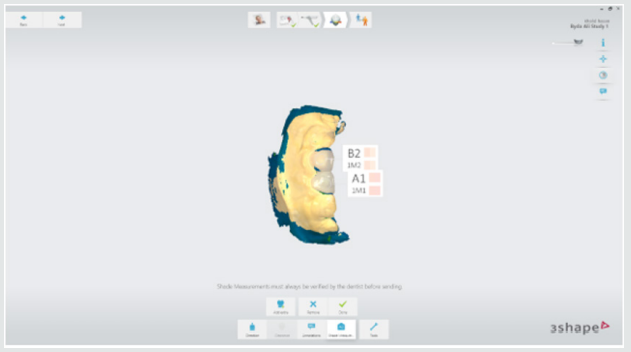
Table 1 demonstrated the color shade of all samples at the baseline and after one-week and two weeks-time intervals. The results of the present study revealed that for all samples, the color measurement demonstrated that in CAD/CAM group even with using A1 shade acrylic block but at the baseline measurement the abutment revealed A0 shade while the pontic revealed A1 shade. Meanwhile, in manual group the abutment measured to be A1 shade and the pontic gave B1 shade. The results demonstrated that for all samples there were no changes in the color shade of CAD/CAM and manually fabricated acrylic bridges after one-week time interval, meanwhile, there were significantly color changes of all abutment and pontic portions of all samples of both bridge types after two weeks-time interval immersed in chocolate solution (Figure 4 & 5).
Figure 4: color shade measurement of CAD/CAM acrylic bridge after two weeks.
lupinepublishers-openaccess-journal-pediatric-dentistry
Figure 5: Color shade measurement of manual acrylic bridge after two weeks.
lupinepublishers-openaccess-journal-pediatric-dentistry
Table 1: Demonstrated the color shade of all samples at the baseline and after one-week and two weeks-time intervals.
lupinepublishers-openaccess-journal-pediatric-dentistry

Discussion

Restorations in the oral cavity are exposed to several factors that make them vulnerable to color changes, such as temperature, humidity, food and beverages. In the oral environment, restorative materials are also subjected to numerous other liquids, to temperature and load stress, and to tooth brushing. The success of restorations depends not only on mechanical and physical properties, but also on the esthetic appearance [9]. The color measurement in this study demonstrated that in CAD/CAM group even with using A1 shade acrylic block but at the baseline measurement the abutment revealed A0 shade while the pontic revealed A1 shade. These occurred because the thickness of the abutment was only about 0.5mm lead to that the color measured of the abutment was lighter than the pontic portion. Meanwhile, in manual group the abutment measured to be A1 shade and the pontic gave B1 shade. These results agreed with other studies those found the thickness of the material significantly affected the color shade of the prostheses [10,11].
The results demonstrated that the color changes demonstrated only after two weeks-time intervals immersed in chocolate solution. Even the color shades recorded in the CAD/CAM group considered to be lighter than in manual fabricated group, the discoloration from chocolate solution was probably due to adsorption of color colorant of chocolate solution at the surface of the prostheses.
The CAD/CAM bridges fabricated from blocks of pre-polymerized acrylic resin those had a hydrophobic surface that repels water [12]. As well as, perfect polishing surfaces of the bridges involved in this study revealed the limited discoloration that occurred agreed with other research [13]. As the duration of immersion increased, the color change values of both types of prostheses were recorded by 3Shape scanner system. Thus, the time is considered to be important factor in the staining of the dental prostheses and these results agreed with others [14,15]. Fabrication of dental prostheses with the help of CAD/ CAM technology is related to the advantages of high-density polymers based on highly cross linked polymethylmethacrylate [16]. Those advantages include; good esthetic, low water solubility and absorption, sufficient strength, low toxicity, easy repair with simple fabrication technique [17]. The using of hot cure acrylic for fabrication of dental prostheses even of some advantages but the main disadvantages include porosity with the presence of residual monomer which is a potential allergen, increased finishing time, brittle and uneven thickness [18]. A limitation of this study is that it was an in vitro study and need to be collected with in vivo study to measure the degree of color changes of the prostheses with presenting the effect of saliva and oral hygiene measures. Further clinical and in vitro studies are necessary to evaluate the susceptibility of CAD/CAM and manually acrylic bridges to discoloration by other beverages and nutrients.

Conclusion

Color considered as the most important factors for aesthetic appearance of dental restorations. In addition to the optimal chemo mechanical properties of acrylic resins, their availability in different color-shades has increased their application in fixed and removable prostheses. Acrylic resins can have acquired discoloration over time because of the process of adsorption and liquid molecules adhere to resin materials which was decreased their effect with using of CAD/CAM technology over conventional methods of acrylic resin prostheses fabrication.

For more Lupine Publishers Open Access Journals Please visit our website:
http://lupinepublishers.us/
For more Open Access Journal on Pediatric Dentistry articles Please Click Here:
https://lupinepublishers.com/pediatric-dentistry-journal/

To Know More About Open Access Publishers Please Click on Lupine Publishers

Follow on Linkedin : https://www.linkedin.com/company/lupinepublishers
Follow on Twitter   :  https://twitter.com/lupine_online

980 nm Diode Laser: A Good Choice for the Treatment of Pyogenic Granuloma

Abstract Pyogenic granuloma is a benign non/neo plastic mococutanous lesion . It is a reactional response to constant minor trauma and ca...