Showing posts with label Journal on Pediatric Dentistry. Show all posts

Saturday, August 26, 2023

Treating Inflammatory Root Resorption Following Endodontic Regeneration – Case Repor


Abstract

Aim: To describe complication management following regenerative endodontics.
Introduction: Regenerative endodontic procedures (REPs) are aimed to treat apical periodontitis and promote root maturation of immature necrotic teeth. Recently it was shown that REP may arrest inflammatory external root resorption (IERR) in replanted avulsed teeth. The purpose of this study is to describe a complication of REP in which IERR had been developed after REP treatment. Root Canal Treatment (RCT) using bio ceramic materials helped to arrest the IERR.
Methods: An eight-and-a-half-year-old girl was referred for treatment after avulsion of tooth #9 which has been splinted in a hospital for 2 weeks. After thorough examination tooth #9 was diagnosed as having necrotic pulp with symptomatic apical periodontitis. The tooth was treated by REP using plasma rich fibrin (PRF) and restored. Eight months later, IERR has begun. RCT using trichloroacetic acid (TCA) and mineral trioxide aggregate (MTA) plug was performed. The resorption area was treated with bio ceramic putty material.
Results: 35 months after the treatment the tooth is functioning without signs and symptoms
Conclusions: The present case demonstrate a method in which a conservative RCT with some modification may arrest IERR using TCA. IERR may develop long time following REP treatment. A thorough long follow-up is needed after REP. An early detection of IERR may overcome this complication.
Key learning points: Although very rare, complication after REP such as IERR may occur, an early diagnosis of such complication may be resolved by modified conservative RCT.

Keywords: Avulsion; Bio ceramics, External inflammatory root resorption; Regenerative endodontics, Trichloroacetic acid

Read More About This Article Please Click on Below Link:
https://lupinepublishers.com/pediatric-dentistry-journal/fulltext/treating-inflammatory-root-resorption-following-endodontic-regeneration-case-report.ID.000261.php

Read More About Lupine Publishers Google Scholar Articles:
https://scholar.google.com/citations?view_op=view_citation&hl=en&user=h1QvhsYAAAAJ&cstart=20&pagesize=80&citation_for_view=h1QvhsYAAAAJ:zA6iFVUQeVQC

Friday, July 7, 2023

Analysis of Marginal Bone Loss and Implant Success Rate After Implant Implantation


Abstract

Purpose: Patients with prosthesis-equipped post-implantation prosthesis performance were continuously observed, revealing the association of marginal bone loss with gender and age implant success and survival rates during a 1year study of clinical prosthesis placement; with results used as a reference for implant procedures and prognosis.

Methods: This study was conducted on 31 patients from January to February 2021 at Y Dental Hospital in Gwangju, with 20 men and 11 women equipped with prosthetics after implant procedure a year earlier. The subjects of the study were carried out with the G-power program, and 31 subjects were extracted. The distance from the shoulder of the implant to the attachment point of the dichotomous bone was measured at the center of the area of atrophy, checking both from the mesial region to the distal region using a radiation ruler taking photos with endocardial radiation as well as panoramic images. The subjects of the implant study were analyzed by age, gender, cause of release, duration of implant, implant length, implant width, implant assembly period, osteoplasty type, shield presence, match with corresponding teeth, implant survival symptom, implant success rate symptoms, and 1mm loss within a year. This study was conducted after approval.

Results: As a result of testing the contribution and statistical significance of individual independent variables to dependent variables, at a significance level of 0.05, the independent variable loses 1 mm at a ratio of (t=5.473, p=.00) in 5.473, p=.000). This loss significantly affects implant success rates.

Conclusions: Recognizing the importance of bone loss management within a year after implant procedure, dental staff and patients can achieve higher success and survival rates after implant procedure if they can be thoroughly managed within a year.

Keywords:Implant formula; marginal bone loss; success rate; survival rate; implant peripheral inflammation; Implant prosthesis

Read More About This Article Please Click on Below Link:
https://lupinepublishers.com/pediatric-dentistry-journal/fulltext/analysis-of-marginal-bone-loss-and-implant-success-rate-after-implant-implantation.ID.000253.php

Read More About Lupine Publishers Google Scholar Articles:
https://scholar.google.com/citations?view_op=view_citation&hl=en&user=h1QvhsYAAAAJ&citation_for_view=h1QvhsYAAAAJ:g5m5HwL7SMYC

Friday, February 3, 2023

Gorlin Goltz Syndrome: Report of A Clinical Case with no Genetic Background


Abstract

First described in 1960 by pathologist Robert Gorlin and dermatologist Robert W Goltz of the University of Minnesota, USA, they provided the basis for the diagnosis of the syndrome, establishing basal cell carcinomas, keratocysts, and skeletal malformations as the primary criteria for the syndrome. Subsequently, authors such as Evans, Kimonis, and Bree established major and minor criteria for the correct diagnosis. According to the International Classification of Applied Diseases in Dentistry and Stomatology (ICD-AO), Gorlin Goltz Syndrome is classified within congenital anomalies and other hamartomatosis. It is caused by a mutation in chromosome 9q22.3 in the tumor suppressor Patched 1. This work presents the clinical case of a 10-year-old female patient with a 13x15cm volume increase in the maseterine region on the left side, diagnosed as Gorlin Goltz Syndrome with no hereditary antecedents.

Keywords:Syndrome; Gorlin-Goltz, Basal Cell carcinoma; Keratocysy; Chromosome

Read More About This Article Please Click on Below Link:
https://lupinepublishers.com/pediatric-dentistry-journal/fulltext/gorlin-goltz-syndrome-report-of-a-clinical-case-with-no-genetic-background.ID.000233.php

Read More About Lupine Publishers Google Scholar Articles:
https://scholar.google.com/citations?view_op=view_citation&hl=en&user=h1QvhsYAAAAJ&citation_for_view=h1QvhsYAAAAJ:hMod-77fHWUC


Friday, January 13, 2023

Palatal Therapy in A Child with Down Syndrome: Case Report


Abstract

Down syndrome (DS) or chromosome 21 trisomy is a genetically provided human condition, it is a more common chromosomal alteration in humans. The presence of the extra chromosome in the genetic makeup determines specific physical characteristics in the oral cavity, such as orofacial muscle hypotonia and lingual protrusion. The aim of this paper is to describe the clinical case of M.S patient, 11 months old, with Down syndrome, admitted to the children’s clinic of UNOESC, accompanied by her mother. In the clinical dental examination, the patient presents with Atresic palate, hypotonia and lingual protrusion, in addition to insufficient lip sealing, with indication for the use of the palatal memory plate (PPM) or Castillo Morales, a device indicated for those associated with functional diagnosis of hypotonia, with lingual protrusion, labial hypotonia, open mouth and craniofacial malformation. PPM promotes neuromuscular balance of the face and neck, and consequently affects the movements of the head and body, has the function of stimulating the tongue and upper lip and the development of nasal breathing. The treatment sequence followed the molding steps, making the model and, from there, the base plate was made in acrylic resin with extension, for adaptation in the child’s pacifier. After making the retentions on the lateral edges to stimulate the musculature of the mucosa and upper lip, the device was polished and adapted to the baby. It is intended that the device cause lingual retraction to the oral cavity, movements of stimulation of the lips and lip sealing. The recommended time for the child to use the device initially is five to ten minutes twice or three times a day, and as the child adapts to the device, the time of use increases.

Keywords: Pediatric dentistry; patients with special needs; jaw orthopedics

Abbreviations: DS: Down Syndrome; PPM: Palatal Memory Plate DS: Down Syndrome; PPM: Palatal Memory Plate

Read More About This Article Please Click on Below Link:
https://lupinepublishers.com/pediatric-dentistry-journal/fulltext/palatal-therapy-in-a-child-with-down-syndrome-case-report.ID.000230.php

Read More About Lupine Publishers Google Scholar Articles:
https://scholar.google.com/citations?view_op=view_citation&hl=en&user=h1QvhsYAAAAJ&citation_for_view=h1QvhsYAAAAJ:M3NEmzRMIkIC

Monday, November 7, 2022

Has the Qovid-19 Epidemic Affected Children’s Oral Health Worldwide?


Editorial

Since the onset of the COVID-19 pandemic, most parents have been afraid to go to dental offices. They don’t feel safe to refer for visits and dental treatments; consequently, after passing one year, we see the prevalence rate of dental decay and increasing dental abscesses, and extraction of teeth which could be preserved by implementing good oral hygiene.

Read More About This Article Please Click on Below Link:
https://lupinepublishers.com/pediatric-dentistry-journal/fulltext/has-the-qovid-19-epidemic-affected-childrens-oral-health-worldwide.ID.000223.php

Read More About Lupine Publishers Google Scholar Articles:
https://scholar.google.com/citations?view_op=view_citation&hl=en&user=h1QvhsYAAAAJ&citation_for_view=h1QvhsYAAAAJ:r0BpntZqJG4C

Monday, November 8, 2021

Lupine Publishers | Mentoring of SDMUPR Students in Pediatric Dentistry and Academics

 Lupine Publishers | Journal of Pediatric Dentistry


Abstract

The American Association for Dental Education (ADEA) is working on several initiatives to foster an interest in academic careers. One of these strategies is a student fellowship aimed at allowing the student at the School of Dental Medicine to explore early experiences in academia. In 2016, the School of Dental Medicine of the University of Puerto Rico (SDMUPR), Medical Sciences Campus, began this fellowship. A pipeline of SDMUPR students with interest in pediatric dentistry and academics has evolved from this initiative.

Keywords:The pipeline of future pediatric dentists; mentoring in pediatric dentistry

Abbreviations: ADEA:American Association for Dental Education; SDMUPR: School of Dental Medicine of the University of Puerto Rico

Introduction

The School of Dental Medicine of the University of Puerto Rico (SDMUPR), Medical Sciences Campus, began in 2016 an ADEA Fellowship Program in Dental Academic Careers (ADEA/ADCFP) to encourage students to discover and experience the options available in the academic work setting[1]. This initiative began a parallel pipeline. Students with interest in academics and pediatric dentistry applied continuously to the ADEA/ADCFP and achieved successful educational projects in pediatric dentistry with their mentor assistance. This article will relate the SDMUPR4 years’ experience with this pipeline project of future pediatric dentists with interest in academics.

Discussion

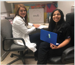
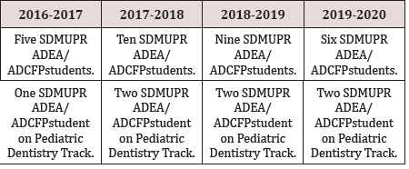
ADEA’s Dental Academic Careers Fellowship Program was founded in 2006 to provide students and residents of the USA and Canadian Dental Schools with the opportunity to venture into academia, providing a structured mentoring to be able to observe and reflect on what an academic career requires[1]. The student is required to complete eight components during the ADEA Fellowship Program in Dental Academic Careers. These components include building an action plan, bi-monthly meetings with a mentor, reflective essays before and after an academic experience, interviews with faculty, academic practice, research practice, presenting a poster, and submitting a portfolio with the evidence of all activities made[1]. In 2016, the SDMUPR began with the ADEA/ADCFP with SDM/UPR students. Since then, every year, at least one student applies to the pediatric dentistry track (Table 1). The mentor is a pediatric dentistry faculty at the SDMUPR, and they work together for eight months on a pediatric educational dentistry track project (Figures 1-3). The number of Applications and First-year Enrollment for Advanced Dental Education Programs continues to rise in the USA[2]. With proper guidance and early experiences, the students can learn and explore their areas of interest in dentistry.

Table 1: SDMUPR ADEA/ADCFP Fellows.

Lupinepublishers-openaccess-pediatric-dentistry-journal

Figure 1: Faculty and student participating in academic activities.

Lupinepublishers-openaccess-pediatric-dentistry-journal

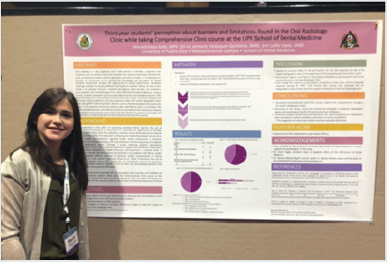
Figure 2: Student presenting a poster at ADEA’s Annual Meeting.

Lupinepublishers-openaccess-pediatric-dentistry-journal

Figure 3: Faculty and student in by-monthly meetings.

Lupinepublishers-openaccess-pediatric-dentistry-journal

Conclusions

Twenty-six students have completed the ADEA Academic Dental Careers Fellowship Program at the UPRMSC School of Dental Medicine and one of the students who participated in the Program in 2016, currently is a pediatric dentist, from the 2017-2018 cohort, two are pediatric dentistry residents at the SDMUPR, and from the 2019-2020 cohort, one applied to pediatric dentistry programs and is waiting for interviews invitations.

Acknowledgments

We want to thank Dr. Jocelyn Medina Paneto, professor and co-liaison of the SDMUPR ADEA/ADCFP, Dr. Carla Rodríguez, main mentor of the pediatric dentistry track, and all the faculty of the School of Dental Medicine of the University of Puerto Rico, Medical Sciences Campus, for their collaboration in this Program.

Conflict of Interest

The author of this article has no conflict of interest to disclose.

Read More Lupine Publishers Pediatric Dentistry Journal Articles:
https://lupine-publishers-pediatric-dentistry.blogspot.com/

Friday, October 8, 2021

Lupine Publishers | Children’s Perception About Dental Care in Patients Attended at Ceulp-Ulbra School Clinic

 Lupine Publishers | Journal of Pediatric Dentistry


Abstract

The dental care of children is an area that requires special attention. The dental visit, even in the early years of life, allows the child to have, early on, greater contact and familiarity with the dental environment, thus having the possibility to learn new habits in addition to positive experiences with regard to oral health. Thus, it is extremely important to know the view of children about the dental care provided by the institution CEUPL-ULBRA. We randomly selected children aged 3 to 11 years, 13 males and 8 females. Data collection was performed through interviews and story-design after the service, as well as analysis of the medical records to record the procedures performed. For the analysis of the drawings and for the interview, four categories were considered:
a) Dental environment
b) Dental treatment
c) Dentist image and
d) Behavioral manifestation
The most frequent categories in storytelling were the environment and dental treatment, with the most cited curative procedures. The operator / dentist’s image according to the drawing was considered technical. According to the interview, the clinical procedure itself was considered a positive point of care, especially when it was associated with pain relief. The most negative point was evidenced at times that led to some kind of discomfort in the child such as anesthesia, taste of the prophylactic paste and noise of high rotation. The perception of the operator’s image was considered humanized in all responses. Most children showed satisfaction with their smile and some reported the need to return to the dental clinic for new procedures. Only a small portion was free of oral problems. It is concluded that: the need for dental follow-up is not consistent with the oral health condition of the children evaluated and that the care process follows the curative model.

Keywords:Pediatric Dentistry;child psychology;health evaluation

Introduction

Pediatric dentistry is the dental specialty that takes care of children’s oral health. It is known that the great fear presented by adult patients in the dentist’s chair originates from the negative experiences of dental treatments that occurred in childhood. For this reason, the role of pediatric dentists is of great relevance in dentistry. Pediatric dentists are responsible for the care of children from infants to adolescence, and their exercise is comprehensive, as it is not limited only to the prevention and solution of oral problems, it also plays an important role with regard to the psychological and educational aspects of the patient [1].The practice of children’s dental clinic shows that children have some peculiarities, such as growth and development, biodynamics, tissue and organic responses, behavior, and personality structure. These peculiarities cause the sociological methods and the techniques of physical examination to have a different approach from that performed in adults, despite having the same diagnostic and therapeutic purpose [2]. According to Melo et al. [3], the dentist’s approach must be in accordance with the child’s age and psychological development, and can be used from a more playful language in early childhood to logical explanations in early adolescence, so that in many situations children are driven to overcome fears and phobias. During clinical practice, it can be observed that lessinvasive procedures do not generate major behavioral reactions, whereas more invasive procedures are directly related to rejection and fear in the face of treatment [3].The negative attitude towards dental treatment is a process that begins in childhood, and the origin and cause must be investigated by the Pediatric Dentist before any behavior control technique is applied [4]. According to Gomes et al. [5], the child may manifest fear and anxiety in several ways, the most frequent symptoms being tachycardia, sweating, palpitations, tremor, flushing and gastrointestinal complications.

Behind a behavior, positive or negative, there are a multitude of characters that exert marked influences on children, such as: age; the socioeconomic class of the parents; temperament; psychological development; the environment in which you live. Through such knowledge, the operator / pediatric dentist will have more baggage for the application of certain measures, and for a better understanding of the types of behavior presented by the children[5]. According to the literature, several control techniques can be used, such as: orders; compliments; reward; suggestions; containment; distraction; restriction; dominance by voice and sayshow- do. The pediatric dentist must, therefore, play an active role in the psychological and educational sectors, allowing the avoidance of possible trauma that almost always determines incompatible relationships with the dentist or with the clinical environment or even with the surgical procedures [6]. The control of fear and anxiety during dental treatment must be performed throughout the service. Thus, it is essential to use basic conducts to control the situation, such as verbalization, associated with pharmacological techniques for muscle relaxation or psychological conditioning, reducing the wear of the professional in relation to the patient. The proper use of these behavioral control techniques is fundamental for the success of the planned treatment and consequent restoration of the child’s oral health. The choice of behavioral approach techniques may vary according to the professional’s criteria, being influenced by factors observed during anamnesis, such as age, child’s behavior, and parental acceptance. In this sense, it is of great relevance to establish which procedures generate more behavioral disorders through specific control protocols and techniques for the care of pediatric patients, because regardless of the procedure, it is clear that they present some type of discomfort such as fear and / or anxiety, proving to be of great importance for professionals to update themselves in offering treatment options and techniques so that this moment becomes more dynamic and comfortable, recognizing, first of all, each child with their particularities.Therefore, knowing the child’s perception about the dental experience is extremely important for understanding the dental practice developed within the different environments that offer this service. Such knowledge allows the dental surgeon to identify possible failures committed and can develop new forms of interaction during care, thus modifying negative behaviors and / or reinforcing positive ones. This will allow the use of more effective methods so that they accept and understand the need for the procedure[7].Based on this principle, this study evaluated the perception of children between 3 and 11 years of age regarding dental treatment, the figure of the dentist and their own oral health condition, through analysis of information obtained by interview, drawing on the topic and analysis of medical records.

Method

The present study is characterized in a cross-sectional analytical and descriptive design, where the bibliographic research took place through the consultation of online publications such as: LILACS, SCIELO, BVS, PubMed. The search strategy occurred through Health Sciences Descriptors (DECs) registered in Portuguese as: Pediatric Dentistry, Child Psychology and Health Assessment, which are terminologies that make up electronic articles and made possible their search, in addition to the terms in English: Pediatric Dentistry, Psychology Child and Health Evaluation. 100 publications were found on the proposed theme, including subjects related to fear, anxiety and behavior management, with 33 articles selected, 3 master’s dissertations, 3 conclusion papers of a specialization course and 2 doctoral theses. For inclusion criteria, articles in Portuguese or English were selected, complete and published from 2009 to 2019, in addition to national books that addressed methods of controlling behavior in pediatric dentistry, considering the reliability of the selected material. As exclusion criteria, articles, monographs, and dissertations that do not fit the research objectives, in addition to those that are not available in full.The research was carried out at the Pediatric Dentistry’s School Clinic of the Lutheran University Center of Palmas, during the second semester of 2019.The object of the study was children assisted by the children’s clinic (I and II) of the institution, and the data were collected in the second semester of 2019. A random sample of 21 children aged 3 to 11 years participated in the research. The variables used in this study relate to those observed by the child, in relation to the procedures and the dental environment, and were adapted using a questionnaire already validated, and a drawingstory about their care.

As inclusion criteria, children in need of care participated in the research, whether for the first time or not at the school clinic. Children with motor difficulties or mental disabilities were excluded. Children who refused to do the drawing and answer the questions even with the parent’s permission, were also excluded. Children who failed to answer just one question were not excluded from the sample. Children who stopped making the drawing, but answered the questions, were not excluded from the sample. Based on previous studies and according to several authors [8- 13]the application of the instrument was carried out through a questionnaire already validated, where the child’s perceptions regarding the situation of care were recorded. All data were collected in the clinic environment, each child was approached individually. This collection was made after the service, with the application of a questionnaire containing 8 questions of the type:

a) What is a dentist?
b) Are you happy with your teeth? why?
c) Do you think you need to take more care of your teeth?
why?
d) While you were with the dentist, how did he treat you?
e) How was your reaction during the consultation?
f) What did you like most about the consultation?
g) What did you like least?
h) Finally, how do you feel now?

After the questionnaire was completed, the child was invited to carry out a drawing-story about his care (each child was provided with crayons and a blank sheet to carry out the drawing.), And afterwards, describe the meaning of the researcher to the researcher. In addition to the interview, the researcher was responsible for collecting data regarding the patient’s clinical history at the school clinic. The answers and the description of the drawing were faithfully transcribed and evaluated. Those responsible were informed of the research, and those who wished toparticipate signed the free and informed consent form. The level of invasion of the procedure to which the children were subjected was also subject to classification where they were divided into groups 1 and 2, being classified as invasive procedures (extraction; endodontics; restorations, which require absolute isolation) and not invasive (prophylaxis; topical application of fluoride and use of sealants that are carried out without absolute isolation) respectively. Only those drawings that met the following requirements were included in the study:

focus on the proposed theme
j) be completed
k) be clear for interpretation
l) in isolation or with the help of the child’s oral description.

Result and Discussion

A fluctuating reading was carried out for the initial knowledge of the material produced. Subsequently, the drawings were systematically observed, and the texts obtained for each of them were read, as well as their responses. Thus, the quantification allows to define the shared thought collectively among the researched group. Twenty-one children aged 3 to 11 years participated in the study, with the frequency of each age as follows: 3 years (1), 4 years (3), 5 years (1), 6 years(1), 7 years (3 ) and 8 years (5), 9 years (4) 10 years (2) and 11 years (1). The male gender had the highest frequency (13).

Analysis of the interview

It was found that the majority of respondents reported to more than one subcategory during the interview. The perception of the operator’s image was considered humanized in all reports and the design of the treatment model was described by most children as a curative, with preventive treatment being little mentioned. In the positive view, objects from the dental environment were highlighted, such as, for example, a dental chair, a Robinson brush, and a dental sucker. In the negative view, the most frequent responses were in relation to the dental procedure itselfwhen the treatment generated pain or discomfort: “I did not like the needle” and “I thought that noise was bad”. In the negative view, discomfort related to other objects was also highlighted, such as a needle and explorer probe.

Design analysis

It was identified that most of the interviewees reported to more than one subcategory during the elaboration of the drawingstory. The dental environment category was most frequently addressed, being the subcategories, operator, and equipment. The other categories demonstrated that, according to the child’s view, the institution’s dental treatment presents itself as a curative and technical model; but humanized, where the interviewees reported having been treated with great empathy. The moment of consultation in the days of the survey was reported by the interviewees as a pleasant moment and the environment was referred to as peaceful. The children’s behavior during the observation made by the researcher was satisfactory (positive). The operator was sometimes mentioned in phrases such as: “nice, nice and polite” as well as “he treated me very well”, “she explained what she was going to do”.The appreciation of the dental treatment received by each one was evaluated by means of a positive view (what he liked best) and a negative view (what he liked least). In the positive view, the dental procedure was the most mentioned, when related to the child’s pain relief, as identified in the excerpts: “I liked it when she passed the ointment, and when she removed the tooth, I didn’tfeel anything”; “I liked to pull the tooth out it because it hurt.” In the negative view, the dental procedure was also the most cited when it caused a sensation of pain or discomfort, as identified in the excerpts: “When she put the needle it hurt” and “I didn’t like that thing to brush my teeth”.

Analysis of the record

In the analysis of the medical record, despite the innumerable invasive procedures, most of the subjects showed positive behaviors, as the drawings and the speeches that showed tranquility, empathy towards the dentist, establishing dialogue were expressive; it was evident in the studied group that there is a relationship of trust and good communication between professionals and patients.The self-perception of the oral condition was evaluated as positive by most children, and the most frequent reasons were demonstration of health (8), absence of pain (6), self-care (4) as observed in the report “I’m happy because they they are beautiful ”. For those who negatively assessed their oral health condition, the presence of pain related to the carious process was the main related reason (5). Regarding the need for dental care, most children believe that they should attend other dental appointments (12), for one or more reasons, and the condition “To treat decayed tooth” was mentioned (9) times and procedures related to prevention were cited (3) times. Children who do not intend to return or only want to return in case of pain.It was found that the child’s behavior in the dental consultation can be determined by a series of factors, such as maturity, relationship with parents, approach to the dentist, past experiences, office environment, this because their handling, in some circumstances, becomes a great challenge for the professional. The professional’s positive interaction with the child brings out the image of a humanized professional. In both cases, the most apparent reaction in the child during and after the interview was joyful and calm. Behaviors related to anxiety, fear and scared were mentioned a few times (3), (1) and (2).In the present study, children who claimed to be happy with their oral health condition, even though they were compromised, demonstrate the inability to recognize oral health as an integral part of systemic health, which demonstrates a deficiency in health actions aimed at this evaluated population.

To demonstrate the view that the child has on the care that receives the drawing technique, it proves to be efficient because it is a pleasant activity and easy to perform. This can be seen in the course of this research. All children, after explaining the research, readily accepted the invitation and expressed satisfaction in drawing and reporting their drawings.The analysis of the story-drawings about the dental care provided by the Clinic of Pediatric Dentistry of CEULP-ULBRA showed very positive aspects regarding the actions developed by academics and teachers. The view of the infant patient that was part of this study reflects the effectiveness of the work performed by the teams of the Pediatric Dentistry Clinic of CEULP-ULBRA. The description of the drawings, through the children’s speeches, denoted a scenario of tranquility and empathy. It was very evident that there is a relationship of trust, good communication between academics and children. Communication between the dentist and the child, aimed at a friendly and friendly relationship during care, is essential for the success of dental treatment and, therefore, for the establishment of healthy behaviors. This condition was perceived in the storydesigns carried out by the patients as being from a cordial setting, with good communication and goodwill.Therefore, it is suggested that further studies be carried out in order to identify the best way of working with health professionals in order to encourage the practice of prevention as a health promotion strategy in addition to raising the awareness of children and their guardians. about the importance of periodic dental consultations for the benefit of your children’s health.

Conclusions

For most of the subjects participating in the research, the context of the dental consultation is revealed to be a pleasant situation, characterized by an educational-curative practice, and permeated by a humanized view of the dental professional, configuring itself in a pleasant situation. It was found that even in the midst of invasive procedures, in the days of the research, the children had behaviors of collaboration and demonstration of interest in the care and tranquility during the consultation. It was found that the evaluation process, through the technique of drawing-story, is rich and authentic. Therefore, the technique can be considered an excellent methodological alternative when compared to the use of questionnaires, which can induce the respondents’ answers, limiting the quality and depth of the evaluation process. According to the interview and the medical record findings, the interviewees’ oral health is not consistent, requiring dental follow-up for both new procedures and for hygiene and oral health care instructions.

Read More Lupine Publishers Pediatric Dentistry Journal Articles:
https://lupine-publishers-pediatric-dentistry.blogspot.com/
Friday, January 8, 2021

Lupine Publishers | Pediatric Dentistry Condition- A Mini Review

 Lupine Publishers | Journal of Pediatric Dentistry


Abstract

Tooth decay is the most common chronic childhood disease and the World Health Organization has identified it as a worldwide problem with 60-90% prevalence among school-age children. Dental caries is a topical contagious infectious disease that affects people of all ages and in any area of the world. Oral hygiene is a part of public health and oral and dental diseases affect different aspects of quality of life. DMFT is one of the indicators that the World Health Organization has introduced to determine the severity and prevalence of caries. One of the goals of the World Health Organization is to keep the DMFT index of students at less than 2. Different factors are effective in occurrence of dental and interdental caries that we discuss about them in this mini review.

Keywords: Decay Missing Filled Index, Dental Caries, Oral Hygiene, Tooth Decay

Abbreviations: WHO: World Health Organization; DMFT: decay-missing-filled index; BSS: Basic Screening Survey

Introduction

Tooth decay is the most common chronic childhood disease and the World Health Organization [WHO] has identified it as a worldwide problem with 60-90% prevalence among school-age children [1]. According to statistics in European countries, 6.1% of children aged 6-12 have at least one decayed or missed tooth, and due to the prevalence of tooth decay in all social classes, this disease can impose heavy costs on society [2]. Also, according to the statistics in Iran, decay-missing-filled index [DMFT] was 0.2% among 6 to 9-year-old children and 0.9% to 1.5% among 12-yearold children. Also, DMFT was 1.7% in 3 to 6-year old children and 3.3 to 4.8% in 9-year old children [3]. Four important factors: host, germs of the oral environment, food and time, has a role in tooth decay, without each of which, tooth decay will not occur [4]. Therefore, oral hygiene is very important in preventing it. Prevalence of dental caries in 6 to 12-year old children is one of the most important health problems. This can directly and indirectly impair the health of children and teenagers, and this problem is common among low-income groups and groups that do not comply with oral hygiene standards, such as not using toothbrushes and floss, dental caries is much more severe and acute [5].

Dental Caries

Dental caries is a topical contagious infectious disease that affects people of all ages and in any area of the world. Oral hygiene is a part of public health and oral and dental diseases affect different aspects of quality of life [6,7]. DMFT is one of the indicators that the World Health Organization has introduced to determine the severity and prevalence of caries. Nowadays, general dental health programs are usually only concerned with determining the prevalence of dental caries. Therefore, to measure the prevalence of dental caries and to determine oral health status in the society, especially in teenagers, appropriate indicators have been introduced by reputable authorities such as the Association of State and Territorial Dental Directors [2011]. This indicator is called Basic Screening Survey. The main purpose of using this indicator is to provide a framework for obtaining cheap and easy oral health information. On the basis of this indicator, people are classified into two parts, and it is ultimately determined whether or not they have caries [8,9].

Dental caries in Iranian students

One of the goals of the World Health Organization is to keep the DMFT index of students at less than 2. There are many studies on the calculation of DMFT in Iran, including Hamisi et al. which studied 323 students in Qazvin and showed that the prevalence of non-caries was 14.3% and their DMFT was 12.1%. [10]. Also, in a study on 12-year-old children in Tehran and Isfahan, DMFT was 11.2, which was the highest rate of caries [11]. In another study, the rate of DMFT in children in Isfahan was reported 3.41, and the conclusion of this study was that it showed a high proportion of DMFT in decay [12]. A study in Sirjan [a city in Kerman province] showed that the prevalence of non-caries state in 12-year-old students was 34.1%, which means that about 60% of 12-yearold students in this city due to various reasons, particularly not using toothbrush and floss, had dental and interdental decay [13]. Also, in the U.S Department of Health and Human Services in New Hampshire, a large evaluation of oral health status among public school students was done using the BSS index and it showed that approximately 2.3% of students had dental caries [14].

Are There Any Differences Between Girls or Boys in Caries Status?

The results of some studies show that there is no significant relationship between the prevalence of caries in male and female students. Among these studies, we can mention Nabipour et al., Who did not report a significant difference in caries status between male and female students [15]. However, in some studies, such as the study by Boroumand et al., Caries in 3-6-year-old boys was less than that of girls [16]. However, in some studies, such as the study by Boroumand et al., Caries in 3-6-year-old boys was less than that of girls [16]. Also, Loyola-Pontigo et al. and Rosado-Casanova et al. reported a higher incidence of caries in girls than boys [17,18].

Some Other Effective Factors of Children’s Caries Status

Another factor affecting dental and interdental caries is the level of parents’ education. In a study by Campus et.al, there was a significant relationship between parents’ education and lower incidence of caries in children [19]. Ismail and Sohn also stated in their study that children whose parents had a college education had significantly lower dental caries than children whose parents had lower educational level [20]. Also, the differences in socioeconomic status of families can lead to a different status of caries in children, as Primosch in a study in this regard observed a changing status of caries in families with different structures and concluded that this difference may be the result of the different socio-economic status of families, which may affect children’s dietary habits as well as hygiene [21].

Acknowledgement

The authors thank Farzanegan Hazrat Zeinab High School of Rey city and student research center of Basirat especially Dr. Salmani for their supports.

Read More Lupine Publishers Pediatric Dentistry Journal Articles:
https://lupine-publishers-pediatric-dentistry.blogspot.com/

Friday, October 30, 2020

Lupine Publishers | Lasers & Pedodontics

 Lupine Publishers | Journal of Pediatric Dentistry


Introduction

The medical terms such as magical and lightening quick are used to represent lasers [1]. Theodore H. Maiman in 1960 coined the term laser, which was initially termed as maser which stands for “microwave amplification by stimulated emission of radiation”. However, the term LASER is an acronym for light amplification by stimulated emission of radiation [2,3]. Three types of lasers used for surgical therapy in the oral cavity are neodymium lasers - YAG (Nd: YAG), of argon (Ar) and carbon dioxide (CO2) [3]. Lasers have largely replaced scalpels and other instruments in the field of medicine because of its advantages [4-6]. Different laser wavelengths have different absorption coefficients wherein laser energy can be absorbed or transmitted based on the structure of the target tissue. The presence of water, which is an essential component of all biologic tissues, is important for the use of lasers [2,6]. For hard tissues, Er lasers are used whereas any laser can be used for soft tissue components [2,6,7].

Applications of Lasers in Pediatric Dentistry

For caries removal

Erbium group of lasers are preferred for deep enamel, dentin, and caries removal, whereas the Nd: YAG laser is designated for superficial pigmented caries removal. The other advantages being the non-requirement of anesthesia and the use of conventional drills, which cause micro-fracture of tooth during preparation [1,2]. During cavity preparation, after the removal of enamel, the settings are adjusted to reduce the energy levels as dentin is less mineralized and has higher water content than enamel [2].

Removal of restorations (including amalgam)

Lasers should never be directed towards amalgam and should be pointed towards the surrounding enamel to create a small trough, and hand instruments are used to elevate the restoration out and later the cavity preparation is completed. Also, other restorations like composite and glass ionomer can be removed/replaced [1,2].

Preventive treatment

At the early stages after tooth eruption, enamel grooves are the site of early caries. This can be treated using lasers by cleaning, sterilizing and restoring the same. Also, many studies have reported that etched enamel by erbium has properties like the acid-etched enamel [2].

Treatment of peri coronal problems in erupting teeth

Lasers are used in non-contact mode to remove the pericoronal tissue covering the newly erupted tooth, which might help in relieving any discomfort, swelling, or infection in the tissue overlying the emerging tooth [2,9].

Gingival re-contouring and orthodontic purposes

Excess gingival growth by the use drugs or by poor oral hygiene, and during other surgical procedures including orthodontics requires removal of tissue in some cases. This can be accomplished by the use of lasers which can be done without the need for a local anesthesia. Use of topical anesthetic can be supplemented for the treatment procedures [8].

Treatment of ankyloglossia

Tongue is stabilized with a hemostat and the frenum is revised, while avoiding any damage to the glands on the floor of the mouth [8].

Treatment of aphthous ulcers and herpetic lesions

Use of low power settings with the laser energy directed at the target tissue in the non-contact mode, for a duration of 15-30 second intervals for three to four times, helps in pain relief. The use of laser in the initial stages in herpes labialis may prevent its further progression and provide a palliative effect for the area and prevent its progression [2,8].

Pulp therapy

The ability of laser to close the dentinal tubules and provide a sedative effect on pulpitis has somewhat encouraged the use of laser in indirect pulp capping [8]. Also, the use of lasers to sterilize the canals and also create a hemostatic environment in adjunct to the conventional procedures has created a stir for the use of lasers.

Other surgical procedures

Other surgical procedures like apicectomies and amputation of impacted teeth underneath the bone also can be performed with the use of lasers. The erbium lasers are ideal for these surgeries and a variety of tips, settings and water sprays can be used. Softtissue ablation does not require water spray whereas removal of bone needs to be done with water [2,9].

Advantages of laser therapy

a) Decreasing inflammation and pain.

b) Reduced healing period [3,4,9].

c) Good & faster healing properties.

d) Reduced chances of infection.

e) Reduced bleeding.

f) Instant hemostatic achievement.

g) Good margins.

h) Patients apprehensive for blade.

Contraindication of Laser Therapy

a) Patients with pacemakers, however it can be used with precautions in some case [9].

b) Patients who are sensible to light.

c) In epileptic patients.

d) In patients with antecedent of arrhythmia or chest pain.

e) Avoided on tumorous tissues or benign tumors with malignant potential.

Conclusion

Natural light is and has been considered as the curator [10]. Lasers have gained tremendously over the years; its advantages far outweigh its disadvantages. However, there still exists some limitations as well as some contraindications, which stop its usage with the cost factor being one of it. Nevertheless, it would be the future instrument of choice for most of the procedures included in all the fields with surgery, periodontics, endodontics and orthodontics being one of them.

Read More Lupine Publishers Pediatric Dentistry Journal Articles:
https://lupine-publishers-pediatric-dentistry.blogspot.com/
Friday, October 9, 2020

Lupine Publishers | Oral Squamous Papilloma on the Tongue of a 12-Year Old Female: Report of a Case with Human Papilloma Virus Literature Review

 Lupine Publishers | Journal of Pediatric Dentistry


Abstract

Human papillomavirus (HPV) infection that causes squamous papilloma is common in the oral cavity of adults but not in children. Although benign, the slow progressive growth is a concern to clinicians and parents as the lesion may clinically appear as an exophytic verrucous carcinoma or squamous carcinoma. This case report describes a squamous papilloma arising on the tongue of a 12-year old child.

Keywords:Child; Human Papillomavirus (HPV); Squamous Papilloma; Oncogenic Potential; Tongue; Koilocytes

Introduction

Human papillomavirus (HPV) are slow, benign proliferations of stratified squamous epithelium frequently observed in the oral cavity that are the viral etiologic agent for squamous papilloma [1,2]. Squamous papillomata are commonly observed in adults 30-50 years of age and is the fourth most common oral mucosal lesion in both children and adults [3]. Although the entire oral cavity may be affected, the viral lesion has a predilection for the laryngotracheobronchial complex in children and the lower lip, hard and soft palate of the maxilla and uvula in adults [1,4-6]. Clinically, the lesion appears as an exophytic mass and may cause anxiety to the clinician and parents, as the lesion can clinically appear like an exophytic verrucous carcinoma, squamous carcinoma or condyloma accuminatum [1,4,7,8]. This case report describes a squamous papilloma arising on the tongue of a 12-year old child.

Virology

The human papillomavirus is a 55nm non-enveloped icosahedral double-stranded deoxyribonucleic acid (DNA) virus that is a member of the papovavirus group [7-14]. There are over 150 genotypically different types and classified as either mucosal or cutaneous.10-12 The viral types with oncogenic potential include HPVs 16, 18, 31, 33, 35, 39, 45, 51, 55, 56, 58, 59, 66 and 68. Oral squamous papilloma infection is associated with HPV subtypes 6, 11 and 16 [7-13]. Papillomas induced by HPV types 6 and 11 are considered to have low oncogenic potential. However, approximately 85% of dysplastic lesions, carcinoma in situ and squamous cell carcinoma involve the DNA sequence of HPV 16 and 18. DNA replication of HPV occurs in the nuclei of epithelial cells. The HPV capsid proteins enter the host cell delivering the viral DNA to the nucleus which allows proliferation of the viral lesion [7,10,11,12,14].

Case Report

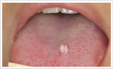
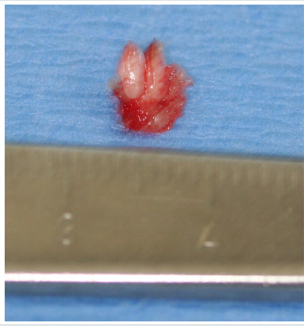
A 12-year old Asian female was referred to the office by her family dentist for evaluation of a soft tissue lesion localized to the dorsal surface of the tongue (Figure 1). The patient stated that the lesion has been present for at least six months. Her past medical history was unremarkable. She was not taking any medications and denied any allergy to medications. The patient also denies being sexually active. Sexual abuse was also ruled-out. Head and neck examination were negative for palpable neck masses and lymphadenopathy. Oral examination of the dorsal surface of the tongue revealed a pink-white colored exophytic lesion that was freely movable. The surface texture had a pebbly appearance that resembled, “cauliflower”. The remaining oral examination was unremarkable. Examination of the upper and lower extremities was negative for any soft tissue lesions resembling HPV. Based on the clinical appearance of the lesion, the differential diagnosis included squamous papilloma, verruciform xanthoma, papillary hyperplasia and condyloma accuminatum of the tongue. The father and patient were informed of the clinical findings and excisional biopsy (Figure 2) for a definitive microscopic diagnosis under local anesthesia was recommended.

Figure 1: Clinical photograph of squamous papilloma on left dorsal surface of the tongue in 12-year old Asian child.

lupinepublishers-openaccess-journal-pediatric-dentistry

Figure 2: Excised squamous papilloma specimen from dorsal surface of tongue of 12-year old Asian patient.

lupinepublishers-openaccess-journal-pediatric-dentistry

Histopathology

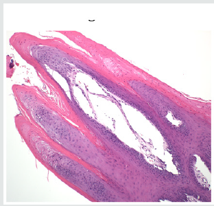
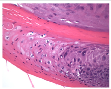
Excisional biopsy was completed under local anesthesia with 1.0mm margins of normal tissue and to the depth of the tongue musculature. The lesion was stained with hematoxylin and eosin for histopathological diagnosis. Histological examination demonstrated long, thin papillary projections of parakeratinized stratified squamous epithelium (Figure 3a). Localized areas of basilar hyperplasia with koilocytes were observed (Figure 3b). The histological findings were consistent with squamous papilloma.

Figure 3(a): Histopathology demonstrating proliferation of hyper keratinized stratified squamous epithelium finger-like projections with thin fibrovascular connective tissue core (Hematoxylin and eosin stain. Original magnification x 40).

lupinepublishers-openaccess-journal-pediatric-dentistry

Figure 3(b): Presence of koilocytes (arrows) in the spinous layer of the epithelium that are characteristic histopathologic findings of oral squamous papilloma (Hematoxylin and eosin stain, x 200).

lupinepublishers-openaccess-journal-pediatric-dentistry

Discussion

In children, viral transmission remains controversial. HPV transmission may occur by any number of different mechanisms, such as autoinoculation, heteroinoculation, perinatal transmission, sexual abuse and contact with fomites [1,4,5,9,10]. However, trauma is one mechanism that should be considered in our patient. Iatrogenic tongue biting may initiate an infection of the basal squamous epithelial cells that allows the entrance of the HPV into the tongue. Viral DNA then enters the nucleus of the infected epithelial stem cells and can replicate causing the HPV infection [15]. Histopathologic features of oral squamous papilloma demonstrate hyperkeratosis in the epithelium, proliferation of the spinous cells that result in long, thin finger-like projections above the mucosa [1- 3,8,14]. The characteristic feature of HPV infection is the presence of koilocytes (Figure 3b) which are virus-infected epithelial cells due to perinuclear cytoplasmic vacuolization of cells of the spinous layer of the epithelium. This results in the nuclei becoming pyknotic and cremated surrounded by an optically clear zone [1-10]. All the described histopathologic findings in our patient are characteristic of HPV infection. Although the prevalence of oral HPV infection is low, a bimodal distribution is observed. The highest prevalence is observed in children less than 1 year of age and the second peak occurs in adolescents, between 13 to 20 years old [9,16]. Despite the bimodal distribution, oral HPV infection is considered low in children [17]. In a study of 4140 children between the ages of 10 to 18 years old, the prevalence of oral HPV infection was 1% and the most common type was HPV 11 [18]. Treatment of HPV is by surgical excision [19]. The United States Food and Drug Administration (2016) approved Gardasil 9 human papillomavirus 9-valent vaccine to prevent infection against HPV types 6, 11, 16 and 18 (Merck & Co., North Wales, Pa). At present, only Gardasil 9 human papillomavirus 9-valent vaccine recombinant has been approved for vaccination in the United States (Center for Disease Control, 2019) [20]. To obtain the greatest clinical efficacy and cost effectiveness of vaccination, the Center for Disease Control (CDC) recommends that both male and female children between the ages of 11-12 years get two HPV vaccinations six months apart before individuals have been exposed to the human papillomavirus. It is also recommended that adult women up to age 26 years and men up to age 21 years also obtain the HPV vaccine [21].

Conclusion

A case of oral squamous papilloma on the tongue of a 12-year old female is presented to create awareness of this soft tissue lesion in the oral cavity of children due to the human papillomavirus. It is only after educating clinicians and parents about the HPV will we observe greater identification, management and treatment in the HPV infected child.

To Know More About Lupine Publishers Pinterest Click on Below Link: https://www.pinterest.com/lupineonlinegroup/

Friday, September 4, 2020

Lupine Publishers | Management of Mesiodens In Mixed Dentition- Molariform and Tuberculate: A Case Report

  Lupine Publishers | Journal of Pediatric Dentistry


Abstract

Timely intervention is the key to any setback in the mixed dentition. Teeth which are supplemental to the normal dentition are supernumeraries, the most common being mesiodens, present in the premaxillary region. Certain pathological consequences may arise due to mesiodens like unaesthetic midline diastema, rotation, displacement, root resorption and cyst formation. The current case report presents the management of developing malocclusion in the anterior region due to the presence of mesiodentes- a molariform and a tuberculate. The molariform mesiodens was impeding the eruption of the maxillary right central incisor, thus the surgical removal of both mesiodentes was planned and executed. In addition to this soft tissue uncovering was done for the unerupted maxillary central incisor. On follow up, uneventful healing was observed successfully.

Keywords:Mesiodens; Mesiodentes; Mixed Dentition; Molariform; Tuberculate

Introduction

Supernumerary teeth, or hyperdontia, is a term that describes teeth that are surplus in number when compared to the normal complement of teeth [1]. The etiology of supernumerary teeth remains unclear and not yet completely understood [2]. Among the various proposed theories that have attempted to explain the causes behind the development of supernumerary teeth, current literature favors the ‘lamina hyperactivity theory’ that states hyperdontia results from independent, locally conditioned hyperactivity of the dental lamina [1]. The most commonly occurring supernumerary tooth is the ‘mesiodens’, a term that was initially coined by Balk in 1917. As the name suggests, the mesiodens is usually located mesial to the central incisors in the premaxillary region [3]. With a frequency of prevalence between 0.15-3% in the permanent dentition and 0.02-1.9% in the primary dentition, it may occur as single or multiple, unilateral or bilateral, may be erupted or impacted and frequently found in conjection with cleft lip and palate and syndromes like Cleidocranial dysostosis, Gardner’s syndrome, Ellis-Van Creveld syndrome, Ehlers- Danlos syndrome, Incontinentia Pigmenti, and Tricia-Rhino- Phalangeal syndrome [4,5]. Also, mesiodentes may vary in shape from simple conical form to a larger, more complicated crown shape with several tubercles [6]. The dysfunctional nature of mesiodens is known to cause a variety of clinical complications such as being unaesthetic, pathological disturbances in the normal eruption and positions of adjacent teeth, altered growth and development in the area, retention of primary teeth, odontogenic cysts, caries, pulp necrosis of the adjacent teeth, dilaceration of developing tooth, nasal teeth, gingival and periodontal problems [2,7]. Accordingly, their early diagnosis and management is vital to waive off complications of such kind.

Case Report

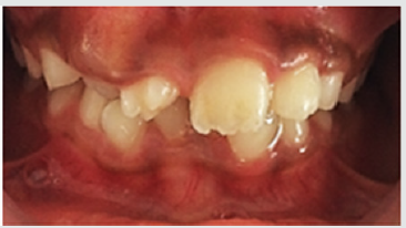
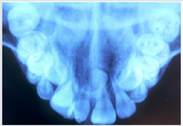
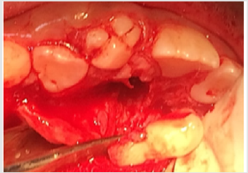
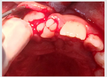
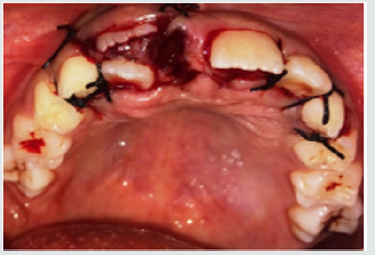
A 9-year old female patient reported to the department of Pedodontics and Preventive Dentistry with the complaint of irregularly placed upper front teeth and wanted it to be corrected. The patient was normal and healthy with non-contributory medical and dental histories. The extra oral examination did not reveal any abnormalities. Intraoral examination revealed a Class I mixed dentition with a missing upper right central incisor and an erupted molariform mesiodens in its place. In addition, there was a firm bulge palpable in the upper right central incisor area which suggests the impediment in the path of eruption of the central incisor by the erupted mesiodens (Figure 1). An occlusal radiograph was taken to rule out the possibility of multiple supernumerary teeth and surprisingly another unerupted and impacted inverted mesiodens with an incomplete root was found mesial to the upper left central incisor (Figure 2). The SLOB technique confirmed that the impacted mesiodens was present palatially. Both informed and written consent was obtained from the parents before initiating the treatment. We decided to extract both mesiodentes under local anesthesia. The erupted mesiodens was extracted by intra- alveolar extraction. The impacted mesiodens was surgically removed by raising a mucoperiosteal flap from maxillary first premolar to contralateral first premolar (Figure 3). Rotary cutting instruments with simultaneous irrigation were used for removing enough bone around the impacted mesiodens for its easy retrieval. Soft tissue uncovering was done for the unerupted right central incisor by placing an elliptical incision over the incisal portion of the palpable bulge (Figure 4). The extraction socket was checked for any pathological tissue and the flap was relocated and sutured with interrupted sutures (Figure 5). The patient was recalled after a week for suture removal and followed-up after 3 months. Uneventful healing with no associated symptoms was observed.

Figure 1: Preoperative view showing erupted molariform mesiodens along with unerupted maxillary central incisor.

lupinepublishers-openaccess-journal-pediatric-dentistry

Figure 2: Occlusal radiograph showing unerupted and impacted inverted mesiodens with an incomplete root.

lupinepublishers-openaccess-journal-pediatric-dentistry

Figure 3: Surgical removal of impacted mesiodens by raising a mucoperiosteal flap from maxillary first premolar to contralateral first premolar.

lupinepublishers-openaccess-journal-pediatric-dentistry

Figure 4: Soft tissue uncovering for the unerupted right central incisor by placing an elliptical incision over the incisal portion of the palpable bulge.

lupinepublishers-openaccess-journal-pediatric-dentistry

Figure 5: Interrupted sutures placed.

lupinepublishers-openaccess-journal-pediatric-dentistry

Discussion

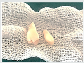
The realm of pediatric dentistry incorporates the practice of interceptive orthodontics, thereby bestowing upon the pediatric dentist, opportunities of providing timely guidance in the development of occlusion. The current case report presents the management of developing malocclusion in the anterior region due to the presence of mesiodens. A mesiodens occurring in the primary dentition is a rarity even though, it being the most common dental abnormality in the permanent dentition [8]. Most cases of mesiodens are discovered during the first decade as maxillary central incisors are erupting and radiographic examinations are performed as an aid to screening for any other malformations and abnormalities [1]. Various theories regarding the etiology of mesiodens have been reported in the literature but the subject remains controversial [3]. Heredity has been suggested to be an etiologic factor based on the observation that supernumeraries are more common in family members; however, it does not follow a simple Mendelian pattern [9]. It was originally postulated that the mesiodens represented a phylogenetic relic of the extinct ancestors who had three central incisors. This is known as phylogenetic theory reversion (atavism) which has now been discarded by the embryologists [10]. The dichotomy theory states that, a mesiodens arises due to the splitting of the tooth bud. On the contrary, Taylor argued that splitting of the tooth bud may either form two equal sized teeth or one normal and one dysmorphic tooth [8]. The hyperactivity theory states that development of mesiodens is due to the hyperactivity of the dental lamina. The ‘field model’, proposes that a tooth bud which is forming at a given location develops according to its position within the field, further determining its shape. The ‘clone model’ postulates that that each tooth class is derived from a clone of ectomesenchymal cells which are programmed by epithelium to produce teeth of a given pattern. Depending upon the specific factors expressed from these ectomesenchymal cells, the shape of the accessory tooth germ forms in the vicinity of the incisors class of teeth becomes evident at the bell stage [11]. This case report presented with an erupted molariform mesiodens and an unerupted tuberculate mesiodens with an undeveloped root (Figure 6). Since the molariform mesiodens was impeding the eruption of the maxillary right central incisor, the surgical removal of both mesiodentes was planned and executed. Timing of interceptive treatment should be as soon as possible following clinical detection of an abnormal eruption pattern. It has been suggested that a tooth delayed in its eruption by more than six months with respect to its antimere should be radiographically investigated. Hogstrum and Andersson [12] suggested two alternatives exist.

The first option involves removal of the supernumerary as soon as it has been diagnosed. This could lead to an unpleasant experience that may have a psychological effect on a very young child and has been said to cause devitalization or deformation of adjacent teeth. Secondly, the supernumerary could be left until root development of the adjacent teeth is complete. The potential disadvantages associated with this deferred surgical plan include; loss of eruptive force of adjacent teeth, loss of space and crowding of the affected arch, and possible midline shifts. In the present case, since the roots of the adjacent teeth were completely formed and the child was at an age where she could sustain a surgical procedure, the surgery was undertaken with utmost attention to detail and caution. Access to mesiodens during surgery must dealt carefully considering the quantity of bone amputation and the possible damage to the adjacent teeth [7]. Follow up is indispensable in such cases since the eruption status should be monitored. The patient revealed satisfactory healing and suitable eruption of the maxillary central incisor. Pediatric dentists are the firsts to usually identify developing malocclusions and thus it is their responsibility to intervene and intercept in an apt manner to prevent future unfavorable sequalae.

Figure 6: Molariform and tuberculate mesiodens.

lupinepublishers-openaccess-journal-pediatric-dentistry

Acknowledgements

Authors are thankful to their colleagues and faculty.

For More About Lupine Publishers Pinterest Please Click on Below Link: 
https://www.pinterest.com/lupineonlinegroup/

Friday, November 29, 2019

Lupine Publishers | Acute Primary Herpetic Gingivostomatitis In A Child: Strategies for Pain Suppression and to Improve Oral Intake

Lupine Publishers | Journal of Pediatric Dentistry

Abstract

This case report describes the management strategies and the evolution of the acute herpetic gingivostomatitis condition in a 3-year-old female child with a focus on suppressing pain and to improve oral intake with approaches to medicine and dentistry.
Keywords: Herpect Stomatitis; Drug Therapy; Child

Introduction

Herpetic gingivostomatitis is a condition that most often results from initial gingiva (gums) and oral mucosa infection with herpes simplex virus type 1 (HSV-1). While herpetic gingivostomatitis is the most common cause of gingivostomatitis in children before the age of 5, it can also occur in adults. The condition is characterized by a prodrome of fever followed by an eruption of painful, ulcerative lesions of the gingiva and mucosa, and often, yellow, perioral, vesicular lesions. HSV-1 is usually spread from direct contact or via droplets of oral secretions or lesions from an asymptomatic or symptomatic individual. Once a patient is infected with the herpes simplex virus, the infection can recur in the form of herpes labialis with intermittent re-activation occurring throughout life [1]. The pathogenesis of herpetic gingivostomatitis involves replication of the herpes simplex virus, cell lysis, and eventual destruction of mucosal tissue. Exposure to HSV-1 at abraded surfaces allows the virus to enter and rapidly replicate in epidermal and dermal cells. This results in the clinical manifestation of perioral blisters, erosions of the lips and mucosa, and eventual hemorrhagic crusting. Sufficient viral inoculation and replication allow the virus to enter sensory and autonomic ganglia, where it travels intraaxonally to the ganglionic nerve bodies. HSV-1 most commonly infects the trigeminal ganglia, where the virus remains latent until reactivation most commonly in the form of herpes labialis [2]. While most children with primary gingivostomatitis will be asymptomatic, some will experience considerable pain and discomfort and are at risk of dehydration. There are no large, well designed studies to clearly determine appropriate therapy for all children [3]. Professionals who treat children in this age group must be able to diagnose and treat common oral manifestations when necessary and should refer the child to a pediatrician for effective treatment if the presence of any systemic alteration is suspected [4]. Herpetic infections commonly affect the dental profession’s anatomical area of responsibility and the diagnosis and management of such infections fall in the purview of oral healthcare providers. To administer competent care to patients with herpetic infections, clinicians must understand the disease, its treatment, the impact the disease or its treatment may have on the patient and the extent to which the presence of a herpetic infection may impact on caregivers in the clinical process [5]. The purpose of this case report was to describe the treatment recommended for a child diagnosed with acute herpetic gingivostomatitis associated with tonsillitis and the ways to suppress pain and to improve oral intake from the perspective of medicine and dentistry.

Case Report

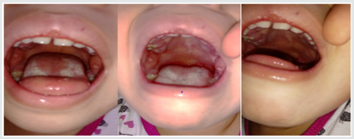
Parents of a 3-year-old and female child sought pediatrician due to inflammation in the throat of their daughter, with fever and irritability for two days, then their child feels pain in the mouth, and the drooling starts with the appearance of diffuse lesions in the oral mucosa, complaining of pain and having difficulty feeding. There was the prescription of antibiotics (amoxicillin and clavulanate potassium for oral suspension), anti-inflammatory and antipyretic. Intraoral cleaning with gauze and saline was recommended and the request for a new consultation, to eliminate the possibility of fungal contamination. The diagnosis of acute and viral primary herpetic gingivostomatitis was established (Figure 1). On intraoral examination, gingiva appeared fiery red in color and multiple vesicles were present on the attached mucosa. Multiple vesicles and ulcers were seen along the lateral border and anterior surface of the tongue. Both sided buccal mucosa revealed multiple vesicles. Her parents also complained about his bad breath during this period due to poor oral hygiene. Submandibular lymphatic glands of the kid were enlarged [6]. The pediatric dentistry was consulted because the child persisted with much pain, unable to sleep or eat (Figure 2). There was then the option of laser applications, with faster healing of ulcers and greater pain relief. There was substantial improvement in food, oral hygiene and sleep. The patient will perform control examinations, with simultaneous evaluation by pediatrician and pediatric dentistry.
Figure 1: Child oral examination two days under antibiotic prescription.
lupinepublishers-openaccess-journal-pediatric-dentistry
Figure 2: Aspect of the child’s tongue on the fourth day of drug treatment.
lupinepublishers-openaccess-journal-pediatric-dentistry

For more Lupine Publishers Open Access Journals Please visit our website:
For more Open Access Journal on Pediatric Dentistry articles Please Click Here:

To Know More About Open Access Publishers Please Click on Lupine Publishers

980 nm Diode Laser: A Good Choice for the Treatment of Pyogenic Granuloma

Abstract Pyogenic granuloma is a benign non/neo plastic mococutanous lesion . It is a reactional response to constant minor trauma and ca...