Showing posts with label Dental and Oral Health Journals. Show all posts

Wednesday, July 13, 2022

Rotary Systems Versus Manual K-File System in Primary Molar Root Canals - In Vitro Study


Abstract

Aim: To evaluate and compare the cleaning ability between two, rotary shaper and manual K-files in primary molar root canals.

Material and method: Twenty extracted maxillary and mandibular primary molars and 10 mm length were cleaned in water and stored in 0.5% sodium hypochlorite for 1 week. Caries excavation was done. Coronal access was made. After irrigation of the root canal with normal saline, a K-file with a compatible diameter was introduced into the root canal and the canal length was determined. A K-file size #15 was introduced into the root canal and 1 ml Indian ink was injected into the orifice. The roots were then randomly divided into 2 groups.

Read More About This Article : https://lupinepublishers.com/pediatric-dentistry-journal/fulltext/rotary-systems-versus-manual-k-file-system-in-primary-molar-root-canals-in-vitro-study.ID.000132.php

Read More Lupine Publishers Google Scholar Articles  : https://scholar.google.com/citations?view_op=view_citation&hl=en&user=h1QvhsYAAAAJ&citation_for_view=h1QvhsYAAAAJ:qxL8FJ1GzNcC


Friday, December 6, 2019

Lupine Publishers | Comparison of the Efficacy of Plaque Removal of Listerine Smart Rinse Kids and Vi– One Junior Fluoridated Mouthwash in Children Aged 6 To 10 Years

Lupine Publishers | Journal of Pediatric Dentistry with High Impact Factor


Abstract

Objectives: In this study, a comparative study was done on the effects of Vi-One and Listerine fluoridated mouthwashes on the reduction of dental plaque in pediatric patients between 7-11 years of age in dental clinics of Sepideh and apple in Shiraz, Iran. Listerine Smart Rinse Kids is a product of the United States and Vi-One Junior mouth wash is the domestic production of the country at Rozhin Corporation. This research was conducted by Mohammad Karimi and Hassan Dehghan in 2018-2019.
Material and Methods: In this study, 100 individuals were selected and divided into two groups of 50. During the study, no other method of controlling the plaque was used. In this method, the first group first used Vi-One mouthwash for 10 days and after two weeks of rest and minimizing dental plaque, they used Listerine for 10 days. While the second group used Listerine first, then they applied Vi- One in the same way. The results of this review were then evaluated.
Results: The mean of plaque index in total mouth and in the posterior teeth area with the use of Listerine Smart Rinse Kids was lower than that of Vi-One Junior mouth rinse. In another words, Listerine had a better effect on plaque removal than the Vi-One mouthwash in the posterior mandibular region.
Conclusion: The results show that although Listerine mouthwash had a better effect on dental plaque removal, none of the two mouthwashes had a significant difference in effects on maxillary and mandibular jaws.
Keywords: Dental Plaque; Vi- One, Listerine, Fluoridated Mouthwash; Periodontal Diseases; Plaque Index

Introduction

Currently, dental caries and gingivitis are common oral and dental diseases in this country. One element that can prevent tooth decay is Fluoride. In the oral health program of the country, fluoride mouth wash 2% was used to prevent dental caries in elementary school students all over the country [1]. In the other hand, dental plaque is an important factor in the formation of dental caries and periodontal diseases. Leo and his colleagues have identified dental plaque as the main cause of gingivitis [2]. With the use of mouthwashes, one can control the dental plaque, chemically [3,4]. In fact, mechanical plaque removal is one of the most common and effective methods for preventing caries and inflammation of the gum [5]. Fluoride mouthwash usage is contraindicated in children younger than six years of age due to the risk of swallowing and causing systemic toxicity and fluorosis [6-8]. Symptoms of acute oral fluoride toxicity in children include severe nausea, vomiting, hyper salivation, abdominal pain, and diarrhea [9]. In severe or fatal cases, these symptoms can be followed by convulsions, cardiac arrhythmias, and coma [10- 12]. Laboratory and animal data have shown that prevention the accumulation of plaque and consequently, reduction in dental plaque can be achieved when fluorides is applied topically which inhibits the bacterial multiplication [13]. The fluoride from mouth rinse is retained in dental plaque and saliva to help prevent dental caries [14]. In one review, the average caries reduction in nonfluoridated communities attributable to fluoride mouth rinse was 31% [15]. Another study in Sweden reported that the use of fluoride mouthwash along with brushing has a significant effect in decreasing of dental caries [16]. Listerine Smart Rinse Kids has been used for the purpose of this study. This product is an alcoholfree mouthwash. The ingredients include Sodium fluoride 0.02% (0.01% w/v fluoride ion), Water, Sorbitol, flavor, phosphoric acid, Sucralose, Cetylpyridinium chloride, disodium phosphate, sodium saccharin, menthol, blue 1 and green 3 [17]. One study reported that use of this mouthwash can strengthen teeth 99% better than brushing alone [18]. Another source indicated that it gives 12- hour cavity protection [19]. Vi-one Junior Mouthwash is specially designed for children. This mouth rinse contains Sodium fluoride 0.05%, Cetylpyridinium chloride 0.05% and Disodium phosphate agents. The respective flavors contain sugar-free and harmless sweetener. This brand also is an alcohol-free product [20]. The purpose of this study was to compare the efficacy of two types of mouthwashes, one the domestic mouthwash (Vi-One Junior) and the other, the brand name Listerine Smart Rinse kids fluoridated mouthwash in the removal of the dental plaque.

Material and Methods

This study was a cross-over clinical trial. The eligibilities for entering in our study were as follow:
a) Children having at least 20 teeth with no large restorative area.
b) No history of periodontal Diseases.
c) Not having any Prosthodontic or Orthodontic appliances.
The condition for withdrawal from the study, if there was any sign of reactions to any of these mouth rinses. There was no obligation to have any food regimen.
The study population consisted of 100 patients who were in a 50-member group. Before taking oral mouthwash, plaque index was minimized, and all subjects underwent tooth scaling at the beginning and, if necessary, teeth polishing were done before taking mouthwash. Oral hygiene was assessed via a plaque index. First, in both groups, the Silness-Löe plaque index was recorded. It is an Index for evaluating the thickness of the plaque in the gingival region, which measures the thickness of plaque on all surfaces (M, B, D, and L) [21].
Coding for the plaque index was carried out according to the criteria [22]:
a) Code 0: No plaque
b) Code 1: A film of plaque is adhering to the free gingival margin and adjacent area of the tooth. The plaque may be seen in situ only after application of disclosing solution or by using the probe on the tooth surface.
c) Code 2: Moderate accumulation of soft deposits can be seen with the naked eye within the gingival pocket, the tooth, or gingival margin.
d) Code 3: Abundance of soft matter can be seen within the gingival pocket and/or on the tooth, and gingival margin.
In this index, each tooth is divided into four surface area but in our purposes in the present study, we modified the surfaces area from 4 to 6; thus, we have three surfaces in the buccal area (Mesiobuccal, Midbuccal, and Distobuccal) and three surfaces in the lingual area (Mesiolingual, Midlingual and Distolingual). The first group used Vi-One & Listerine mouthwash (kids mouthwash), for 10 days in the following way. Needless to say, this process was supervised by parents at home. The kids have to gargle 5 cc ’s of Vi-One mouthwash 2 times per day for 30 seconds, and during this period of time, no other plaque control methods and tooth brushing should be used. At the end of the period of 10 days, the plaque index was recorded again. Then, the subjects were given a week to rest and stop using the mouthwash while they had permission to start brushing like before. Again, the plaque index was minimized for patients with polishing the teeth, and they used Listerine mouthwash for 10 days. In the same way, 5 cc ‘s of the mouthwash twice daily was used for 30 seconds, and at the end of the one-week period, the plaque was recorded. For the second group, in the first 10 days, mouthwash. Listerine was prescribed and in the second 10 days, the Vi-One mouthwash was applied. All procedures were performed according to the above pattern.

Results

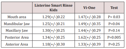
Paired T-test was used for statistical analysis of the findings. The findings showed when Listerine Smart Rinse Kids was used; the mean of plaque index in all area of the mouth (especially in the mandibular jaw and the posterior region) was significantly less than the time Vi-One was applied. However, there was no significant efficacy difference between the use of both types of mouthwash in the upper jaw and the anterior region.
There was no significant difference between the mean plaque index in Vi-One mouthwash between upper and lower jaw, and there was no significant difference between the maxillary and lower jaw in the case of Listerine Smart Rinse Kids either. The presence of this indicator in both types of mouthwash in the anterior region was significantly less than the posterior region. The mean and standard deviation of the plaque index in both groups, as well as in different regions of the mouth, are listed in Table 1.
Table 1: The amount of dental plaque in terms of area and type of Mouthwash.
lupinepublishers-openaccess-journal-pediatric-dentistry

Discussion

In general, the anti-plaque properties of mouthwashes are completed through bactericidal and bacteriostatic effects, separation of microorganisms from dental surfaces, loosening of joints to these surfaces or lowering of the surface tension of the tooth [2,21,23]. Some mouthwashes can be useful for preventing tooth decay or periodontitis [6,24]. Furthermore, mouthwashes are recommended for children and adolescents with orthodontic appliances or adults who need deep cleansing (such as curettage) [6]. These types of mouthwashes were generally used before and after surgery (especially Chlorhexidine) and have a very positive effect on the treatment of gum and ulcerative inflammation [6]. Use of this type of mouthwash should not last longer than 2 to 3 weeks due to some side effects such as staining the teeth and soft tissue staining, increased calculus deposition, unpleasant taste, burning sensation, and mucosal irritation [6]. It’s time to use this mouthwash after brushing and before bedtime, and it’s best not to eat anything after half an hour after use. Fluoride-containing mouthwashes are another type of mouth rinse that has a fairly large use. These mouthwashes have a significant effect on teeth strengthening. Fluoride in the mouthwash cause bonding with enamel and dentin, and with bonding with calcium and phosphorus, they form Fluorapatite, which is more resistant to caries than Hydroxyapatite. Fluorides also accelerate the mineralization, repair the decayed teeth surfaces, and help to increase the reverse processing of damaged tooth surface area [25,26]. Fluoride also reduces the effect of oral bacteria on teeth. It is done by interfering with the function and formation of the microorganisms. The best fluoride mouthwash protects the teeth against the acids which are produced by dental plaques. “Neglecting the oral hygiene of children leads to the accumulation of plaque and as a consequent the formation of dental calculus which will have a devastating effect on the both child’s gums and teeth” [27].
In one study, the statics showed an alcohol-free mouthwash containing a combination of 0.075% CPC and 0.05% Na F produces statistically significant reductions in dental plaque and gingivitis after three and six months compared to baseline [28]. In another research, Jessica E. Koopman, et al argued that the oral microbial community displayed remarkable resilience towards the disturbances it was presented with. The effects of the fluoride mouthwash on the microbial composition were trivial [29]. On the other side, in another study, the research showed that all four fluoride mouth rinses were effective in decreasing the plaque levels of S. Mutans [30]. In this study, we investigated the effect of two mouthwashes of Listerine Smart Rinse Kids and Vi-One in which Vi-One mouthwash in the posterior region was less efficient than the Listerine mouthwash, and the interesting point that most kids mentioned the taste of Listerine was more acceptable. Given that the contents of sodium fluoride were equal in both mouthwashes, due to the fact that Listerine mouthwash was more acceptable than the mouthwash, it could be related to the other materials present in this product which can be a part of the manufacturer’s secrets. This difference in taste can be a factor in the effect of improving Listerine’s efficacy in the posterior regions.

Conclusion

Listerine Smart Rinse Kids had a better effect on plaque removal than the Vi-One mouthwash in the posterior mandibular region. Both types of mouthwash had a better effect on the anterior region than the posterior region, but none of the two mouth rinses had a different effect on the maxillary and lower jaw. Although many popular types of mouthwash may help to control dental plaque and gingivitis, they should only be used as an adjunct to other oral hygiene measures such as brushing and flossing. Fluoride mouthwashes should be encouraged in children above the age of 6 with a high risk of caries.

For more Lupine Publishers Open Access Journals Please visit our website:
http://lupinepublishers.us/
For more Open Access Journal on Pediatric Dentistry articles Please Click Here:
https://lupinepublishers.com/pediatric-dentistry-journal/

To Know More About Open Access Publishers Please Click on Lupine Publishers


Follow on Linkedin : https://www.linkedin.com/company/lupinepublishers

Follow on Twitter   :  https://twitter.com/lupine_online

Saturday, August 31, 2019

Lupine Publishers | Bilateral Dens in Dente in Maxillary Laterals: A Case Report

Lupine Publishers | Journal of Pediatric Dentistry

Abstract

Dens invaginates is a defect categorized by a prominent lingual cusp and centrally located fossa. It occurs due to early invagination of the enamel epithelium into dental papilla of the underlying tooth germ. The affected teeth show a deep invagination of enamel as well as dentin initiating from foramen caecum or tip of the cusps and may extend even into the root. The teeth that are most frequently involved teeth are the maxillary lateral incisors, there might also occur a bilateral involvement. In this anomaly there can be seen several morphologic variations and it may lead to early pulpal involvement from the caries progressing into the pulp from lingual pit. The treatment varies from a preventive restoration to endodontic therapy, depending on the severity of the case. The present case report refers to one such case having a deep lingual pit bilaterally in both the maxillary permanent lateral incisors.

Introduction

Dens invaginatus is a rare malformation of teeth, showing various morphological variations. Radiographically the teeth show an invagination of enamel and dentin extending up to the pulp cavity or the roots. This malformation was described earliest by Ploquet [1], who discovered this anomaly in a whale’s tooth [2]. Dens invaginatus in a human tooth was first described by a dentist Socrates’ in 1856 [3]. ‘Anomalous cavities in human teeth were reported by Mühlreiter [4] in 1873, Baume [5] in 1874 and Busch [6] in 1897 reported about this anomaly as well. In 1887 Tomes [7] described the dens invaginatus as: The enamel of the coronal portion is generally well developed but we tend to find a small depression in its centre that appears to be a dark spot. On division of the tooth longitudinally, there occurs a dark centre of depression that is a blocked orifice within the tooth. If the section be a fortunate one, we shall be able to trace the enamel as it is continued from the exterior of the tooth through the opening into the cavity, the surface of which is lined completely with this tissue’ [7]. There are various reports on cases of dens invaginatus malformation in the dental literature [8-11]. Synonyms for this anomaly include:
a) Dens in dente,
b) Invaginated odontome,
c) Dilated gestant odontome,
d) Dilated composite odontome,
e) Tooth inclusion,
f) Dentoid in0 dente.

Case Report

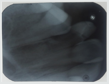
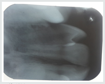
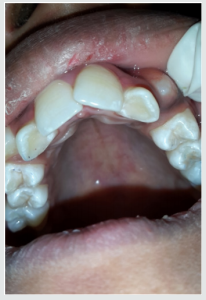
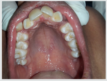
A 12-year-old female patient referred to the Pedodontics department with the complaint of irregularly arranged teeth. General health of the patient was normal and medical history was not relevant. Clinical examination revealed teeth 12 and 22 with a lingual pit. The pre-operative photographs were taken for the same (Figure 1). An intraoral Periapical radiograph was also taken for both the teeth, which revealed deep lingual pits involving the coronal part of teeth (Figure 2). As the teeth showed only incisal invaginations, and no pulpal involvement preventive treatment was selected. Also, if the pits were left untreated, it would harbor irritants and microorganisms further leading to caries. And as these pits were deep enough, the caries would progress rapidly into the pulp and cause pulpal infection leading to the need for endodontic treatments.
Figure 1: Pre-operative photograph.
lupinepublishers-openaccess-journal-pediatric-dentistry
Figure 2a: IOPA.
lupinepublishers-openaccess-journal-pediatric-dentistry
Figure 2b: IOPA.
lupinepublishers-openaccess-journal-pediatric-dentistry

Treatment Done

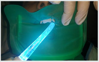
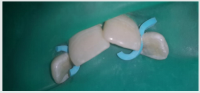
Following the oral prophylaxis, rubber dam placement was done for 12 and 22 (Figure 3). The teeth were initially washed, cleaned and dried. After that acid etching was performed using 37% phosphoric acid for 20 seconds. It was then washed and dried. Following that dentin bonding agent was applied with the applicator tip and cured for 20 seconds. The pits were then restored with flowable composite bilaterally (Figure 4). The teeth were then examined for any excess material and later finishing was done. The occlusion was also checked for any interference followed by post-operative photographs (Figure 5). Furthermore, the child was referred for orthodontic consultation.
Figure 3: IOPA.
lupinepublishers-openaccess-journal-pediatric-dentistry
Figure 3: Rubberdam placement.
lupinepublishers-openaccess-journal-pediatric-dentistry
Figure 4: Composite restoration.
lupinepublishers-openaccess-journal-pediatric-dentistry
Figure 5: Post-operative view.
lupinepublishers-openaccess-journal-pediatric-dentistry

Discussion

Dens Invaginatus has several variations of it. Oehlers gave a classification for various types of dens invaginatus [12]:
a) Type I: an enamel-lined minor form occurring within the confines of the crown not extending beyond the amelocemental junction.
b) Type II: an enamel-lined form which invades the root but remains confined as a blind sac. It may or may not communicate with the dental pulp.
c) Type III: a form which penetrates through the root perforating at the apical area showing a ‘second foramen’ in the apical or in the periodontal area. There is no immediate communication with the pulp. The invagination may be completely lined by enamel, but frequently cementum will be found lining the invagination.

Prevalence of Dens Invaginatus

The teeth mainly affected are maxillary lateral incisors and bilateral occurrence is not rare, they occur in almost 43% of all cases [13]. Swanson & McCarthy (1947) were the foremost to present bilateral dens invaginatus malformation, Conklin (1968) presented a patient with both maxillary central and lateral incisors affected bilaterally, and Burton et al. (1980) reported a case with six teeth involved, the maxillary incisors as well as the maxillary canines. Krolls (1969) also detected dental invaginations in maxillary central incisors and in many maxillary and mandibular premolars in a single patient. Conklin (1978) found the malformation in four mandibular incisors in one patient. There are many theories described in the literature to explain about the dental coronal invaginations some of them being:
a) The bulking of the enamel organ was thought to be caused due to the growth pressure of the arches [14,15].
b) Focal failure of the growth of the inner enamel epithelium leads to the invaginations; however, the surrounding epithelium continues to grow and engulfs the static area [11].
c) The invagination is due to the aggressive proliferation of a part of the inner enamel epithelium invading the dental papilla. It was thought as a ‘benign neoplasm of limited growth [16].
d) Protrusion and distortion of the enamel organ is thought to cause an enamel lined channel terminating at the cingulum or at the incisal tip. This might also lead to an irregular crown formation [17,18].
e) The ‘twin-theory’ [18] suggested a fusion of two toothgerms to be the causative factor.
f) Infection was also considered to be responsible for the anomaly [19].
g) Trauma was thought to be a reason, but they could not adequately explain why just maxillary lateral incisors were affected and not central incisors [20].
h) Most authors consider dens invaginatus as a deep folding of the foramen coecum during tooth development which may even result in a second apical foramen [3].
i) On the other hand, the invagination also may start from the incisal edge of the tooth. Genetic factors cannot be expelled (Grahnen 1962, Casamassimo et al. 1978, Ireland et al. 1987, Hosey & Bedi 1996).

Clinical Features

Due to the invagination there is just a thin layer of enamel and dentin left, which thus allows several microbes into the pulpal space easily. In some of the cases there is a complete enamel lining present. Also, channels may be present between the invagination and the pulp chamber. Pulp necrosis hence occurs in the earlier stages, within a few years of eruption and may be even before the root closure. The sequelae of untreated coronal invaginations can be as follows:
a) Abscess formation.
b) Retention of neighboring teeth.
c) Displacement of teeth.
d) Cysts, or Internal resorption.

Treatment Modalities

Preventive and restorative treatment should be opted for teeth with deep palatal or incisal invaginations and should be treated by fissure sealing [21]. Also, composite resin restoration followed by pit and fissure sealant can be done in the required cases [21]. Root canal treatment, surgical treatment and extraction of teeth should be done in severe cases wherein there is radicular invagination and pulpal involvement.

Conclusion


The clinician should be aware of occurrence of such tooth anomalies and should do a thorough clinical diagnosis as these anomalies are not rare. Earlier diagnosis and treatment of such cases can prevent further complications such as endodontic therapies and extractions. Hence the needed treatment should be done as early as possible and a periodic follow up should be maintained.

For more Lupine Publishers Open Access Journals Please visit our website:
http://lupinepublishers.us/
For more Open Access Journal on Pediatric Dentistry articles Please Click Here:
https://lupinepublishers.com/pediatric-dentistry-journal/

To Know More About Open Access Publishers Please Click on Lupine Publishers

 Follow on Linkedin : https://www.linkedin.com/company/lupinepublishers
 Follow on Twitter   :  https://twitter.com/lupine_online

Thursday, July 11, 2019

Analysis of Reasons for Extraction of Permanent Teeth in Children in Senegal: A Retrospective Study | Lupine Publishes

Lupine Publishers - Journal of Pediatric Dentistry


Abstract

Background: Loss of permanent teeth can have negative functional, psychological and social consequences, especially for children and adolescents with growing bone structures. Knowledge of their causes is of interest in the development of comprehensive dental public health programs. The objective of this study was to evaluate the reasons for extractions of permanent teeth in children in public oral structures in Dakar.
Material and method: A retrospective descriptive study, based on consultation registers and patient records, was performed. Patients between the ages of 6 and 15 who had permanent tooth extractions between January 2014 and August 2018 were included. A questionnaire including socio-demographic data, reason for consultation, reason for extraction and extracted teeth was included allowed to collect the data.
Results: A total of 321 patients aged 6 to 15 years received 375 permanent tooth extractions (1.16 teeth / child). Dental caries and its complications were the main reasons for extractions (94.7%).
Conclusion: Dental caries remains a real public health problem in developing countries. Decision-makers need to focus on strategies for the prevention and management of early childhood oral conditions to avoid the extraction of permanent teeth.
Keywords: Reasons for Extraction; Permanent Teeth; Child, Tooth Decay; Senegal

Introduction

The extraction of permanent teeth should not be an insignificant act, especially in children, because of the negative repercussions on eruption phenomena, the harmony of the arches, the primary functions of chewing, swallowing, breathing and phonation. Indeed, the first permanent molar, which is the first permanent tooth to erupt, is the keystone of the occlusion; it determines the shape of the lower part of the face and conditions the position and health of the other permanent teeth [1]. The decision to extract permanent teeth must be reasoned and integrated into a global treatment plan that often requires collaboration with other dental specialties. The analysis of the causes of permanent tooth loss is of interest to practitioners and decision-makers in order to develop control strategies to be integrated into overall dental public health programs. It is from this perspective that an indirect method based on the search for reasons for these permanent tooth losses by extraction has been developed and used in many countries. Numerous studies on the causes of permanent tooth extraction in children, adolescents and adults have been conducted in industrialized countries [2-5] and caries and periodontal disease have been the main causes of extractions. In Africa, studies are rare. The main objective of this study was to evaluate the reasons for extractions of permanent teeth in children in public oral structures in Dakar.

Materials and Methods

This was a descriptive retrospective study of patients consulting the oral structures of Aristide Dantec Hospital (HALD), Albert Royer National Children’s Hospital of Fann (CHNEAR), Dakar Institute of Odonto-Stomatology (IOS) and Grand Yoff General Hospital (HOGGY). Patient selection was based on consultation records. All records of patients aged 6 to 15 years who received permanent tooth extractions were included in the study. A “reasoned choice” sampling was conducted. A data sheet was used to collect information on socio-demographic data, the reason for consultation, the reasons for extraction, and the type of teeth extracted. The collected data were analysed with the SPSS 20.0 IBM software. The quantitative variables were described by their means and standard deviations. The qualitative variables were described by their numbers and percentages.

Results

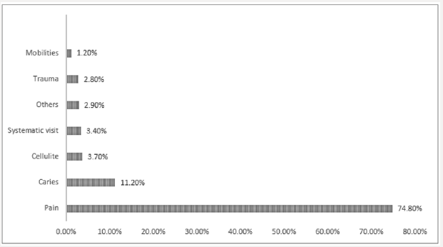
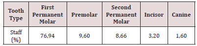
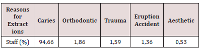
Among 26362 children consulted, 321 patients (1.21%) aged 6 to 15 years had received permanent tooth extractions. The number of permanent teeth extracted was 375 (1.16 teeth/child). Girls had received 55.2% of extractions. The number of permanent teeth extracted was 375 (1.16 teeth / child). Girls had received 55.2% of extractions. The 12-15 age group had benefited from 77.88% (Table 1). The distribution of extractions by health centre was 33.64% in the CHNEAR and 31.77% in HOGGY (Table 2). The first molars accounted for 76.94% of the extracted teeth (Table 3). Dental caries and its complications were the main reasons for extracting permanent teeth in 94.16% of cases (Table 4). Pain was the reason for consultation for 74.8% of extracted teeth (Figure 1).
Figure 1: Breakdown by reasons for consultation.
lupinepublishers-openaccess-journal-pediatric-dentistry
Table 1: Distribution of extractions by age group.
lupinepublishers-openaccess-journal-pediatric-dentistry
Table 2: Distribution of extractions according to the host structure.
lupinepublishers-openaccess-journal-pediatric-dentistry
Table 3: Distribution of extractions according to tooth type.
lupinepublishers-openaccess-journal-pediatric-dentistry
Table 4: Distribution by extraction reasons.
lupinepublishers-openaccess-journal-pediatric-dentistry

Discussions

The health structures selected in this study have the particularity of being centres receiving many patients for general health and oral health care. In addition, the CHNEAR and the IOS have services exclusively oriented towards the oral care of children and adolescents. The prevalence of permanent tooth extractions seems low but not negligible given the key role played by these teeth and in particular the first permanent molar. This result is lower than that reported by Murray et al [6] and Johansen et al [7]. This could be explained by the difference in the study population, which was made up of adults and elderly subjects (20-50 years and >70 years). Children in the 12-15 age group received more extractions than others. This shows that the number of permanent teeth extracted increases with age. Pain was the main reason for consultation and concerned the majority of patients. Studies by several authors [8,9] have produced similar results. This is linked to an economic situation and/or a lifestyle that is not compatible with a “preventive-conscious” approach. Difficulties in access to dental care, lack of dental facilities, lack of financial resources or lack of information and education on oral health may constitute a barrier to systematic visits or consultations as soon as the first signs of dental problems appear [10]. Dental caries and its complications were the most common reasons for extraction of permanent teeth. The first permanent molars were the most frequently extracted teeth. These data support the results of Shammari et al [11] who reported that dental caries was the leading cause of permanent tooth extractions in patients under 40 years of age and that the first permanent molars were the most affected teeth. Other studies [12-17] have shown that the first permanent molar was the most extracted tooth with prevalence ranging from 11.7% to 86.2%. A study by Safadi et al [18] in subjects aged 13 to 20 years showed that the prevalence of extractions of the first permanent molars was 31.3%, of which 76.5% concerned the first lower molars and that dental caries and its consequences were the main reasons for extraction. The greater susceptibility of the first molars to extraction can be explained by several factors. Their eruption, usually around 6 years of age, is silent and usually goes unnoticed, resulting in defective brushing. They have an occlusal surface whose morphology is more favourable to the retention of soft cariogenic deposits [19]. However, given their important roles, especially in children and adolescents, their extraction should be the ultimate therapeutic choice.

Conclusion

It should be noted that dental caries and its complications are the main reasons for the extraction of permanent teeth in children. It is important to implement a policy of promotion, prevention and early management of oral diseases in children aged 12 to 15 years in order to reduce dental loss.


For more Lupine Publishers Open Access Journals Please visit our website:

Wednesday, June 19, 2019

Lupine Publishers - Journal of Pediatric Dentistry

Oral Status of Diabetic Children in Casablanca (Case-Control Study) by Benkirane L in IPDOAJ - Lupinepublishers

Objectives: This study aims to assess the oral health status of diabetic children in comparison with a control group, to appreciate the impact of diabetes mellitus and its treatment on oral and dental health and to test their knowledge regarding oral hygiene.
Material and Methods: This transversal epidemiological study with comparative purpose was conducted on 100 diabetic children aged 2 to 16 years followed in the service of the University Hospital of Pediatrics II HAROUCHI and within the association “Hayat” to support children with diabetes, and 100 healthy model children aged 3-15 years from two public schools and one private school. Through a questioning and an oral examination, the parameters of diabetes, oral and dental hygiene, and the dental status were determined.
Results: The DMF index was more pronounced in the patient group (2.933) than in the control group (1.798) with a statistically significant difference (p = 0.002). The mean Plaque index was 1.338 for the patient group versus 1.542 for the control group. In the group of sick children and tartar was present in 62% of cases.
Conclusion: In the waning of this study, it was shown that diabetes has a significant impact on teeth and oral health of children. Following these results, the oral care needs are high among the population of children with diabetes.


For more Lupine Publishers Open Access Journals Please visit our website: 
For more Open Access Journal on Pediatric Dentistry articles Please Click Here: 

Wednesday, May 22, 2019

Lupine Publishers - Open Access Journal of Pediatric Dentistry


The aim of this article is to highlight an uncommon eruption sequence of deciduous maxillary central incisors, create awareness and its early management if indicated. There might be underlying systemic cause and a prompt visit to a dentist might help in arriving on a proper diagnosis, and thus improve prognosis.


For more Lupine Publishers Open Access Journals Please visit our website: http://www.lupinepublishers.com/


Wednesday, May 1, 2019

Lupine Publishers - Dental and Oral Health Journals Impact Factor


Effects of Fluoridated and Non- Fluoridated BleachingAgents on Bovine Surface Enamel by Prashant Babaji in IPDOAJ - Lupinepublishers

In the modern era, aesthetic dentistry is becoming famous. The demand of tooth bleaching is increasing day by day and it has become the treatment of choice for tooth discoloration. If there is deposition of chromatogenic material into dentin and enamel during the tooth development stage or after eruption, it leads to intrinsic tooth discoloration [1]. Among various bleaching techniques for intrinsic tooth discoloration, vital and non-vital bleaching techniques are common one.


For more Lupine Publishers Open Access Journals Please visit our website: http://www.lupinepublishers.com/

Wednesday, April 17, 2019

Lupine Publishers - Dental and Oral Health Journals Impact Factor


Damage to the teeth of a young child can leave serious and prolonged periods of time, leading to a change in color, deformity or possible loss of it. The psychological impact of such damage can be very wide. Therefore, the pediatric dentist should have the following characteristics such as: having the knowledge of the treatment of the traumatized tooth and being available to provide the necessary treatment and services at any time of the day.


For more Lupine Publishers Open Access Journals Please visit our website: http://www.lupinepublishers.com/

Wednesday, March 13, 2019

Lupinepublishers - Journal of Pediatric Dentistry


This study was carried out to determine the prevalence of HBsAg among pregnant women attending antenatal clinic at General Hospital in Agba, Ekwulobia, Anambra State. Two hundred pregnant women who consented voluntarily after thorough explanation of the purpose of the study were recruited for this study. The samples were tested for the presence of antibodies using rapid Dia Spot HBsAg rapid test strip developed by Dia Spot Diagnostics, USA and ClinotechHBsAg (Clinotech diagnostics, Richmond, Canada). Analysis of the result showed that 23(11.5%) of the pregnant women had HBsAg.


For more Lupine Publishers Open Access Journals Please visit our website: http://www.lupinepublishers.com/

Wednesday, February 20, 2019

Dentistry Journals - Lupinepublishers


Strategies for Reducing Dental Stress in Children by Karimi in IPDOAJ - Lupinepublishers

The dental office for children has always been scary and eerie. The reason for this fear is the lack of mental fitness of children towards the dental health environment. Unknown instruments, scary sounds of dental devices, and sometimes seeing others’ fears during dental procedures would affect the child’s mind; and creates fear within them. No research has pointed to the definitive causes of anxiety and stress, but some studies have shown that the bad experience of dental treatments associated with pain is one of the most common in this case.


For more Lupine Publishers Open Access Journals Please visit our website: http://www.lupinepublishers.com/

Friday, January 11, 2019

Posterior Indirect Adhesive Restorations: for Vital Posterior Teeth: Clinical Report (IPDOAJ) - Lupinepublishers


This article describes a case of compromised posterior vital teeth treated by indirect bonded ceramic restorations. A 24 year old female patient presented to the department of Prosthodontics. He was complaining about sensitivity in the posterior region. A comprehensive examination revealed failing amalgam restoration in the first molar with compromised cusps in the first molar. The treatment plan included indirect bonded ceramic restorations consisting of inlay and on lay on the concerned teeth.


For more Lupine Publishers Open Access Journals Please visit our website: http://www.lupinepublishers.com/

Monday, December 17, 2018

Effect of Twin Block Appliance on a Growing Patient – A Case Report (IPDOAJ) - Lupinepublishers


Twin block appliance is very effective in a growing patient. The successful use of this appliance in the treatment of skeletal Class II malocclusion is based upon factors such as; age of patient, compliance of the patient and other case selection criteria. This case report includes a 14-year-old boy who was treated with twin block appliance. If treated timely, growth modification and orthopedic effects with this appliance can be achieved in a growing individual. This appliance is very successful in a patient with retruded mandible and well aligned arches in whom VTO is positive. This brings the mandible forward and improves the profile immediately.

https://www.lupinepublishers.com/pediatric-dentistry-journal/abstracts/effect-of-twin-block-appliance-on-a-growing-patient-a-case-report.ID.000117.php

https://www.lupinepublishers.com/pediatric-dentistry-journal/fulltext/effect-of-twin-block-appliance-on-a-growing-patient-a-case-report.ID.000117.php


For more Lupine Publishers Open Access Journals Please visit our website: http://www.lupinepublishers.com/

For more peer reviewed dentistry journals Please Click Here: 

Friday, December 7, 2018

Oral Hemangioma Management in Children: A Concise Review (IPDOAJ)- Lupinepublishers


Head and neck region bears the burden of approximately 60% of all the vascular anomalies diagnosed, affecting about 1 in 22 children. These lesions, irrespective of the size, when present on the face are a major cause of esthetic concern and patient usually seek treatment for the same. Various treatment modalities are available and sclerotherapy with intra/peri-lesional injections of 03% Sodium tetra decylsulphate remains the mainstay of treatment in most cases. This article provides a concise review regarding various management options available for smaller vascular lesions especially emphasizing the role of sodium tetradcy sulphate.

https://www.lupinepublishers.com/pediatric-dentistry-journal/abstracts/oral-hemangioma-management-in-children-a-concise-review.ID.000114.php

https://www.lupinepublishers.com/pediatric-dentistry-journal/fulltext/oral-hemangioma-management-in-children-a-concise-review.ID.000114.php


For more Lupine Publishers Open Access Journals Please visit our website: http://www.lupinepublishers.com/

Friday, November 30, 2018

An Unusual and Aggressive Ewing Sarcoma Involving Parapharangeal Space: A Case Report and Review of Literature (IPDOAJ) - Lupinepublishers


Ewing’s sarcoma (ES) is a malignancy primarily affecting bone tissue that is commonly diagnosed in adolescents and young adults. Its occurrence in the head and neck region is unusual and generally involves the mandible and maxilla. We report here a case of Extraskeletal Ewing’s sarcoma (EES) of the Parapharangeal space in a 20 year-old female who presented with blurred vision, dysphasia. CT examination showed a large, seemingly well-defined soft tissue mass in the left side of face involving parapharyngeal space with intracranial extension. The patient undergone chemotherapy and radiotherapy. A review of the literature revealed only one previous reports of EES occurring in the parapharyngeal space.

https://www.lupinepublishers.com/pediatric-dentistry-journal/abstracts/an-unusual-and-aggressive-ewing-sarcoma-involving-parapharangeal-space-a-case-report-and-review-of-literature.ID.000112.php

https://www.lupinepublishers.com/pediatric-dentistry-journal/fulltext/an-unusual-and-aggressive-ewing-sarcoma-involving-parapharangeal-space-a-case-report-and-review-of-literature.ID.000112.php

For more Lupine Publishers Open Access Journals Please visit our website: http://www.lupinepublishers.com/

980 nm Diode Laser: A Good Choice for the Treatment of Pyogenic Granuloma

Abstract Pyogenic granuloma is a benign non/neo plastic mococutanous lesion . It is a reactional response to constant minor trauma and ca...