Showing posts with label Pediatric Dental Care Journal. Show all posts

Friday, April 28, 2023

Pallor and Whitening of the Gums in Children


Abstract

Whitening of the gums often indicates that there is a problem with the health of the mouth. Many conditions can lead to gingival whitening, from Aphthous ulcers to long-term inflammatory disease. This condition may be accompanied by some signs and symptoms. In some rare cases, whitening of the gums can be a sign of oral cancer; hence, there should be a concern of the parents to see a pediatric dentist for a correct diagnosis.

Keywords: Gingival whitening; Aphthous ulcer; inflammatory disease; oral cancer

Read More About This Article Please Click on Below Link:
https://lupinepublishers.com/pediatric-dentistry-journal/fulltext/pallor-and-whitening-of-the-gums-in-children.ID.000245.php

Read More About Lupine Publishers Google Scholar Articles:
https://scholar.google.com/citations?view_op=view_citation&hl=en&user=h1QvhsYAAAAJ&citation_for_view=h1QvhsYAAAAJ:lSLTfruPkqcC

Tuesday, December 21, 2021

Lupine Publishers | The Applied Importance of Pediatric Dentistry (An Overview)

 Lupine Publishers | Journal of Pediatric Dentistry


Abstract

Today, with the promotion of community awareness, fortunately, the importance of oral health and maintenance of primary teeth for parents is clear. Due to the important roles of primary teeth in the oral system, these teeth should be maintained until the eruption of the permanent teeth. For this reason, it seems the parent’s perspective has changed, and children’s dentistry is now a priority for most families. Pediatric dentists should be specialized in different fields of pediatric dentistry. They will be training in the fields of pediatric psychology, child anesthesia and sedation, and age-appropriate behavior management so that they can manage children’s treatments in the best way. This group of dentists is specialized in eliminating child concerns and anxiety too. They can distract children’s minds by talking to them from what they plan to do. This friendship makes the child’s fears go away and the treatments are accomplished positively so that the child would not feel any bad dental experiences at all. Pediatric dentists provide the parents with the necessary training information to protect their children’s teeth from the very beginning of their childhood.

Keywords: Pediatric dentists; pediatric psychology; child anesthesia; sedations; elimination of anxiety; child behavioral management

Introduction

Pediatric Dentistry is one of the branches of dentistry in which the dentist works with children and adolescents. In 1909, Minnie Evangeline Jordon established the first dental practice in the United States devoted only to pediatric patients [1]. This discipline was first recognized by the American Dental Association in the United States [2] and subsequently graded in other countries as a separate specialty. In general, for a general dentist to receive a child’s specialty, he must complete two to three years of special training. In this area, the dentist not only focuses on oral and dental health, but he will also be interlinked with children and the psychological behavior of the children. Children have their mental characteristics and do not tolerate oral examination and dental treatments; consequently, they may not cooperate well with the general dentist. In pediatric dentistry, the dental practitioner thoroughly performs examination and treatment with the use of applied techniques, so that the child will become fully cooperative. To ease the process of dental treatment, they will use the appropriate and fun tools for the child to accept the dental procedures. The dental office environment is also one of the factors that can be effective in improving the treatment in the child [3]. Entertaining the child before and during treatment with toys and cartoons is one of the things that play significant effects [3-5]. The pediatric dentist offers a range of therapeutic approaches that can help the improvement of the oral health status of the child. The pediatric dental specialization has given special attention to dentistry in children from birth to adolescence. Due to the importance of pediatric dentistry, a pediatric dentist must have the experience, expertise, and the ability to maintain the health of teeth, gums. In general, the oral conditions of children can be monitored by a pediatric dentist at different stages of the child’s development [4]. If oral health is neglected, the likelihood of developing dental caries and other illnesses that cause permanent complications for the teeth of the baby will be very high.

Pediatric Dentistry Features

In this article, we intend to introduce some of the most important features of pediatric dentistry.

Oral health education

Tooth decay, as well as non-compliance with health in children, reduce their school performances and create poor social relationships [6-9]. Children’s dentistry with providing a pleasant and educational environment for children and adolescents not only reducing their stress but also can greatly increase the awareness of children and prevent the occurrence of various types of oral and dental diseases [7].

Specialized children’s dental services

Primary teeth are very important in childhood and adolescence, which plays an important role in their correct chewing of foods. Not only does this contribute to the health of children, but it also helps children to speak the words correctly. All services such as primary teeth restoration, pulpotomy and pulpectomy procedures, SS crowns, fluoride therapy, and permanent teeth services in children are provided by children’s dentistry.

General anesthesia

One of the causes that prevent children from visiting the dentist is the fear and anxiety in referring to pediatric dentistry [5,10]. This fear causes the child to suffer from oral and dental illness due to a lack of regular referral. In a general dental office, due to a lack of knowledge of the psychology of the child, it is not possible to provide a warm and comfortable environment for children. Referring the child to the pediatric dentist, in addition to addressing the problems caused by the child’s lack of health, allows the child to address the dental and oral problems in a happy and intimate environment. Today, for children with anxiety and fear of referring to dentistry, general anesthesia is used during examination and dental procedures to increase the accuracy and quality of the work of the pediatric dentist.

Application of Hypnosis (artificial sleep)

Oral and dental hygiene is one of the things parents should consider from childhood. Since children are sensitive and fearful of the dental problem, many solutions have been proposed to counteract it. Sometimes children’s dental conditions are in a special situation where pediatricians turn to the use of hypnosis in children’s dentistry [11]. Dissatisfied children with a weak collaboration attitude could delay the treatment in most cases. This artificial sleep in dentistry is done by an anesthetist and is one of the newest dental care services. Although artificial sleep does not make the child feel unfamiliar with the office environment the advantage is the dental work is accomplished while they are sleeping.

Promoting the right habits

While a child may not need a dentist at the age of two, it is important to start training oral hygiene habits as soon as possible [12-14]. During dental examinations, the pedodontist can help the child learn how to use a toothbrush, or how to use dental floss to prevent dental caries and dental diseases. Considering that good habits can be formed from childhood and adolescence, so constant referral to the dentist can enhance that.

Features of Pedodontist

a) Pediatric dentists must be an expert in child psychology and know how to deal with children; they have to able to get the patient’s confidence in the shortest possible time [4].
b) Pediatric dentist should be able to interact with the parents; should not speak harshly and should not blame parents for the delayed treatment processes [4].
c) Because the child’s patience is very limited, the dentist should be on time, and the patient should not wait for a long time in the waiting room [4].
d) During the examination, the Pedodontist must show a high degree of accuracy and focus on the problem [4].
e) During the dental procedure, he has to avoid talking about things that are not suitable for a child’s age [4].
f) Treating adolescents like adults, should be another critical criterion that a Pedodontist must pay close attention to that.

Benefits of Pediatric Dentistry

Perhaps some parents think about what the difference between dentists is. All dentists are informed about dentistry, so they can refer to any dentist for treatment. But parents may not be fully aware of the benefits of pediatric dentistry. We review these advantages.

Maintaining oral and dental health

Perhaps one of the most important benefits of pediatric dentistry is that these professionals try to keep the child’s mouth clean and healthy by using the best training and educational tools, special features, and appropriate accessories. Constant and ongoing periodic examinations can help parents alleviate the child’s oral and dental illnesses, as well as help them to take care of regularly, and identify potential problems that may appear in the future.

Preventing early teeth loss

When parents think of the health of kids’ teeth, it is better to start their job right now! Pediatric dental care ensures that the child’s teeth will be healthy, free from decay and other dental diseases in the future. Dental caries is the most common chronic infectious disease of childhood [15]. Streptococcus Mutans, Lactobacilli, Actinomyces spp. and some other anaerobic bacteria are the most primary etiologic agents of dental caries [16]. Dental services and periodic examinations of children regularly can help prevent this complication and other types of other diseases of the mouth, teeth, and gums. Diagnosis of cavities in the teeth of a child is a critical issue insomuch as the lack of continuous care and periodic examinations can lead to more acute problems, such as tooth loss shortly.

Gaining awareness of the important issues of oral health of children

Oral health has to be started at the very beginning of the baby’s birth. Even if mothers are nursing or feeding with a bottle of milk, it is necessary to start a good oral hygiene program from the very beginning and as soon as possible. Before teething, mothers should keep the gums clean with soft and wet cloth. When teeth begin to erupt, they should also be cleaned. It is essential to use the right toolset for this daily work. For example, it is necessary to buy a special toothbrush for a baby that is more suitable for his little mouth. A Pedodontist keeps them healthy by removing microbial plaques and impacted food which may be stuck in between the teeth in a routine dental examination. This is an important part of pediatric dental care because it prevents the growth of bacteria that can lead to dental diseases such as tooth decay or inflammation of the gum tissue.

Children’s dentistry and dental care

The most important background for children’s dentistry is the prevention of dental decay and intensive care for their teeth. This care should be continued under the supervision of a pediatric dentist which starts at the age of one when the baby’s teeth begin to erupt and gradually would be replaced with permanent teeth until the age of ten to twelve. It is best to take the child to a pedodontist at the earliest age and repeat this procedure every six months. This prompts not only make the child become familiar with the dental environment but also it causes the child’s fears to diminish. More importantly, in both childhood and early youth, the kid’s oral health will be under the supervision of the pediatric dentist.

The Importance of Referring to the Pediatric Dentist

Oral health is an important part of public health that plays a significant role in the quality of life of people in society, especially children. Unfortunately, the frequency of teeth decay is often high due to the lack of attention paid by both parents and children. One of the most effective measures to improve the oral and dental health of children is conducting examinations by pediatric dentists [4,12]. To refer to a pedodontist office, parents should not wait for the pain and dental problems of the child because this delay can cause more severe problems. The primary teeth are very important and depend on the evolutionary stages necessary for the health of the children’s permanent teeth. Pedodontist can help parents to provide the necessary health care for children’s oral health and give them the necessary advice. A pediatric dentist has some special skills in oral health and dental care from childhood to adolescence. He has the expertise and experience to protect and maintain and treat the teeth, gums, and oral tissues of a child of all ages. Since the first teeth have erupted, regular consultation with a pedodontist is mandatory. This visit should be done every six months. These referrals help both parents get the right information and advice on how to protect the health of the child’s teeth. Furthermore, the child will get familiar with the office environment and its staff, and get some pleasant dental experience before having caries, pain, or dental abscesses. In this case, the child will be more cooperative in future treatment sessions.

Why are Periodic Dental Referrals Essential?

Nowadays, parents should be aware of the need for periodic examinations before further destruction of children’s teeth is taken into consideration. Regarding the principle of the existence of a problem, faster treatment will ensure longer teeth life. Moreover, it will increase the function of the teeth and will include a lower cost. Because if they close their eyes to the problems, those problems will get worse every day, and these will not ever stop. Problems can be accumulated and result in tooth loss, fabricating of space maintainers and apace retainers for the edentulous area, troublous and costly orthodontic treatments, implant-prone treatments, and so on. In other words, if parents fail to take the advantage of these referrals, they will be damaging to their children’s oral health which is hard to compensate and consequently, its effects remain until the end of their lives. Effects that are being handled on time definitely would be crucial in terms of cost, pain, and the length of the period of recovery for their parents and their children.

Conclusion

Pediatric dentistry is a branch of dentistry which concerns with dental and oral health in children. Since oral and tooth injuries start in childhood and adolescence ‘prevention and treatment of these injuries are very important in this period. For this reason, pediatric dentists not only can increase the level of knowledge of children and adolescents in oral health but also can prevent many abnormalities in the mouth and teeth. On the other hand, a pedodontist in addition to knowledge of dentistry has learned the principles of behavioral control of children. The dentist will provide attractive and pain-free behavioral therapy techniques for children. It is interesting to know that some dentists manage the treatment of children with cerebral palsy, mentally retarded, or even autism. The difference between children’s dentistry and other branches of dentistry is how to deal with children.

Read More Lupine Publishers Pediatric Dentistry Journal Articles:
https://lupine-publishers-pediatric-dentistry.blogspot.com/

Friday, July 2, 2021

Lupine Publishers | Long-Term Treatment of Early Childhood Caries (ECC)

 Lupine Publishers | Journal of Pediatric Dentistry


Abbreviations: ECC: Early childhood caries; CDC: Center for Disease Control; NHANES: National Health and Nutrition Examination Surveys

Introduction/Literature Review

Early childhood caries (ECC) is an infectious disease affecting the primary dentition in infants and children younger than 6 years of age. Despite being a largely preventable condition, ECC remains a major public health problem in both developing and developed countries, with nearly 1.8 billion new cases diagnosed each year globally [1,2]. In the United States, Center for Disease Control and Prevention (CDC) reported a 15.2% increase in the prevalence of dental caries among the nation’s youngest children, aged 2-5, comparing the findings of two National Health and Nutrition Examination Surveys, NHANES II, 1988–1994 and NHANES III, 1999–2004 [3,4]. A comprehensive epidemiological review of ECC showed that although the overall ECC prevalence varies from population to population, the socio-economically disadvantaged and minority groups are most at risk [5]. Numerous studies indicate that Hispanic and Native American children are among the high-risk North American populations [6]. ECC is multifactorial in its etiology and has been associated with a multitude of risk factors including inappropriate and prolonged bottle or breast feeding, frequent consumption of fermentable carbohydrates, and poor oral hygiene [6-8]. Vertical transmission of Streptococcus mutans from caregiver to child has been demonstrated [9]. A previous study has shown that S. mutans or S. sobrinus alone may not be the only indicators for high risk of caries, but the prevalence of S. oralis in saliva may be a risk factor for increased caries activity in severe ECC forms [10]. Being born preterm or small for gestational age [11], improper oral health practices, higher birth order, low fluoride in drinking water, poor saliva buffering capacity and behavioral factors are other risk factors [12,13]. The initial clinical presentation of ECC appears as white-spot lesions in the upper primary incisors along the margin of the gingiva [14]. Complete destruction of the crown can potentially follow if the disease continues [15]. The spread of caries to primary maxillary molars is seen in moderate cases and primary mandibular molars are involved in severe stages [16]. Greater probability of subsequent caries in both the primary [17] and the permanent18 dentitions is observed among the children who experienced caries as infants or toddlers [18]. Missing anterior teeth causes poor phonetics, unpleasant appearance, hardship in sociological and psychological adjustment, and nonefficient mastication [19]. Several studies have demonstrated the considerable negative impact ECC has on the health-related quality of life of children [20].

Maintaining the primary dentition in a healthy condition has innumerable benefits, therefore dental caries management in children is pivotal. Treatment options include the restoration or extraction of the carious teeth although the importance of preventative and preservative measures cannot be overstated. Construction of orthodontic appliances such as space maintainers or prosthetic management may be needed in some cases. The following case demonstrates the use of the Nance appliance and the Hyrax expander in addition to conventional orthodontic treatment over the course of nine years in order to obtain a Class I (Angle) canine and molar relationship in a child with severe early childhood caries.

Diagnosis

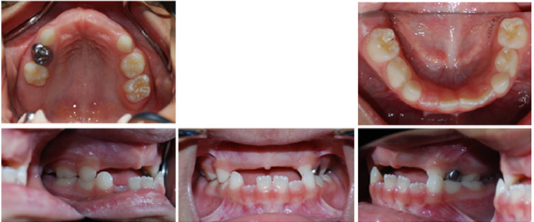
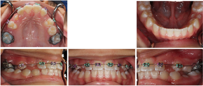
The patient was a four-year-old Hispanic female that presented with the chief complaint of “missing frontal and some back teeth having difficulty chewing and speaking.” No remarkable medical findings were noted. A facial evaluation revealed a symmetric face. She had a unilateral posterior cross bite with flush terminal deciduous molar relationship (Figure 1).

Figure 1.

Lupinepublishers-openaccess-pediatric-dentistry-journal

Treatment objectives

The primary objectives of treatment were to maintain the space and replace the missing anterior teeth, to obtain a Class I (Angle) canine and molar relationship, with ideal overjet and overbite, and to improve facial esthetics. The complementary treatment objectives were to establish good functional and stable occlusion, to avoid crowding and lack of space of the upper incisors and premolars, while improving smile characteristics and eliminating lip incompetence.

Treatment progress

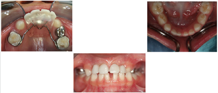
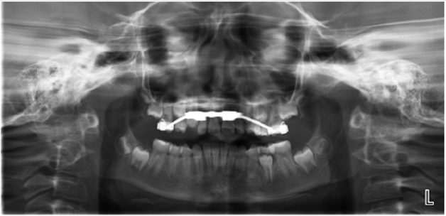
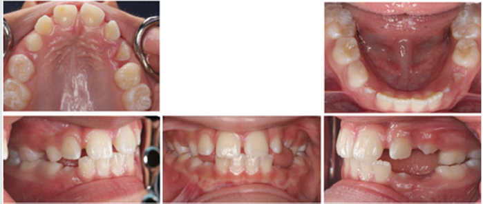
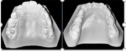
The treatment of choice was utilization of Nance appliance followed by conventional orthodontic treatment. After consultation with the patient and her parents, informed consent was obtained and treatment was begun. All components of the appliances were submitted to a dental laboratory for fabrication. Modified Nance appliances was fit on deciduous maxillary first molars at the age of four and a half Figure 2. The Hyrax appliance was fixed on teeth #3 and #14 when the patient was 11 years old Figure 3. The appliance’s screw was turned twice a day until proper palatal expansion was achieved Figure 4. Treatment was followed by comprehensive orthodontic treatment lasting approximately 30 months Figure 5(A&B). Immediate Essix retainers were placed at the deboning appointment Figure 6.

Figure 2.

Lupinepublishers-openaccess-pediatric-dentistry-journal

Figure 3.

Lupinepublishers-openaccess-pediatric-dentistry-journal

Figure 4.

Lupinepublishers-openaccess-pediatric-dentistry-journal

Figure 5a.

Lupinepublishers-openaccess-pediatric-dentistry-journal

Figure 5b.

Lupinepublishers-openaccess-pediatric-dentistry-journal

Treatment Results

Excellent facial and occlusal results were achieved with the nine-year treatment rendered. The most significant changes were the correction of the unilateral cross-bite malocclusion of the deciduous teeth and prevention of mesial shifting of posterior teeth in order to achieve both a molar and canine Class I (Angle) relationship of permanent dentition. The change in appraisal posttreatment also demonstrated normal occlusion with proper alignment of anterior teeth.

Discussion

Utilization of Nance appliance to avoid any further mesial shifting of posterior teeth was a pivotal part of the treatment. Reconstruction of anterior teeth helped the patient improve her speech and have a more desirable smile. Construction of a removable partial denture could be a different way to approach treatment in this case. It must be noted, however, that removable partial dentures have certain disadvantages such as presence of extra material in the mouth and increased speech difficulty for the child. They may be broken or lost easily and are ineffective to maintain space if not used properly. Since children are not competent to wear removable appliances, the ideal treatment option was a fixed non-functional appliance. The Hyrax appliance is a fixed functional appliance which was used during the treatment to correct the unilateral posterior cross bite and to expand the narrow maxillary arch. The alternative choice of appliance was the Haas expander. The latter option was not chosen due to requiring a more demanding oral hygiene regimen while offering the same treatment outcome as the Hyrax appliance in this case. Conventional orthodontic treatment was an inevitable part of the treatment plan to obtain Class I (Angle) canine and molar relationship, with ideal overjet and overbite which helps the patient to have proper tooth position in jaws, improve facial esthetics and reduce dental caries risk and future complications. Relying on conventional orthodontic treatment alone without the use of an expansion would not have been the proper approach since the Hyrax appliance helped with the expansion of the maxillary arch prior to the complete closure of the mid-palatal suture.

Read More Lupine Publishers Pediatric Dentistry Journal Articles:
https://lupine-publishers-pediatric-dentistry.blogspot.com/


Friday, March 19, 2021

Lupine Publishers | The Time Has Come for Pediatric Dentistry and Medicine to Develop A New Thought Process, Rather Than Just Thinking Outside the Box When Caring for Newborn Babies and Infants, We Need A Totally New Box

 Lupine Publishers | Journal of Pediatric Dentistry


Introduction

The concept of thinking outside the box is no longer valid, we now need to create a new box. The volumes of research showing the merging of medicine and dentistry is growing rapidly and those who fail to grasp its importance will be left behind. Our primary goal in caring for newborn infants and babies should be to allow them to grow and develop to their maximum potential. It’s time for the old, outdated conservative ideas, to be shed and have the Dental and Medical community understand that the separation of medicine and dentistry is rapidly disappearing.

Collaboration does not turf concerns

The primary example of this is in the diagnosis and treatment of tethered oral tissues also known as TOTS. In the early 1970’s less than 22 % of US women were breastfeeding their newborn infants, many who were, did so because they could not afford the cost of infant milk formulas [1]. The formula industry has always advertised the benefits of their product over mother’s milk and since so few mothers were nursing, difficulties of nursing due to tongue or lip ties were dismissed or just plain ignored by the medical providers. Today, we have over 80% of mothers breastfeeding , we also are aware of the many long-term benefits for both the infant and mother. We also know that mothers who wish to breastfeed include all socioeconomic levels of society. It is not a fad as many in the medical community refer to treatment of TOTs.
Yet, the medical community still all too often rejects the idea that TOTS do prevent a baby from achieving a good secure attachment to a mother’s breast, resulting in a plethora of symptoms for both mother and infant, often resulting in the mother giving up and using a bottle. Some explain this failure of the Medical community to embrace the release of these TOTS as overstepping our scope of practice and consider it their turf, yet they do nothing. The effects of TOTS begin in the oral cavity, where we as pediatric dentists, are the experts. Improving the health care of mothers and infants and should not involve turf wars and protectionism for physicians and dentists. We need to effectively join forces to create a successful breastfeeding outcome. Dentistry and medicine treat the same entity, where often one or many health problems can overlap into both professions’ territory. This is a key reason why Medicine needs to develop a cooperative team approach to breastfeeding issues with Dentists [2,3]. The understanding in treating infant oral health is growing and changing, this presents an excellent opportunity for physicians and dentists to find ways in to effectively join forces to create a successful breastfeeding outcome.

Infant- Mother Bonding

It is time to look beyond just the infant’s latch, but the actual potential complication that a poor latch creates beyond the act of breastfeeding comfortable for the Mother. Medicine recognizes the term attachment theory, which states that the babies initial bonding with the mother lasts a lifetime, if it is short circuited, the mother may develop signs of post-partum depression, which rather than treating symptoms pharmacologically, in many cases this may be resolved if we , as pediatric dentists can fix the inability of the infant to latch onto the breast by a simple in office surgical release, not just a simple snip, known as a frenectomy [4]. Breastfeeding plays an integral role in forming the deep attachment between mother and baby.

Failure to thrive

Infants are far too often placed in hospitals for failure to thrive, parents are occasionally subjected to Child abuse accusations because their child appears malnourished, and in severe cases have had nasal-gastric or tubes inserted directly into the stomach surgically to literally force feed infants [5]. All this has happened because the physician, pediatrician, ENT or GI doctors never even consider the ankylosed tongue as the primary cause.

Brain growth and development

The window of opportunity to help many babies may close with-in the first 90 days after the infant’s birth [6]. Studies indicate that a baby’s brain grows by 1 percent each day beginning right after birth. A newborn brain grows extraordinarily fast right after birth but slows down to a growth rate of 0.4 percent per day by the end of three months. An infants’ brains grow by 64 percent in the first 90 days, according to the study. The average brain size was 20 cubic inches (341 cubic centimeters) at birth, and 34 cubic inches (558 cubic cm) at 90 days. Simply stated, the brains of newborns grew from about 33 percent of the average adult brain doubles in size in the first year, and by age three it has reached 80 % of its adult volume.

Air induced reflux

It was those hellish hours between darkness and dawn when my husband and I would take turns walking around the house with our baby inn our arms, praying and, mostly in my case, sobbing because we couldn’t console our baby. Infants suffering from reflux are still being treated with adult anti-reflux drugs rather than looking a t the most likely source of gas build up, the swallowing of air during the infant’s latch due to a poor latch and seal onto the mother’s breast. Adult pharmaceuticals such as Peracid and Nexium are not approved drugs for children under age one [7-10]. Studies show they are not effective. Yet the use is skyrocketed in recent years. Simply releasing TOTS will allow a good secure latch to occur in most infants, quickly resolving the issue. Medications like ranitidine (Zantac) or omeprazole (Prilosec) can prevent absorption of calcium and iron and increase the risk of certain intestinal and respiratory infections. Children who used PPIs had a 22% increased likelihood of fracture, while children who used both PPIs and H2-blockers had a 31% increased likelihood of fracture.

Sleep disordered breathing

Tethered oral tissues (TOTS) is also a common undiagnosed source of obstructive airway problems, obstructive sleep apnea and reflux in infants and newborns [11-16]. The American Academy of Pediatric Dentistry (AAPD) recognizes that obstructive sleep apnea (OSA) occurs in the pediatric population. In order to reduce such complications, AAPD encourages healthcare professionals to routinely screen their patients for increased risk for OSA and to facilitate medical referral when indicated The most common form of Pediatric OSA is a disorder of breathing characterized by prolonged, partial upper airway obstruction and or intermittent/ complete obstruction (obstructive apnea) that disrupts normal ventilation during sleep and normal sleep patterns. These cycles of awakening prohibit the infant as well as toddlers having apnea from reaching deep, restful sleep. For this reason, children with untreated OSA may be inappropriately diagnosed as having ADHD. Often the ankylosed tongue is again ignored and not considered as a part of a differential diagnosis. Findings from studies that used complementary research methods have converged to strongly suggest that inadequate sleep quality and quantity are causally linked to sleepiness, inattention, and probably other cognitive and behavioral deficits that impact daytime functioning, with potential implications for long-term development.

Conclusion

It has been stated that it can take a 17-year lag to change and understand traditional research. This lack of knowledge puts those responsible for enabling new research at a disadvantage [17]. A staggering 36,000 randomized controlled trials (RCTs) are published each year, on average, and it typically takes about 17 years for findings to reach clinical practice [18]. We do not have this time to waste in treating our infants and newborns. We need to establish a good collaboration and trust between our two professions and develop a good understanding of how tethered oral tissues can have a significant impact on the overall growth and development of newborns and infants [19].

Read More Lupine Publishers Pediatric Dentistry Journal Articles:
https://lupine-publishers-pediatric-dentistry.blogspot.com/


Friday, March 12, 2021

Lupine Publishers | Anterior Open Bite Using Simões Network in Growing Patient: A Case Report

 Lupine Publishers | Journal of Pediatric Dentistry


Abstract

The anterior open bite is characterized by the negative vertical overhang occurring in the anterior region. It consists of a discrepancy in the vertical direction and is one of the malocclusions with greater aesthetic-functional impairment, besides dental and skeletal alterations. It has a high prevalence in the deciduous and mixed dentition and its etiology is multifactorial, highlighting the deleterious oral habits as the most prevalent. The objective of this study was to present the clinical case of a growing female patient presenting an anterior open bite associated with thumb sucking, by means of treatment with the functional orthopedic device Simões Network (SN3). During the first 12 months follow-up, we observed facial and intraoral oral changes and forward, the improvement of functional and craniofacial relationships, observed through complementary tests.

Keywords: Open Bite; dentition, mixed; orthopedics

Introduction

The balance of soft tissue growth and facial changes are important for the craniofacial development. Mineralized bone is formed through a process known as ossification by the membrane activity providing the function of remodeling and displacement. Breath, chewing, phonation and swallowing functions are prior to the regular growth development [1]. Thumb or pacifier sucking, and tongue thrusting may cause a disorder knowing as an anterior open bite. The severity of the malocclusion will be according to the magnitude, frequency and time of the habit [2]. Prolonged breastfeeding will be recommended to avoid nonnutritive sucking habits, as the sucking of fingers, pacifiers and bottle feeding [3,4]. Anterior open bite (AOB) is defined as the lack of incisal contact between anterior teeth in centric relation. AOB creates aesthetics problems, speech disorders and tongue thrusting habit [5]. This malocclusion requires early treatment due to all the etiological factors mentioned before. The stability will be achieved in a long term; thus, the pediatric dentist must be alert and minimize the problem as soon as possible in attempt to decrease the time of the treatment and to maintain the stability 5. The auto correction index is low when the correct habits are achieved [6,7]. The prevalence in the population ranges from 1,5% to 11%. Some authors also describe that 17% to 36% of those seeking orthodontic treatments feature AOB [8-11]. This malocclusion may also occurs due to a skeletal component classified open bite into dental and skeletal, associated with excessive molar height, divergent upper and lower occlusal planes, steep mandibular plane angle, increased gonial angle, short mandibular ramus, downward rotation of posterior part of the maxilla or palatal plane tipped up anteriorly, increased lower anterior facial height and decreased upper anterior facial height. According to severity, modalities of treatment are required: growth modulation; orthodontic mechanotherapy and the combination with orthognathic surgery [12-14]. Orthopedics devices is a therapy to readapt the muscular system which is very efficient in growing patients resuming the facial balance [15]. This article presents a clinical case of growing female patient, with anterior open bite treated with the functional orthopedic device Simões Network (SN3) [16].

Case Report

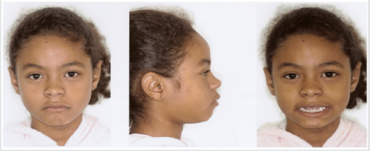
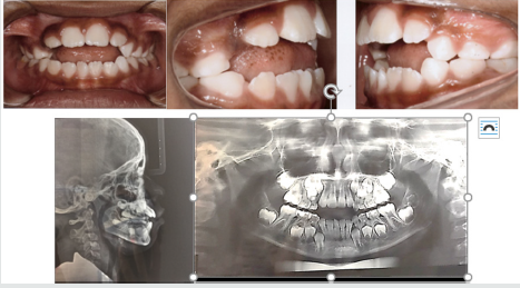
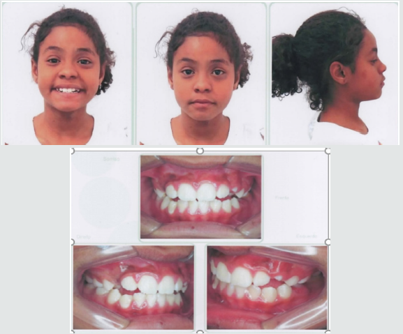
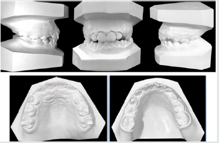
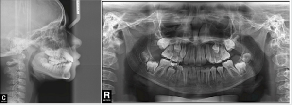
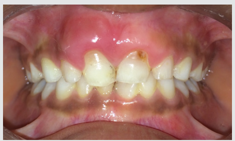
A female patient, 8 years and 4 months of age, melanoderma, came for treatment at the Postgraduate Course in Orthodontics of Brazil University (São Paulo, SP, Brazil). A facial analysis detected convex facial profile, lack of lip closure, and a decrease in nasolabial angle (Figure 1). The patient exhibited thumb sucking habit, mixed breathing, atypical swallowing and speech. shows angle class II malocclusion, 6 millimeters of an anterior open bite, mild crowding and a supernumerary Figures 2&3 tooth in the anterior lower jaw with mandibular midline deviation to the right. Cephalometric Rx shows proclined upper incisors due to the thumb sucking the objetives

Figure 1: Convex facial profile, a decrease in nasolabial angle and the upper lip covering the incisor.

Lupinepublishers-openaccess-pediatric-dentistry-journal

Figure 2: Anterior open bite, mild crowding and a supernumerary tooth in the anterior lower jaw and proclined upper incisors.

Lupinepublishers-openaccess-pediatric-dentistry-journal

Figure 3: Angle class II malocclusion, supernumerary tooth in the anterior lower jaw and an 6mm open bite.

Lupinepublishers-openaccess-pediatric-dentistry-journal

for the first phase of the treatment were to eliminate the thumb sucking, the open bite, dental deviations, provide arch expansion and the extraction of the supernumerary tooth.

Treatment Progress

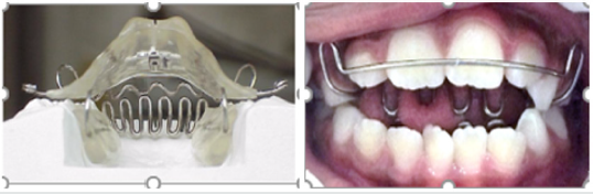
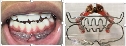
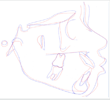
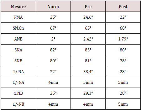
A removable appliance known by Simões Network (SN3) composed with a stainless steel bimaxillary grid (“lower winglets model”) that simulates the incisors occlusion and provides the correct tongue position [15] (Figure 4). The screw was expanded with one-quarter turn biweekly. After 3 months of the treatment beginning o, we added a lip bumper to improve lip seal (Figure 5). We recommended the use for 10 or 12 hours a day. The supernumerary extraction was performed 7 months of the treatment beginning. Figure 6 shows the final of the first stage. Figure 7 shows Angle class I malocclusion and Figure 8 shows the Cephalometric and panoramic Rx after 23 months with the orthopedic appliance. Cephalometric superimposition (Figure 9) and analysis (Table 1) indicated dentoalveolar open bite pretreatment and the correction posttreatment.

Figure 4: SN3 appliance, bimaxillary anchorage with “lower winglets model”.

Lupinepublishers-openaccess-pediatric-dentistry-journal

Figure 5: A lip bumper was added to improve lip seal.

Lupinepublishers-openaccess-pediatric-dentistry-journal

Figure 6: The correction of open bite and the improvement of the alignment.

Lupinepublishers-openaccess-pediatric-dentistry-journal

Figure 7: The arch expansion and molar Class I.

Lupinepublishers-openaccess-pediatric-dentistry-journal

Figure 8: Cephalometric and panoramic Rx after 23 months of treatment.

Lupinepublishers-openaccess-pediatric-dentistry-journal

Figure 9: Cephalometric superimposition revealed maxillary incisor retrusion and mandibular incisor in normal bite.

Lupinepublishers-openaccess-pediatric-dentistry-journal

Table 1: Summary of cephalometric measures.

Lupinepublishers-openaccess-pediatric-dentistry-journal

Discussion and Conclusion

According to the authors, AOB is a challenge daily faced because and it can result speech and swallows’ problems, tongue posture and imbalance between jaw postures [17-19]. Bonna 2016 alerts that the orthodontics or orthopedic devices are fundamental but without family support the habit suppression will not be achieved [20]. The objective for the first phase of treatment were to eliminate thumb sucking, open bite and arch expansion with orthopedic appliance Simões Network SN3 and after a lip bumper was included to improve seal lip. These goals were achieved during the first stage. Graphic 1 shows best fit reduction open bite from May to November 2017. Even pubertal increments offer best time for orthopedic treatment helping determine the predictability, growth direction, patient management and total treatment time, we did not wait to treat because the disadvantages of the open bite [21]. This reported case was successfully treated with SN3 remained stable after the AOB correction. For the second phase with fixed orthodontic treatment will be necessary [22-29].

Graphic 1: Closure open bite variation during time.

Lupinepublishers-openaccess-pediatric-dentistry-journal


Friday, January 22, 2021

Lupine Publishers | Subluxation Injury in Primary Teeth: A Case Report

 Lupine Publishers | Journal of Pediatric Dentistry


Abstract

Subluxation, also called central dislocation, is often the result of an axial impact in the apical direction, moving the tooth within the alveolar bone. This is perhaps one of the most traumatic injuries, since the displacement of a tooth within its socket implies extensive and acute involvement of the periodontal ligament, bone damage and rupture of the neurovascular bundle. The close relationship between the apexes of the primary teeth and the developing permanent successor in turn can lead to multiple complications, which are greater when the permanent tooth is affected in the early stages of development.

Keywords: Subluxation; Dental Trauma; Primary Teeth

Introduction

Trauma to the oral hard and soft tissues is commonly seen in children. Among all facial injuries, dental injuries are the most common. As much as 18% of all injuries in children up to 6 years of age are seen in the oral region [1]. Injuries to the primary dentition are common, occurring with a significantly higher annual incidence than in the permanent dentition [2]. One third of all children in the primary dentition stage suffer from traumatic injuries to the mouth. This is possibly related to poor motor coordination and is sometimes due to the child’s inability to evaluate risks [3]. Resilience of the alveolar bone in young children causes dental luxations of the intrusive type to be more common than crown fractures [4-6]. Traumatic injuries to the primary dentition are often overlooked by parents mainly because less attention is given to the primary dentition and to the child’s inability to cope with the situation.

Case Report

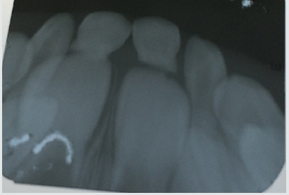
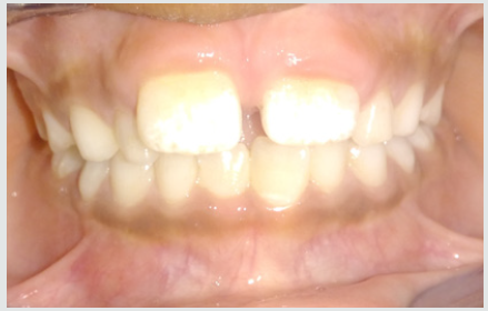
A male patient of 6 years old reported to the dental hospital along with his parents. The chief complaint of the patient was pain in upper anterior teeth. His height was 4 ft. 8 inch and weighed 32 kg. No relevant medical history was recorded. Family medical history was also not significant. It was his first dental visit. No other oral habits were present. On further questioning patient gave history of brushing by self once a day with adult size toothbrush using fluoridated toothpaste. He was well built, and his gait was normal with straight posture and mesomorphic body type. His facial profile was convex with competent lips.eth traumatized. On functional examination patient had nasal breathing and adult type of swallow pattern. He had 24 teeth present in his oral cavity. The parents informed that the child fell from a bicycle and got his teeth traumatized. On intra oral examination of the maxillary arch, the central incisors were found to be tender and mobile (Figure 1). Radiograph was recorded and it showed more than 50% root resorption of primary central incisors in the maxillary arch (Figure 2). Also, the soft tissue around 51 and 61 was swollen due to the injury. Hence, the diagnosis of subluxation was made.

Figure 1:

Lupinepublishers-openaccess-pediatric-dentistry-journal

Figure 2:

Lupinepublishers-openaccess-pediatric-dentistry-journal

Treatment Regimens

The overall principle of treatment is not to take any risk of damage to the permanent successor, which usually implies a very conservative approach. Use of topical anesthetics, local anesthesia, and sedation should be considered. Analgesics may improve the quality of care when pain is anticipated. The administration of a single dose of analgesic 1 h before injection and the use of a topical anesthetic will reduce the discomfort of a local anesthetic [7]. Adequate oral hygiene and a soft diet should be prescribed. Management of an intruded primary incisor depends on the following variables:

a) Direction of intrusion.

b) Degree of intrusion.

c) Presence of alveolar bone fracture.

Figure 3:

Lupinepublishers-openaccess-pediatric-dentistry-journal

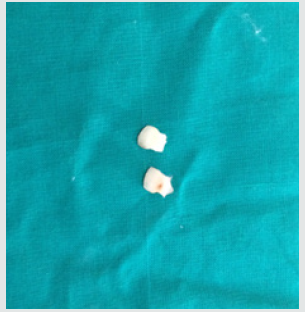
For the present case we did the extraction of 51 and 61 as already there was resorption of them (Figure 3). Extraction was done using local anesthesia. The soft tissues were cleansed and irrigated with betadine solution. The patient was recalled every 3 months for regular follow up until the permanent incisors were erupted in normal condition (Figure 4).

Figure 4:

Lupinepublishers-openaccess-pediatric-dentistry-journal

Discussion

Injury to the child patient is a traumatic experience on a physical as well as emotional and psychological level. In view of the patient’s tender age and the likelihood that the dental visit will be the patient’s first, managing the child’s traumatic injuries is a demanding task. The close relationship between the apex of the injured primary tooth and the underlying permanent tooth germ must be kept in mind. In the present case, we extracted the tooth and gave post extraction instructions. Antibiotic therapy was prescribed to prevent the spread of inflammation to the permanent tooth germ, as advocated by Kenny and Yacobi , Andreasen and Andreasen [3], Wilson [8], and Andreasen et al. [9]. After extraction, the irregularity of the resorbed root surface was evident, postextraction intra-orally. It was due to external inflammatory root resorption, confirming the radiographic diagnosis [10-15].

Conclusion

Special care should be taken during the evaluation and followup of traumatized immature teeth. The long-term prognosis of immature teeth is dependent on continued root formation. In cases of subluxation of primary teeth, it is essential to diagnose the direction of the displaced tooth to rule out injury to the successor and thus prevent the occurrence of sequelae in the permanent dentition. Regular follow up should be mandatory to observe the successors [15-18].

Read More Lupine Publishers Pediatric Dentistry Journal Articles:
https://lupine-publishers-pediatric-dentistry.blogspot.com/

Saturday, January 16, 2021

Lupine Publishers | Pediatric Oral Systemic Health: From Fetus to Adolescence

 Lupine Publishers | Journal of Pediatric Dentistry


Abstract

The oral systemic health link has now become well recognized and numerous publications have discussed the importance of the oral microbiome in the overall health of any individual. This is likely even more important in children and this influences their future health as they mature. What is particularly interesting is that the microbiome of a child is developed prior to birth and is related to the maternal oral microbiome. Early intervention to the mother prior to the child’s birth, with proper sleep, exercise and dietary adjustments such as the limitation of added sugars and use of coconut oil, for example, will influence the developing child’s microbiome. Direct influence with the polyols xylitol and erythritol to reduce pathogens, and probiotics to boost levels of commensals would also be required. Due to the extent of oral disease, and its tremendous associated costs, urgent action is required by all health professions.

Introduction

Every species depends on adaptation to survive. Humans have managed to survive and flourish while directly influencing the environment of the entire planet, encompassing all other forms of life. While there may be philosophical disagreements as to the inadvertent harm to our natural surroundings, the human species, homo sapiens, has been the only survivor of the hominins [1]. The biology of this success is intertwined in the coevolution of homo sapiens and the associated holobiome [2,3]. Chronic illnesses and debilitations appear to be increasing, requiring reflection into the evolutionary process, and the perturbations that have recently occurred creating this environment of now-declining health [4]. Current research would point to the “Hygiene hypothesis”, overuse of anti-microbials, dietary shifts and the resultant decrease in human microbiome diversity [5,6]. The old model of looking for an increase in pathogens is flawed. Indeed, the fault lies with the decrease in commensals that not only compete directly with the pathogens, but also modulate the immune response of the host [7]. To improve the health of children, we must first improve the microbiome of the mother. The maternal microbiome sets the stage for the child’s microbiome [8,9].

Pre-natal intervention has been studied with positive results reported by the supplementation of the mother with probiotics or polyols [10,11]. Published studies using xylitol that involve the nursing mother and child have demonstrated the decrease in the maternal transmission of mutans streptococci [12]. Certainly, intervention may be desired even earlier, preferably before pregnancy because it is also reported that antecedent use of antibiotics by the mother will influence the maternal microbiome [13]. The placental microbiome is most closely related to the maternal oral microbiome [14]. The presence of commensal bacteria in the placenta and developing fetus is essential to fetal immunological maturation [15]. The oral health of the expectant mother should then be considered primarily important to the oral systemic health of the fetus and later, the child. In addition, the placental microbiome appears to be developed quite early in the pregnancy, by maternal imprinting [14]. This maternal imprinting involves the transportation of viable commensals via circulating monocytes, properly creating a fetal microbiome to program the developing child [16]. Animal studies have demonstrated the transmission of maternal breast commensals into the amniotic fluid [17]. All this depends upon the mother actually having a healthy microbiome [18]. The maternal microbiome can be influenced in numerous ways including diet, exercise and probiotic supplementation [19-22]. Limiting added dietary sugar and the regular addition of polyols can help decrease the prevalence of pathogens before they are passed on to the child [12,23-25]. In the case of Early Childhood Caries, the reduction of maternal Candida albicans will reduce the biofilm formation by Streptococcus mutans potentially reducing the incidence of dental caries [26,27]. Some Lactobacilli, all probiotics such as Lactobacilli rhamnosus, have been demonstrated to inhibit Candida albicans [28-30]. Other supplements, such as N-acetyl cysteine, also have a reducing effect on Candida albicans levels [31-32]. Coconut oil in the form of Medium Chain Triglycerides (MCT) supplements has also been used to reduce levels of Candida albicans and is reportedly as effective as ketoconazole [33-35]. But it has also been reported that coconut oil has more beneficial components than just MCT, giving pause as to why whole coconut oil isn’t utilized more [36]. Regardless, MCT may increase exercise endurance and encourage weight loss [37]. In addition, another natural product, propolis has also been demonstrated to inhibit Candida albicans and other oral pathogens [38-40]. If the expectant mother increases exposure to coconut oil or N-AC, the inhibitory effects may be beneficial in preventing the onset of ECC or, possibly Candida albicans systemic disease if prematurely born [41].

Vitamin K2 has also been reported as being very beneficial as an anti-caries agent and for activation of proper bone and dentin formation in concert with vitamins A and D [42]. This research isn’t new, but recently furthered explored and reported [43]. Insufficient levels of Vitamin D have been linked to S-ECC in a number of studies [44-47]. With increased publications of the beneficial properties of these supplements, it is surprising that the dental profession has not enthusiastically adopted a more fully energetic policy on their role in preventing ECC, especially considering the lack of important micronutrients in the typical American fast food diet [48]. The method of birth has been greatly researched demonstrating that C-section results in an increase in childhood allergies and asthma [49]. The research implicates the lack of exposure to the bacteria of the birth canal and anus as being causative with the neonate’s microbiome lacking maternal commensals [50]. After birth, either by vaginal delivery or C-section, breast feeding provides the infant with Human Milk Oligosaccharides which are much more than just food for bacteria as originally proposed for the child [51]. The HMOs also are antiadhesive antimicrobials that serve as soluble decoy receptors, preventing pathogens attachment to the infant’s mucosal surfaces and thereby lowering the risk for viral, bacterial and protozoan parasite infections [52,53].

HMO’s also reportedly modulate epithelial and immune cell responses, reducing excessive mucosal leukocyte infiltration and activation, lowering the risk for necrotizing enterocolitis and providing the infant with sialic acid, a potentially essential nutrient for brain development and cognition [54,55]. Formula does not have the same protective properties that breastmilk does and sadly, many pediatric dentists criticize breast feeding as being cariogenic, even though published research links the associated dental caries to additional carbohydrate intake and night feeding [56,57]. The benefits of breast feeding have been well documented, and the need to adjust the preventive dentistry protocol to accommodate breast feeding should be evident [58]. Although the World Health Organization recommends two years, mothers probably should breast feed their infants for a least a year, the time interval reported to be the found in early hominins, Australopithecus africanus [59]. Another benefit from breast feeding, besides developing the microbiome and immune modulation, could be regulation of metals, especially zinc and copper, protecting the neurological development of the infant [60-62].

Streptococcus mutans has long been considered the key pathogen for the development of dental caries, the most prevalent chronic disease of humans [63-65]. Efforts to reduce the levels of Streptococcus mutans in infants and children with xylitol and preventing dental caries have been successful, raising the question as to why this is not standard dental practice. 66-67 However, other bacterial and fungal organisms have now been closely identified with the development of dental caries [68]. Scardovia wiggsiae is a Bacillus bacterium found extensively associated with Severe- Early Childhood Caries [69]. Scardovia wiggsiae and Slackia exigua have been reported to be involved in the early caries development [70]. Candida albicans, a fungal organism, helps with the biofilm production by increasing the extracellular polysaccharide matrix which protects Streptococcus mutans from anti-microbials and commensals such as Streptococcus oralis [71]. Lactobacilli inhibit the colonization of Candida albicans, hence decreasing the polysaccharide matrix, exposing the Streptococcus mutans to the bactericins or hydrogen peroxide of its natural competitors, other Streptococcus species [72]. Streptococcus oralis produces hydrogen peroxide that inhibits the anaerobic Streptococcus mutans growth [73,74]. Indeed, Probiora probiotic, a commercially available probiotic product, contains Streptococcus oralis, uberis and rattus, and claims to inhibit several key dental pathogens [75- 77]. Probiotics have been reported to be an important adjunct in preventive dental care [78-80].

Erythritol and xylitol are polyols that have been extensively researched and demonstrated to have notable anti-cariogenic and anti-periodontal disease properties [81,82]. Polyols (particularly the non-hexitol alditols or sugar alcohols erythritol and xylitol) have been found effective in inhibiting the transition to and maturation of biofilms from planktonic cells [83]. Xylitol clearly inhibited the formation of mixed species biofilms, which included Porphyromonas gingivalis in vitro [84]. Erythritol suppressed the maturation of gingivitis biofilms and contributed to a healthier oral ecosystem [85]. Porphyromonas gingivalis takes advantage of early colonizers (Streptococci and Candida) to provide attachment and protection within the biofilm matrix. Polyols can reduce extracellular polysaccharide production and interfere with biofilm matrix elaboration, thereby reducing adherence and biofilm development [86-88]. Streptococci and Candida utilize common dietary sugars sucrose and D-glucose for preferred energy sources, as well as for polysaccharide production. Higher glucose concentrations stimulate Candida growth. Compared with common D-sugars, xylitol induced the lowest adhesion and biofilm formation on either Streptococcus mutans or Candida albicans [89]. In addition, xylitol has been demonstrated to decrease the levels of cariogenic bacteria while having little effect on beneficial bacteria [90]. The discovery of bacteriophages specific for certain strains of Streptococcus mutans also show great promise in the management of pediatric oral health [91]. With the costs of dental disease rapidly escalating, now (2010) estimated at 442 billion US dollars, all effective measures to prevent oral disease should be urgently started in the pediatric population [92].

The Airway evaluation of the infant/toddler is of paramount importance during the first Age One examination [31]. Airway issues in children have been linked to future obesity, diabetes and behavioral issues [94,95]. Mouth breathing increases the oral microbiome pathogenic potential, as the incoming air will reduce the protective nature of the saliva [96]. Studies have demonstrated the correlation between oral disease and airway pathology [97,98]. Sleep Disturbed Breathing in children has been extensively reviewed in the literature, describing an ever-increasing pathologic chain of events [99,100]. Amongst the deleterious effects of mouth breathing are lower and mid-facial adaptations, orthodontic malocclusions, potential speech issues, esthetic concerns, sleep disturbed bruxism, and future temporomandibular joint dysfunctions [101-104]. The key to the future health of children is effective preventive care. What becomes a serious morbidity in adulthood started in childhood. Now more than ever, pediatric health care providers need to emphasize the connection between the oral health of children and their systemic health, with all the future ramifications now clearly reported in the scientific literature. The importance of the oral microbiome, its role as a “gateway” microbiome, and the systemic connection need to be more fully explained to patients, parents and all health care professionals.

Interestingly, the oral health care of the child starts before birth, requiring the participation of all involved in pre-natal care. It is now obvious that what is most important may be the microbiome, and how it is affected by the environment, diet, sleep, exercise, antibiotics, polyols and probiotics. The microbiome then modulates the immune system, allergies, resistance to pathogens, autoimmune responses, and ultimately patient health and longevity. At last, there seems to be great interest in the importance of pediatric and general oral health due to the crisis that poor oral health is bringing upon us [105]. We should be concerned that research studies from several countries have all reported neurotoxicity effects from relatively low levels of fluoride in children [106- 111]. Our over reliance on fluoride to create fluorapatite to inhibit decay seems inadequate at best. Perhaps this means that the time has come to treat a bacterial disease, as a bacterial disease. After all, dental caries and periodontal disease, and to a great extent downstream comorbidity including atherosclerosis, diabetes, strokes, inflammatory Alzheimer’s, diabetes, and many systemic illnesses, can be traced back to a “dysbiosis” started in infancy.

Read More Lupine Publishers Pediatric Dentistry Journal Articles:
https://lupine-publishers-pediatric-dentistry.blogspot.com/

Friday, January 1, 2021

Lupine Publishers | Mouth Guards: Guardians of the Dontium

 Lupine Publishers | Journal of Pediatric Dentistry


Abstract

Children and teens are more prone to injuries while playing, with most of the impact taken by the face especially the jaws and the teeth. The most significant factor in preventing sports-related or recreational orofacial injuries is wearing basic protective devices such as properly fitting helmets, face masks, or mouth guards. A mouth guard, custom-fitted by your dentist and worn every time you play or train, will protect against dental injury. Mouth guards are available in 3 different variants which are stock, self-adapting, custom-made mouth guards. The benefits far exceed the expenditure when considering the fees and discomfort associated with a traumatic dental injury

Keywords: Mouth Guard; Orofacial Structures; Teeth; Custom; Sports; Injuries

Introduction

Physical activity forms a very important part of life. There have been constant reports of sedentary lifestyle being associated with disease processes and over the time a lot of importance has been emphasized on physical activities be it the gym or the sports. Children and teens are more prone to injuries while playing, with most of the impact taken by the face especially the jaws and the teeth. Hence, it becomes very important to wear protective gear as these injuries might not only cause temporary impairment but also cause hindrance to the growth and development of the facial structures and also may lead to an unaesthetic appearance of the child at the later stage. The injuries occurring in sports can range from a simple ball-hit to a serious impairment of the head, face or the mouth. It is very important to wear protective gear when you are actively participating in any recreational outdoor events and also in some instances in the indoor activities with the risk of injuries. The most significant factor in preventing sports-related or recreational orofacial injuries is wearing basic protective devices such as properly fitting helmets, face masks, or mouth guards. The use of the mouth guard forms the basic minimal requirement for protecting your mouth, which should form an essential piece of the athletic equipment that the athlete should use as the standard gear equipment from a very early age.

A mouth guard, custom-fitted by your dentist and worn every time you play or train, will protect against dental injury. Well-fitted mouth guards prevent violent contact between the maxillary and mandibular dentition, which can result in soft tissue lacerations, tooth avulsions, tooth or bone fractures, endodontic injuries, and concussions [1]. This being said it has been found that injury to teeth are 60 times more likely when the athlete is not wearing the mouth guard than when he/she is wearing it. Statistically, sporting activities contribute to nearly one-third of all dental injuries [2-4]. Mouth guards help buffer an impact or blow that otherwise could cause broken teeth, jaw injuries or cuts to the lip, tongue or face. Mouth guards also may reduce the rate and severity of concussions Consequently, the dentist plays an important role in informing patients, athletes and their parents, and coaches of the importance of prevention, diagnosis, and treatment of orofacial injuries in sports and recreational activities [1].

A mouth guard should be able to fulfill the following basic requirements:

a) Encompass all maxillary teeth extending up to the distal surfaces of the second molars in class I and class II patients [4-7].

b) Encompass all mandibular teeth extending up to the distal surfaces of the second molar on class III patients [4-7].

c) Mouth guard may be abridged to cover until the distal surfaces of the first molars, in case it is known to trigger the gag reflex of the patient [4].

d) The labial flange should range to within 2mm of the sulcus [5].

e) The palatal flange should range around 2mm above the gingival margin [5].

f) The margins of the labial flange should be rounded.

g) The margins of the palatal edge should be tapered [5].

h) Be easy to clean [8,9].

i) Not impede with breathing or speech activity [4].

j) Be fabricated from a material approved by the U.S. Food and Drug Administration that can reduce the impact force to teeth, surrounding soft tissues, and bone [4,9].

k) Be comfortable and retentive and fit properly [9].

Mouth guards are available in different variants

A. Stock

B. Self-adapting

C. Custom-made

A. Stock

These are readily available over the counter in different sizes, made from polyurethane, a copolymer of vinyl acetate, or ethylene. As they are produced in bulk and are of standard sizes, they remain inexpensive, however they offer a low level of protection with little retention and is not so easily accepted by the athlete. The need to hold the mouth guard in place by clenching his or her teeth together is another disadvantage.

B. Self-Adapting

Also known as the “boil-and-bite” type mouth guard. It is readily available over the counter and made from ethylene-vinyl acetate (EVA). Herein, it is heated in hot water and then placed in the mouth to be adapted to the teeth by biting down. It is relatively inexpensive and can be replaced frequently in athletes with a mixed dentition or by individuals who are experiencing rapid growth. It has the property of re-adaptability. However, it is often bulky and does not retain its shape over time.

C. Custom-Made

The custom-made mouth guard is fabricated in a dental laboratory on a cast taken from an impression made by a dentist. The custom-made mouth guard offers the best fit and the most protection of any of the protective devices [4,5,9]. It is usually made of a thermoplastic material that is heated and adapted to the cast under pressure or with a vacuum form machine. Due to the laborious work involved in the fabrication of this type of appliance, it is expensive but retentive. The American College of Prosthodontists (ACP) recommends the use of custom mouth guards for all contact sports and for any recreational activities that may hypothetically end in orofacial injuries. Accurate maxillary and mandibular alginate impressions in centric occlusion registration recorded by a qualified dentist at approximately 5-mm opening anteriorly shall be used for fabrication of the custom trays as recommended by the ACP.4,5 The standard thickness is 4-mm, however 5- or 6-mm thickness is recommended as it will be able to protect the athlete better in case of extreme sports [4].

Mouth guards can be either single-layered or multi-layered. Currently, the most commonly used materials in the construction of custom mouth guards are EVA copolymer, soft acrylic resin, polyvinyl chloride, polyvinyl acetate-polyethylene (pEVA), and elastomers [1,8,9]. Many different designs of multi-layered materials are available. The most frequently used is a double layer made of similar materials. Dual laminated mouth guards possess an outer hard shell of styrolbutadiene co-polymerisate, and a soft inner layer of ethylene copolymer and vinyl acetate. This design of a more rigid outer material with an inner softer material will reduce the impact force transferred to the teeth due to the shock-absorbing capability of the softer layer [4,5]. The critical areas in terms of energy absorption and transmitted forces are the incisal edges of the anterior teeth and the attached (marginal) gingiva. Therefore, an optimal thickness of the device is achieved by the application of vacuum forming pressure-lamination technique of two layers of a thermoplastic sheet (EVA copolymer) and if necessary, by placing two layers of protective air-cells against the critical areas. An acrylic-resin-based elastomer may be processed over the thermoplastic sheet to improve protection for the athlete [4]. Custom-made mouth guards have proved to be the most effective means of prevention of injuries to the orofacial structures [1,4,5]. They are superior in quality, comfort, retention, and prevention of injuries when compared to stock or self-adapting devices. Although custom-made mouth guards are the most expensive type of protective oral device, they are the most highly recommended.

Conclusion

The benefits far exceed the expenditure when considering the fees and discomfort associated with a traumatic dental injury. Further, it becomes the duty of the dentist to create awareness among the athletes and make it a habit for them to wear mouth guard as a part of their equipment for sports.

Read More Lupine Publishers Pediatric Dentistry Journal Articles:
https://lupine-publishers-pediatric-dentistry.blogspot.com/

Friday, December 25, 2020

Lupine Publishers | Comparison of the Efficacy of Plaque Removal of Listerine Smart Rinse Kids and Vi– One Junior Fluoridated Mouthwash in Children Aged 6 To 10 Years

 Lupine Publishers | Journal of Pediatric Dentistry


Abstract

Objectives: In this study, a comparative study was done on the effects of Vi-One and Listerine fluoridated mouthwashes on the reduction of dental plaque in pediatric patients between 7-11 years of age in dental clinics of Sepideh and apple in Shiraz, Iran. Listerine Smart Rinse Kids is a product of the United States and Vi-One Junior mouth wash is the domestic production of the country at Rozhin Corporation. This research was conducted by Mohammad Karimi and Hassan Dehghan in 2018-2019.

Material and Methods: In this study, 100 individuals were selected and divided into two groups of 50. During the study, no other method of controlling the plaque was used. In this method, the first group first used Vi-One mouthwash for 10 days and after two weeks of rest and minimizing dental plaque, they used Listerine for 10 days. While the second group used Listerine first, then they applied Vi- One in the same way. The results of this review were then evaluated.

Results: The mean of plaque index in total mouth and in the posterior teeth area with the use of Listerine Smart Rinse Kids was lower than that of Vi-One Junior mouth rinse. In another words, Listerine had a better effect on plaque removal than the Vi-One mouthwash in the posterior mandibular region.

Conclusion: The results show that although Listerine mouthwash had a better effect on dental plaque removal, none of the two mouthwashes had a significant difference in effects on maxillary and mandibular jaws.

Keywords: Dental Plaque; Vi- One, Listerine, Fluoridated Mouthwash; Periodontal Diseases; Plaque Index

Introduction

Currently, dental caries and gingivitis are common oral and dental diseases in this country. One element that can prevent tooth decay is Fluoride. In the oral health program of the country, fluoride mouth wash 2% was used to prevent dental caries in elementary school students all over the country [1]. In the other hand, dental plaque is an important factor in the formation of dental caries and periodontal diseases. Leo and his colleagues have identified dental plaque as the main cause of gingivitis [2]. With the use of mouthwashes, one can control the dental plaque, chemically [3,4]. In fact, mechanical plaque removal is one of the most common and effective methods for preventing caries and inflammation of the gum [5]. Fluoride mouthwash usage is contraindicated in children younger than six years of age due to the risk of swallowing and causing systemic toxicity and fluorosis [6-8]. Symptoms of acute oral fluoride toxicity in children include severe nausea, vomiting, hyper salivation, abdominal pain, and diarrhea [9]. In severe or fatal cases, these symptoms can be followed by convulsions, cardiac arrhythmias, and coma [10- 12]. Laboratory and animal data have shown that prevention the accumulation of plaque and consequently, reduction in dental plaque can be achieved when fluorides is applied topically which inhibits the bacterial multiplication [13]. The fluoride from mouth rinse is retained in dental plaque and saliva to help prevent dental caries [14]. In one review, the average caries reduction in nonfluoridated communities attributable to fluoride mouth rinse was 31% [15]. Another study in Sweden reported that the use of fluoride mouthwash along with brushing has a significant effect in decreasing of dental caries [16]. Listerine Smart Rinse Kids has been used for the purpose of this study. This product is an alcoholfree mouthwash. The ingredients include Sodium fluoride 0.02% (0.01% w/v fluoride ion), Water, Sorbitol, flavor, phosphoric acid, Sucralose, Cetylpyridinium chloride, disodium phosphate, sodium saccharin, menthol, blue 1 and green 3 [17]. One study reported that use of this mouthwash can strengthen teeth 99% better than brushing alone [18]. Another source indicated that it gives 12- hour cavity protection [19]. Vi-one Junior Mouthwash is specially designed for children. This mouth rinse contains Sodium fluoride 0.05%, Cetylpyridinium chloride 0.05% and Disodium phosphate agents. The respective flavors contain sugar-free and harmless sweetener. This brand also is an alcohol-free product [20]. The purpose of this study was to compare the efficacy of two types of mouthwashes, one the domestic mouthwash (Vi-One Junior) and the other, the brand name Listerine Smart Rinse kids fluoridated mouthwash in the removal of the dental plaque.

Material and Methods

This study was a cross-over clinical trial. The eligibilities for entering in our study were as follow:

a) Children having at least 20 teeth with no large restorative area.

b) No history of periodontal Diseases.

c) Not having any Prosthodontic or Orthodontic appliances.

The condition for withdrawal from the study, if there was any sign of reactions to any of these mouth rinses. There was no obligation to have any food regimen.

The study population consisted of 100 patients who were in a 50-member group. Before taking oral mouthwash, plaque index was minimized, and all subjects underwent tooth scaling at the beginning and, if necessary, teeth polishing were done before taking mouthwash. Oral hygiene was assessed via a plaque index. First, in both groups, the Silness-Löe plaque index was recorded. It is an Index for evaluating the thickness of the plaque in the gingival region, which measures the thickness of plaque on all surfaces (M, B, D, and L) [21].

Coding for the plaque index was carried out according to the criteria [22]:

a) Code 0: No plaque

b) Code 1: A film of plaque is adhering to the free gingival margin and adjacent area of the tooth. The plaque may be seen in situ only after application of disclosing solution or by using the probe on the tooth surface.

c) Code 2: Moderate accumulation of soft deposits can be seen with the naked eye within the gingival pocket, the tooth, or gingival margin.

d) Code 3: Abundance of soft matter can be seen within the gingival pocket and/or on the tooth, and gingival margin.

In this index, each tooth is divided into four surface area but in our purposes in the present study, we modified the surfaces area from 4 to 6; thus, we have three surfaces in the buccal area (Mesiobuccal, Midbuccal, and Distobuccal) and three surfaces in the lingual area (Mesiolingual, Midlingual and Distolingual). The first group used Vi-One & Listerine mouthwash (kids mouthwash), for 10 days in the following way. Needless to say, this process was supervised by parents at home. The kids have to gargle 5 cc ’s of Vi-One mouthwash 2 times per day for 30 seconds, and during this period of time, no other plaque control methods and tooth brushing should be used. At the end of the period of 10 days, the plaque index was recorded again. Then, the subjects were given a week to rest and stop using the mouthwash while they had permission to start brushing like before. Again, the plaque index was minimized for patients with polishing the teeth, and they used Listerine mouthwash for 10 days. In the same way, 5 cc ‘s of the mouthwash twice daily was used for 30 seconds, and at the end of the one-week period, the plaque was recorded. For the second group, in the first 10 days, mouthwash. Listerine was prescribed and in the second 10 days, the Vi-One mouthwash was applied. All procedures were performed according to the above pattern.

Results

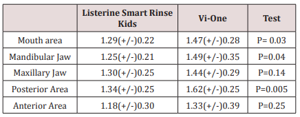
Paired T-test was used for statistical analysis of the findings. The findings showed when Listerine Smart Rinse Kids was used; the mean of plaque index in all area of the mouth (especially in the mandibular jaw and the posterior region) was significantly less than the time Vi-One was applied. However, there was no significant efficacy difference between the use of both types of mouthwash in the upper jaw and the anterior region.

There was no significant difference between the mean plaque index in Vi-One mouthwash between upper and lower jaw, and there was no significant difference between the maxillary and lower jaw in the case of Listerine Smart Rinse Kids either. The presence of this indicator in both types of mouthwash in the anterior region was significantly less than the posterior region. The mean and standard deviation of the plaque index in both groups, as well as in different regions of the mouth, are listed in Table 1.

Table 1: The amount of dental plaque in terms of area and type of Mouthwash.
lupinepublishers-openaccess-journal-pediatric-dentistry

Discussion

In general, the anti-plaque properties of mouthwashes are completed through bactericidal and bacteriostatic effects, separation of microorganisms from dental surfaces, loosening of joints to these surfaces or lowering of the surface tension of the tooth [2,21,23]. Some mouthwashes can be useful for preventing tooth decay or periodontitis [6,24]. Furthermore, mouthwashes are recommended for children and adolescents with orthodontic appliances or adults who need deep cleansing (such as curettage) [6]. These types of mouthwashes were generally used before and after surgery (especially Chlorhexidine) and have a very positive effect on the treatment of gum and ulcerative inflammation [6]. Use of this type of mouthwash should not last longer than 2 to 3 weeks due to some side effects such as staining the teeth and soft tissue staining, increased calculus deposition, unpleasant taste, burning sensation, and mucosal irritation [6]. It’s time to use this mouthwash after brushing and before bedtime, and it’s best not to eat anything after half an hour after use. Fluoride-containing mouthwashes are another type of mouth rinse that has a fairly large use. These mouthwashes have a significant effect on teeth strengthening. Fluoride in the mouthwash cause bonding with enamel and dentin, and with bonding with calcium and phosphorus, they form Fluorapatite, which is more resistant to caries than Hydroxyapatite. Fluorides also accelerate the mineralization, repair the decayed teeth surfaces, and help to increase the reverse processing of damaged tooth surface area [25,26]. Fluoride also reduces the effect of oral bacteria on teeth. It is done by interfering with the function and formation of the microorganisms. The best fluoride mouthwash protects the teeth against the acids which are produced by dental plaques. “Neglecting the oral hygiene of children leads to the accumulation of plaque and as a consequent the formation of dental calculus which will have a devastating effect on the both child’s gums and teeth” [27].

In one study, the statics showed an alcohol-free mouthwash containing a combination of 0.075% CPC and 0.05% Na F produces statistically significant reductions in dental plaque and gingivitis after three and six months compared to baseline [28]. In another research, Jessica E. Koopman, et al argued that the oral microbial community displayed remarkable resilience towards the disturbances it was presented with. The effects of the fluoride mouthwash on the microbial composition were trivial [29]. On the other side, in another study, the research showed that all four fluoride mouth rinses were effective in decreasing the plaque levels of S. Mutans [30]. In this study, we investigated the effect of two mouthwashes of Listerine Smart Rinse Kids and Vi-One in which Vi-One mouthwash in the posterior region was less efficient than the Listerine mouthwash, and the interesting point that most kids mentioned the taste of Listerine was more acceptable. Given that the contents of sodium fluoride were equal in both mouthwashes, due to the fact that Listerine mouthwash was more acceptable than the mouthwash, it could be related to the other materials present in this product which can be a part of the manufacturer’s secrets. This difference in taste can be a factor in the effect of improving Listerine’s efficacy in the posterior regions.

Conclusion

Listerine Smart Rinse Kids had a better effect on plaque removal than the Vi-One mouthwash in the posterior mandibular region. Both types of mouthwash had a better effect on the anterior region than the posterior region, but none of the two mouth rinses had a different effect on the maxillary and lower jaw. Although many popular types of mouthwash may help to control dental plaque and gingivitis, they should only be used as an adjunct to other oral hygiene measures such as brushing and flossing. Fluoride mouthwashes should be encouraged in children above the age of 6 with a high risk of caries.

Read More Lupine Publishers Pediatric Dentistry Journal Articles:
https://lupine-publishers-pediatric-dentistry.blogspot.com/

980 nm Diode Laser: A Good Choice for the Treatment of Pyogenic Granuloma

Abstract Pyogenic granuloma is a benign non/neo plastic mococutanous lesion . It is a reactional response to constant minor trauma and ca...