Friday, July 23, 2021

Lupine Publishers | A reflective approach for pediatric dental care at the university clinical school, in the time of COVID-19

 Lupine Publishers | Journal of Pediatric Dentistry


Opinion

We are experiencing a unique time in everyone’s life, globally, with an indefinite length of stay, as well as the extent of its impacts; the Coronavirus Disease (COVID-19) pneumonia pandemic. This disease is caused by the Severe Acute Respiratory Syndrome Coronavirus (SARS-CoV-2). As health workers, there are mixed feelings. On the one hand, hope, the frantic and urgent search for scientific evidences, prevention and efficient strategies for treatment of so many infected individuals with different levels of symptom severity. In another sense, the fear of contracting and acting as a vector for the transmission and dissemination of this disease. When analyzing the COVID-19 Worldwide Dashboard, the contamination and the number of deaths among health professionals is alarming; mainly if they work on the “front line” or with a risk of exposure to a high viral load, during management of the exposed person. And dentistry is no exception. It is necessary to prepare adjustments of dental practices; especially with undergraduate degrees treat children under the close supervision on faculty members on the Pediatric Dental Team. The participants in dental practice have a higher potential risk of infection, due to the face-to-face communication and the exposure to saliva, blood, and other body fluids, because the procedures that are believed to generate aerosols and droplets and the handling of sharp instruments. There was a suspension of dental care services or a restriction on urgent or emergency cases, which comprise circumstances associated with pain, trauma or infectious processes; these bringing discomfort, impaired performance of oral functions, interventions necessary for critical medical procedures or that increase the risk of death to the patient. Gradually there is a greater openness to elective procedures

At first, the sensation probably experienced by dentists would be related to not being considered as an essential professional when it comes to fighting together to “save lives”. Is the oral health not associated with general health? If the oral cavity constitutes the “gateway” for many microorganisms and there are some oral manifestations of infections and diseases with systemic involvement in individuals; why were dentists not considered as essential workers? Here we do not question the professional skills of each specialty in the health area, but changes should be viewed as developments in multiprofessional health groups working together. Each university is preparing plans for reopening consistent and safe: restart and recovery research, education, and clinical services missions. In this context, the return to dental practice activities still represents a great challenge; a “new reality”. And this perspective expands, particularly when we consider the dental care for children and adolescents. Here we are focused on children, because most of the time their possibility of understanding and clarification is still in development. Children need to interact to people and they should be “freedom” to play, explore and interact with environmental. Teaching experience, singularly in clinic-school for children in the University of Pernambuco College of Dentistry, in the Northeast of Brazil, needs a dynamics challenge. But how not to lose its essence? Playful and humanistic principles and procedures based on scientific evidence and technical improvement. We adopted some reinforces as positive facial expression, handshake, smile, hug and others. Everything supported by bioethics and human rights, in order to develop a better perception about dentists and dental care; to reduce tensions.

Understanding of children and families, and their relationship to providing optimal pediatric dental care. We should construct strong interactions that allow healthy habits, cooperative behaviors, the reduction of fears and anxieties, good oral health conditions, adequate performance of orofacial functions and growth and craniofacial development with characteristics within a “normality” spectrum. Nowadays we are all (dental students, pediatric dental patients /family and staff) living in a world “in transformation”, where insecurity, demands, competitions and hopelessness, associated with isolation or distancing from the other, can lead to high psychological distress. We need to implement measures to ensure the development of dental education and dental treatment, preserving the integrity of all those involved and to build the resilience during the COVID-19 pandemic. The demands for care have not stopped and there is a growing sense of anguish due to lack of dental care.

Read More Lupine Publishers Pediatric Dentistry Journal Articles:
https://lupine-publishers-pediatric-dentistry.blogspot.com/

Saturday, July 10, 2021

Lupine Publishers | Effect of Surface Treatment and Type of Resin Cement on Marginal Fit of Zirconia Crowns

 Lupine Publishers | Journal of Pediatric Dentistry


Abstract

This in vitro study was conducted to evaluate effect of surface treatment and two resin cements on marginal integrity of zirconia crowns.

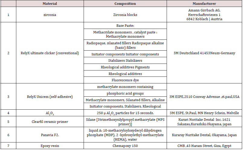
Materials and Methods: Forty zirconia crowns (Amann Girrbach AG,Austri) were fabricated and divided into2 groups according to type of resin cement, group 1 Rely x ultimate clicker (3M Deutschland ,Germany ) and group 2 Rely x unicem (3M ESPE,USA) , each group was then subdivided into 4 groups according to type of surface treatment , no surface treatment, sandblasting, sandblasting with silane, sand blasting with MDP . After cementation each specimen was photographed using USB Digital microscope with a built-in camera (Scope Capture Digital Microscope, Guangdong, and China) for measuring and evaluation of gap width.

Results: group 2 showed higher significant mean of marginal gap than group 2.

Conclusion: group 1 Rely x ultimate clicker resin cement showed less marginal gap than group 2 Rely x unicem resin cement.

Keywords: Surface treatment; resin cement; zirconia

Introduction

As esthetic restorative materials for crowns and fixed partial dentures (FPDs), all-ceramic systems can be used as alternatives to metal ceramic systems. During the last decade, zirconium dioxide (zirconia, ZrO) ceramics, which have superior mechanical properties [1], have been used increasingly for copings and frameworks of fixed restorations. An ideal all-ceramic dental material should exhibit excellent esthetic characteristics, like translucency, natural tooth color, outstanding light transmission and, at the same time, optimal mechanical properties, like flexural strength, fracture toughness and limited crack propagation at the functional and parafunctional load conditions, in order to ensure long-term service. To date, zirconia has been considered a suitable choice for dental restorations due to its good mechanical properties, toothcolored and natural appearance and low plaque accumulation [2- 4] . Zirconia (zirconium oxide) was introduced by Martin Heinrich Klaproth in 1789. This material is a non-cytotoxic metal oxide, is insoluble in water and has no potential of bacterial adhesion. In addition, it has radio-opacity properties and exhibits low corrosion [5]. There are 3 crystalline shapes of this material at different temperatures are as follows: cubic (c) tetragonal (t) monoclinic (m). Increasing the crystal volume, constrained by the surrounding ones, leads to a favorable compressive stress. This limits growth of cracks. Transformation toughening or “phase transformation toughening” (PTT) is the reported mechanism for exceptional flexural strength and fracture toughness of zirconia among all the other ceramics , hence Zirconia is considered as one of the most reliable ceramic materials [6,7]. Zirconia based fixed partial dentures (FPDs) have the potential to withstand physiological occlusal forces applied in the posterior region, and therefore provided interesting alternatives to metal-ceramic restorations. Although combined surface treatment and specific adhesives with a hydrophobic phosphate monomer such as methacryloyl oxydecyl dihydrogen phosphate (MDP), are currently considered reliable for bonding to zirconia ceramics, but further clinical and invitro studies are still needed to obtain long-term clinical information on zirconia-based restorations [8] (Figure 1).

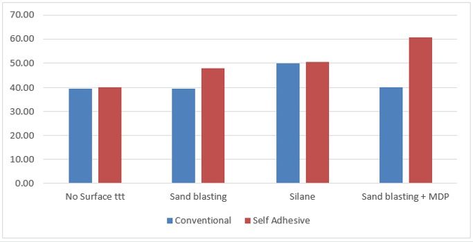
Figure 1: Showing marginal gap values according to surface treatment and type of resin cement.

Lupinepublishers-openaccess-pediatric-dentistry-journal

Materials and Methods

Teeth selection, cleaning, storage and preparation

Atotal 40 extracted caries free human teeth were collected from clinics of faculty of Dentistry Al- Azhar University, Assiut branch, after taking permissions from patients for using their extracted teeth in research purposes. Caries free human upper first premolars extracted for orthodontic purposes from patient 18-20 years old were collected for standardization. Extracted teeth stored in 0.5% thymol not more than 2 months following esthetic protocol of faculty of dentistry Al- Azhar University, Assiut branch.

Specimens Preparation

A machined standard stainless steel was used with 5 mm height and 5 mm diameter with 10 degrees convergent axial walls, the dye was prepared with 50° shoulder finish line (1 mm thickness), forty polyvinyl siloxane impressions (silibest via m.Bonarroti ,cappanoli ,Italy).

Fabrication of zirconia crowns

Crowns made of a partially sintered zirconia ceramic material by using CAD/CAM technology optical impression was made to the epoxy specimen after being sprayed , using model scanning via : desktop extra-oral scanner map 400 Amann girrbach)
a) Crowns were designed via (Exocad software ) with the following parameters,
b) 0.05mml cement gap starting 1mml from the restoration margins, Milling the designed restoration with (ceramill motion 25 axix machine manufactured by Amann girrbach).

Testing procedures

Each specimen was photographed using USB Digital microscope with a built-in camera (Scope Capture Digital Microscope, Guangdong and China) connected with an IBM compatible personal computer using a fixed magnification of 45X. A digital image analysis system (Image J 1.43U, National Institute of Health, USA) was used to measure and qualitatively evaluate the gap width. Within the Image J software, all limits, sizes, frames and measured parameters are expressed in pixels. Therefore, system calibration was done to convert the pixels into absolute real world units. Calibration was made by comparing an object of known size (a ruler in this study) with a scale generated by the Image J software. Specimens were held in place over their corresponding dies using a specially designed and fabricated holding device. Shots of the margins were taken for each specimen. Then morphometric measurements were done for each shot (3 equidistant landmarks along the cervical circumference for each surface of the specimen) (Mesial, buccal, distal, and lingual) [4]. Readings of each surface of specimen were taken, then the data obtained were collected, tabulated then statistically described in terms of mean and standard deviation (SD) (Table 1).

Table 1.

Lupinepublishers-openaccess-pediatric-dentistry-journal

Results

Rely x unicem showed the higher insignificant mean of marginal. According to following Table 2.

Table 2.

Lupinepublishers-openaccess-pediatric-dentistry-journal

Discussion

Zirconia ceramics have superior strength, toughness, fatigue resistance and enhanced long-term durability than other ceramics Oilo et al. [9], Della Bona [10]. CAD/CAM system crowns demonstrated more accurate marginal fit than the traditional fabrication methods Mart´ınez et al. [11]. The treatment of zirconia ceramic surfaces with abrasive alumina enhances the surface roughness resulting in the increase of the effective contact surface between ceramic and resin cement which leads to the increase of the bond strength [12]. also improves micromechanical interlocking resulting to a strong and durable bond between resin/zirconia [13]. Silane coupling primers have been reported to increase the wettability of the surface but there is no chemical reaction occurs between the silane and surface of zirconia which may explain the low bond strength resulting in comparison to the other surface treatment methods [14]. In the present study maximum gap found in zirconia crowns cemented by self-adhesive resin cement after sand blasting with MDP surface treatment, this finding was in agreement with Yuksel et al. [15] which may be due to short setting time of conventional cement as opposed to that of the self- cure resin bonding system [16]. This finding disagreed with Kern et al. [17] which reported that selfadhesive resin cement (RelyX Unicem) produced better marginal continuity, and high adaptation to substrate.

Read More Lupine Publishers Pediatric Dentistry Journal Articles:
https://lupine-publishers-pediatric-dentistry.blogspot.com/


Friday, July 2, 2021

Lupine Publishers | Long-Term Treatment of Early Childhood Caries (ECC)

 Lupine Publishers | Journal of Pediatric Dentistry


Abbreviations: ECC: Early childhood caries; CDC: Center for Disease Control; NHANES: National Health and Nutrition Examination Surveys

Introduction/Literature Review

Early childhood caries (ECC) is an infectious disease affecting the primary dentition in infants and children younger than 6 years of age. Despite being a largely preventable condition, ECC remains a major public health problem in both developing and developed countries, with nearly 1.8 billion new cases diagnosed each year globally [1,2]. In the United States, Center for Disease Control and Prevention (CDC) reported a 15.2% increase in the prevalence of dental caries among the nation’s youngest children, aged 2-5, comparing the findings of two National Health and Nutrition Examination Surveys, NHANES II, 1988–1994 and NHANES III, 1999–2004 [3,4]. A comprehensive epidemiological review of ECC showed that although the overall ECC prevalence varies from population to population, the socio-economically disadvantaged and minority groups are most at risk [5]. Numerous studies indicate that Hispanic and Native American children are among the high-risk North American populations [6]. ECC is multifactorial in its etiology and has been associated with a multitude of risk factors including inappropriate and prolonged bottle or breast feeding, frequent consumption of fermentable carbohydrates, and poor oral hygiene [6-8]. Vertical transmission of Streptococcus mutans from caregiver to child has been demonstrated [9]. A previous study has shown that S. mutans or S. sobrinus alone may not be the only indicators for high risk of caries, but the prevalence of S. oralis in saliva may be a risk factor for increased caries activity in severe ECC forms [10]. Being born preterm or small for gestational age [11], improper oral health practices, higher birth order, low fluoride in drinking water, poor saliva buffering capacity and behavioral factors are other risk factors [12,13]. The initial clinical presentation of ECC appears as white-spot lesions in the upper primary incisors along the margin of the gingiva [14]. Complete destruction of the crown can potentially follow if the disease continues [15]. The spread of caries to primary maxillary molars is seen in moderate cases and primary mandibular molars are involved in severe stages [16]. Greater probability of subsequent caries in both the primary [17] and the permanent18 dentitions is observed among the children who experienced caries as infants or toddlers [18]. Missing anterior teeth causes poor phonetics, unpleasant appearance, hardship in sociological and psychological adjustment, and nonefficient mastication [19]. Several studies have demonstrated the considerable negative impact ECC has on the health-related quality of life of children [20].

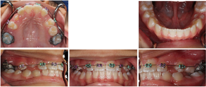
Maintaining the primary dentition in a healthy condition has innumerable benefits, therefore dental caries management in children is pivotal. Treatment options include the restoration or extraction of the carious teeth although the importance of preventative and preservative measures cannot be overstated. Construction of orthodontic appliances such as space maintainers or prosthetic management may be needed in some cases. The following case demonstrates the use of the Nance appliance and the Hyrax expander in addition to conventional orthodontic treatment over the course of nine years in order to obtain a Class I (Angle) canine and molar relationship in a child with severe early childhood caries.

Diagnosis

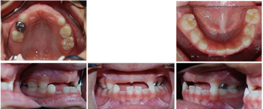
The patient was a four-year-old Hispanic female that presented with the chief complaint of “missing frontal and some back teeth having difficulty chewing and speaking.” No remarkable medical findings were noted. A facial evaluation revealed a symmetric face. She had a unilateral posterior cross bite with flush terminal deciduous molar relationship (Figure 1).

Figure 1.

Lupinepublishers-openaccess-pediatric-dentistry-journal

Treatment objectives

The primary objectives of treatment were to maintain the space and replace the missing anterior teeth, to obtain a Class I (Angle) canine and molar relationship, with ideal overjet and overbite, and to improve facial esthetics. The complementary treatment objectives were to establish good functional and stable occlusion, to avoid crowding and lack of space of the upper incisors and premolars, while improving smile characteristics and eliminating lip incompetence.

Treatment progress

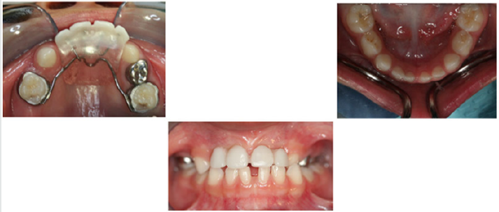
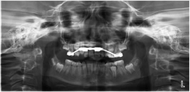
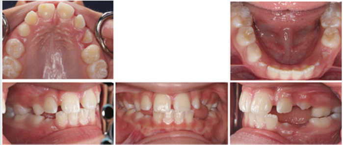
The treatment of choice was utilization of Nance appliance followed by conventional orthodontic treatment. After consultation with the patient and her parents, informed consent was obtained and treatment was begun. All components of the appliances were submitted to a dental laboratory for fabrication. Modified Nance appliances was fit on deciduous maxillary first molars at the age of four and a half Figure 2. The Hyrax appliance was fixed on teeth #3 and #14 when the patient was 11 years old Figure 3. The appliance’s screw was turned twice a day until proper palatal expansion was achieved Figure 4. Treatment was followed by comprehensive orthodontic treatment lasting approximately 30 months Figure 5(A&B). Immediate Essix retainers were placed at the deboning appointment Figure 6.

Figure 2.

Lupinepublishers-openaccess-pediatric-dentistry-journal

Figure 3.

Lupinepublishers-openaccess-pediatric-dentistry-journal

Figure 4.

Lupinepublishers-openaccess-pediatric-dentistry-journal

Figure 5a.

Lupinepublishers-openaccess-pediatric-dentistry-journal

Figure 5b.

Lupinepublishers-openaccess-pediatric-dentistry-journal

Treatment Results

Excellent facial and occlusal results were achieved with the nine-year treatment rendered. The most significant changes were the correction of the unilateral cross-bite malocclusion of the deciduous teeth and prevention of mesial shifting of posterior teeth in order to achieve both a molar and canine Class I (Angle) relationship of permanent dentition. The change in appraisal posttreatment also demonstrated normal occlusion with proper alignment of anterior teeth.

Discussion

Utilization of Nance appliance to avoid any further mesial shifting of posterior teeth was a pivotal part of the treatment. Reconstruction of anterior teeth helped the patient improve her speech and have a more desirable smile. Construction of a removable partial denture could be a different way to approach treatment in this case. It must be noted, however, that removable partial dentures have certain disadvantages such as presence of extra material in the mouth and increased speech difficulty for the child. They may be broken or lost easily and are ineffective to maintain space if not used properly. Since children are not competent to wear removable appliances, the ideal treatment option was a fixed non-functional appliance. The Hyrax appliance is a fixed functional appliance which was used during the treatment to correct the unilateral posterior cross bite and to expand the narrow maxillary arch. The alternative choice of appliance was the Haas expander. The latter option was not chosen due to requiring a more demanding oral hygiene regimen while offering the same treatment outcome as the Hyrax appliance in this case. Conventional orthodontic treatment was an inevitable part of the treatment plan to obtain Class I (Angle) canine and molar relationship, with ideal overjet and overbite which helps the patient to have proper tooth position in jaws, improve facial esthetics and reduce dental caries risk and future complications. Relying on conventional orthodontic treatment alone without the use of an expansion would not have been the proper approach since the Hyrax appliance helped with the expansion of the maxillary arch prior to the complete closure of the mid-palatal suture.

Read More Lupine Publishers Pediatric Dentistry Journal Articles:
https://lupine-publishers-pediatric-dentistry.blogspot.com/

980 nm Diode Laser: A Good Choice for the Treatment of Pyogenic Granuloma

Abstract Pyogenic granuloma is a benign non/neo plastic mococutanous lesion . It is a reactional response to constant minor trauma and ca...