Friday, October 16, 2020
Lupine Publishers: Lupine Publishers | Bilateral Dens in Dente in Max...
Lupine Publishers: Lupine Publishers | Bilateral Dens in Dente in Max...: Lupine Publishers | Journal of Pediatric Dentistry Abstract Dens invaginates is a defect categorized b...

Lupine Publishers | Board Certification in Pediatric Dentistry: Once it Represented the Pursuit of Excellence
Lupine Publishers | Journal of Pediatric Dentistry

Abstract
The scope and changes to the examination format for Board Certification by the American Board of Pediatric Dentistry has successfully achieved the goal of enticing and attracting significantly greater numbers of AAPD members to pursue and complete the board process. Unlike earlier times (prior to 2002), when only 15% of its membership secured Diplomate status, changes in the examination format that reduced the process from four extensive and demanding components to two much abbreviated elements, approximately 80% of its membership has now pursued and achieved certification. For better or worse, standards have been radically modified from that of elite and prestigious to that of minimally acceptable. Movement in the direction of making the examination process more attractive and simplified has become the rule rather than the exception. The bar has been sufficiently lowered by the nature of the examination changes. In so doing the demand and requirements for pursuit of knowledge has diminished. A priority for expanded membership at a significant cost has overshadowed the pursuit of excellence among the masses. The old school of thought envisioned inherent motivations to continually seek excellence and the most comprehensive level of knowledge. Its opposing school determined that inclusion of those unwilling or unable to master the highest level of competency remained deserving of recognition. Among the dilemma created by this shift in priorities is that once one fractionates the degree of difficulty and challenge to a lesser level, it is unlikely the direction can be reversed any time soon.
Introduction
What it once meant to achieve Diplomate status from the American Board of Pediatric Dentistry appears no longer to be what it once represented. In the early 1980’s approximately 15% of the membership of the American Academy of Pediatric Dentistry sought and achieved this prestigious credential. Those achieving this lofty accolade were generally recognized as among the best and brightest in the field. Aspirations to secure Diplomate status were essentially limited to those with the broadest range of knowledge and commitment to excellence and lifelong learning. For whatever reasons, it became apparent that the rigorous nature of the existing four part format remained a deterrent for many to pursue Diplomate status. It became clear that examination format changes represented the only course of action for the board to increase its membership. During the following decade or so, changes in the perception of what this credential represented underwent an evolutionary change foreign to what purists think of today as a movement in an upward direction. The compass appears to have deviated in the direction of lowering rather thana raising of standards, let alone the pursuit of excellence. This was not limited to pediatric dentistry; rather instead, most medical and dental disciplines acknowledged that there was a greater need to secure and encourage larger numbers of their constituents to demonstrate this credential. In so doing, practicality and economics dictated that the ultimate attraction to accomplish this objective was that certification examinations must be abbreviated, and the bar lowered as needed to include rather than limit greater numbers from achieving certification.
Amidst the rationale for change included the fact that hospital bylaws in the late 1980’s began adopting the position that if a specialty had an American Board, that its members would need to demonstrate certification at the time of re-appointment. Significance for pediatric dentists was their ability to make use of hospital operating rooms for general anesthesia. For those unwilling to pursue board certification, the use of itinerant anesthesiologists and free-standing surgical centers was and has become an alternative, without consequences of not having achieved board certification. Nevertheless, until the format of the examination was reduced, from a rigorous four part process to a significantly abbreviated two part format, the gross numbers electing to pursue certification was not increased. To demonstrate that a significant portion of its members pursued Diplomate status, the ABPD was compelled to re-examine its criteria for certification. Perceptions of its existing degree of difficulty, time commitment, and ultimate outcome gave rise to recognition that without a substantial revision in format, increase in those pursuing certifications was unlikely. Revisions of the four part examination to an abbreviated two part exam became inevitable in not only pediatric dentistry but included orthodontics and other specialty areas. In doing so, a complete renovation in thinking took hold in that redefinition of standards was needed to permit increasing numbers to qualify as diplomates within their field. The pursuit of excellence gave way to what constituted for some, mediocrity or minimally adequate. Frank recognition that requirements for current day certification markedly differ from those of before goes largely unacknowledged. Historically, the original format comprised an all-day written examination, generated from a defined list of 200 carefully selected and identified classical as well as contemporary articles spanning all areas of pediatric dentistry. Each multiple choice question was referenced to a specific article. Successful completion of this preliminary component granted eligibility to participate in Section 2 (a no holds barred one hour oral examination before two examiners). Section 3 consisted of the submission of 5-6 specific types of cases treated which included detailed and meticulous write-ups, pre- and post-treatment radiographic evaluation, with follow-up care to demonstrate diagnostic, technical skill and clinician judgment. Upon successful completion, applicants qualified for part 4 an all-day clinical site visit in which any aspect of clinical care and practice was evaluated by two examiners, in one’s clinical or academic setting.
Effective in 2001, the four section format was reduced to two sections. Part one consisted of a half-day multiple choice written examination, based on an unidentified list of texts and papers from the literature. Satisfactory completion permitted candidates to pursue section 2 which served as a 1 1/2 hr clinical simulation on a small range, in essence replacing sections 2,3 and 4. During this period, for one year, till 2002, candidates had the option of choosing between the four part format and the abbreviated two part formats. In recognition that the new format constituted an abbreviated revision, concerns emerged with respect to a potential need for development of a Re-Certification component within a ten year span. Over the next decade, consistent with the concept of Re-Certification developed by the ABPD and other disciplines, a protocol with intent to demonstrate a propensity for Diplomates to continue to manifest a course of lifelong learning resulted in the formulation of a 50 question, open-book multiple choice recertification examination mandatory for those having passed the two part format (granted limited Diplomate status every ten years). 80% correct responses were needed to pass. Unlimited Diplomates (those having completed the more rigorous four part format) today remain exempt or voluntarily excused from recertification unless they chose to participate in any capacity related to examination development or appointment to the board.
Differences in Qualification for those having completed the more comprehensive examination format
At present, there is no distinction amongst which format was undertaken and Diplomate status rewarded other than the subtlety of limited vs unlimited status and a requirement for Re-Certification for those with limited status. Questions remain whether the fifty question open book multiple choice re-certification exam constitutes a valid comparison between examination formats. An argument might be made that individuals who subjected themselves to the four part format possess a sufficient commitment to lifelong learning and keep abreast of new and changing literature. If this premise is accepted, would it not be appropriate to provide distinction in Diplomate status in some recognizable way. The AAPD has reviewed and re-assessed the granting of Fellowship status in the last few years and as such, might consider re-assigning qualification acknowledgment of differential levels of Diplomate status.
For those having completed the four part format, the pursuit of excellence applies; from the perspective of the shortened format, adequacy is implied. The two accolades are distinctly different from one another, granting equal recognition however, seems rather inappropriate. In the course of the past decade, this author has interviewed numerous board certified candidates in search of both academic and private practice positions. Query of knowledge and competency across many fundamental areas of pediatric dentistry including pediatric medicine, recognition and management of medical emergencies, simplified as well as sophisticated sedation techniques, thorough understanding of transitional dentition analysis, cephalometric and orthodontic diagnosis and mechanics, and clinical experience can often at best be described as weak. It should not be surprising that a significantly abbreviated certification process does not possess the latitude to include some or all of these areas in detail. Helpful to analysis of the merit of the different schools of thought along with respective format changes, the ABPD might seek to secure a survey of its the membership who have experienced both sides of the examination format. Similarly, it may wish to explore perceptions of the value and validation of the current Re-Certification requirement that impacts on candidates who have completed the contemporary reduced examination format. A survey to accomplish such is currently underway by this author.
Summary
Lupine Publishers: Lupine Publishers | Impact of Insomnia on Optimism...
Lupine Publishers: Lupine Publishers | Impact of Insomnia on Optimism...: Lupine Publishers | Journal of Otolaryngology Abstract Recent research studies ha...
Wednesday, October 14, 2020
Lupine Publishers: Lupine Publishers| The Left Common Carotid Artery ...
Lupine Publishers: Lupine Publishers| The Left Common Carotid Artery ...: Lupine Publishers | Advancements in Cardiovascular Research Abstract A young female patient of 15y.o presented at my hospital by d...
Friday, October 9, 2020
Lupine Publishers | Oral Squamous Papilloma on the Tongue of a 12-Year Old Female: Report of a Case with Human Papilloma Virus Literature Review
Lupine Publishers | Journal of Pediatric Dentistry

Abstract
Human papillomavirus (HPV) infection that causes squamous papilloma is common in the oral cavity of adults but not in children. Although benign, the slow progressive growth is a concern to clinicians and parents as the lesion may clinically appear as an exophytic verrucous carcinoma or squamous carcinoma. This case report describes a squamous papilloma arising on the tongue of a 12-year old child.
Keywords:Child; Human Papillomavirus (HPV); Squamous Papilloma; Oncogenic Potential; Tongue; Koilocytes
Introduction
Human papillomavirus (HPV) are slow, benign proliferations of stratified squamous epithelium frequently observed in the oral cavity that are the viral etiologic agent for squamous papilloma [1,2]. Squamous papillomata are commonly observed in adults 30-50 years of age and is the fourth most common oral mucosal lesion in both children and adults [3]. Although the entire oral cavity may be affected, the viral lesion has a predilection for the laryngotracheobronchial complex in children and the lower lip, hard and soft palate of the maxilla and uvula in adults [1,4-6]. Clinically, the lesion appears as an exophytic mass and may cause anxiety to the clinician and parents, as the lesion can clinically appear like an exophytic verrucous carcinoma, squamous carcinoma or condyloma accuminatum [1,4,7,8]. This case report describes a squamous papilloma arising on the tongue of a 12-year old child.
Virology
The human papillomavirus is a 55nm non-enveloped icosahedral double-stranded deoxyribonucleic acid (DNA) virus that is a member of the papovavirus group [7-14]. There are over 150 genotypically different types and classified as either mucosal or cutaneous.10-12 The viral types with oncogenic potential include HPVs 16, 18, 31, 33, 35, 39, 45, 51, 55, 56, 58, 59, 66 and 68. Oral squamous papilloma infection is associated with HPV subtypes 6, 11 and 16 [7-13]. Papillomas induced by HPV types 6 and 11 are considered to have low oncogenic potential. However, approximately 85% of dysplastic lesions, carcinoma in situ and squamous cell carcinoma involve the DNA sequence of HPV 16 and 18. DNA replication of HPV occurs in the nuclei of epithelial cells. The HPV capsid proteins enter the host cell delivering the viral DNA to the nucleus which allows proliferation of the viral lesion [7,10,11,12,14].
Case Report
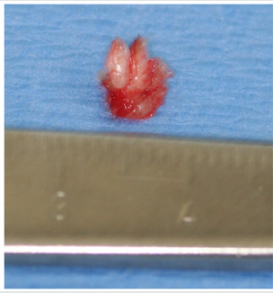
A 12-year old Asian female was referred to the office by her family dentist for evaluation of a soft tissue lesion localized to the dorsal surface of the tongue (Figure 1). The patient stated that the lesion has been present for at least six months. Her past medical history was unremarkable. She was not taking any medications and denied any allergy to medications. The patient also denies being sexually active. Sexual abuse was also ruled-out. Head and neck examination were negative for palpable neck masses and lymphadenopathy. Oral examination of the dorsal surface of the tongue revealed a pink-white colored exophytic lesion that was freely movable. The surface texture had a pebbly appearance that resembled, “cauliflower”. The remaining oral examination was unremarkable. Examination of the upper and lower extremities was negative for any soft tissue lesions resembling HPV. Based on the clinical appearance of the lesion, the differential diagnosis included squamous papilloma, verruciform xanthoma, papillary hyperplasia and condyloma accuminatum of the tongue. The father and patient were informed of the clinical findings and excisional biopsy (Figure 2) for a definitive microscopic diagnosis under local anesthesia was recommended.
Figure 1: Clinical photograph of squamous papilloma on left dorsal surface of the tongue in 12-year old Asian child.
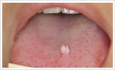
Figure 2: Excised squamous papilloma specimen from dorsal surface of tongue of 12-year old Asian patient.
Histopathology
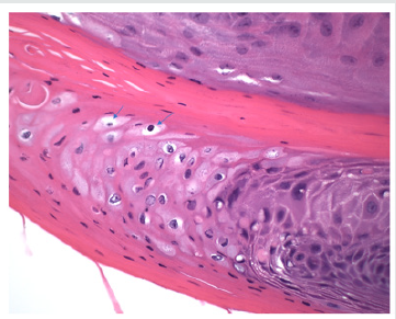
Excisional biopsy was completed under local anesthesia with 1.0mm margins of normal tissue and to the depth of the tongue musculature. The lesion was stained with hematoxylin and eosin for histopathological diagnosis. Histological examination demonstrated long, thin papillary projections of parakeratinized stratified squamous epithelium (Figure 3a). Localized areas of basilar hyperplasia with koilocytes were observed (Figure 3b). The histological findings were consistent with squamous papilloma.
Figure 3(a): Histopathology demonstrating proliferation of hyper keratinized stratified squamous epithelium finger-like projections with thin fibrovascular connective tissue core (Hematoxylin and eosin stain. Original magnification x 40).
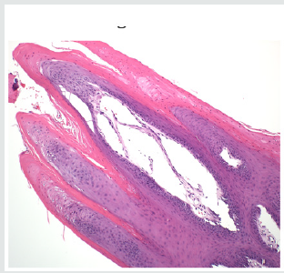
Figure 3(b): Presence of koilocytes (arrows) in the spinous layer of the epithelium that are characteristic histopathologic findings of oral squamous papilloma (Hematoxylin and eosin stain, x 200).
Discussion
In children, viral transmission remains controversial. HPV transmission may occur by any number of different mechanisms, such as autoinoculation, heteroinoculation, perinatal transmission, sexual abuse and contact with fomites [1,4,5,9,10]. However, trauma is one mechanism that should be considered in our patient. Iatrogenic tongue biting may initiate an infection of the basal squamous epithelial cells that allows the entrance of the HPV into the tongue. Viral DNA then enters the nucleus of the infected epithelial stem cells and can replicate causing the HPV infection [15]. Histopathologic features of oral squamous papilloma demonstrate hyperkeratosis in the epithelium, proliferation of the spinous cells that result in long, thin finger-like projections above the mucosa [1- 3,8,14]. The characteristic feature of HPV infection is the presence of koilocytes (Figure 3b) which are virus-infected epithelial cells due to perinuclear cytoplasmic vacuolization of cells of the spinous layer of the epithelium. This results in the nuclei becoming pyknotic and cremated surrounded by an optically clear zone [1-10]. All the described histopathologic findings in our patient are characteristic of HPV infection. Although the prevalence of oral HPV infection is low, a bimodal distribution is observed. The highest prevalence is observed in children less than 1 year of age and the second peak occurs in adolescents, between 13 to 20 years old [9,16]. Despite the bimodal distribution, oral HPV infection is considered low in children [17]. In a study of 4140 children between the ages of 10 to 18 years old, the prevalence of oral HPV infection was 1% and the most common type was HPV 11 [18]. Treatment of HPV is by surgical excision [19]. The United States Food and Drug Administration (2016) approved Gardasil 9 human papillomavirus 9-valent vaccine to prevent infection against HPV types 6, 11, 16 and 18 (Merck & Co., North Wales, Pa). At present, only Gardasil 9 human papillomavirus 9-valent vaccine recombinant has been approved for vaccination in the United States (Center for Disease Control, 2019) [20]. To obtain the greatest clinical efficacy and cost effectiveness of vaccination, the Center for Disease Control (CDC) recommends that both male and female children between the ages of 11-12 years get two HPV vaccinations six months apart before individuals have been exposed to the human papillomavirus. It is also recommended that adult women up to age 26 years and men up to age 21 years also obtain the HPV vaccine [21].
Conclusion
A case of oral squamous papilloma on the tongue of a 12-year old female is presented to create awareness of this soft tissue lesion in the oral cavity of children due to the human papillomavirus. It is only after educating clinicians and parents about the HPV will we observe greater identification, management and treatment in the HPV infected child.
To Know More About Lupine Publishers Pinterest Click on Below Link: https://www.pinterest.com/lupineonlinegroup/
Monday, September 7, 2020
Lupine Publishers | Management of Internal Root Resorption with Bioceramic Material on Permanent Tooth-A Case Report
Lupine Publishers | Journal of Pediatric Dentistry

Abstract
Internal root resorption (IRR) is a category of pulp disease characterized by the loss of dentine as a result of the action of clastic cells stimulated by pulpal inflammation. The objective of this case report was to account for the diagnosis and management of an internal root resorption without perforation. The patient, a 26-year-old male, came to Guru Nanak Institute of Dental Sciences and Research, West Bengal, without having symptoms in the tooth. Endodontic treatment was performed using the following methods: irrigation of the root canal with 2.5% of sodium hypochlorite, then calcium hydroxide (CH) was applied as intracanal medicament for one month. Complete instrumentation was done with Hyflex One File (Coltene) and obturation with corresponding guta-percha and Roeko Guttaflow Bio seal sealer (Coltene). The patient was checked after one week and then after six months. He did not have any symptoms and IOPA radiograph did not show any further progression of the lesion.
Keywords:Internal Root Resorption; Calcium Hydroxide (CH); Sodium Hypochlorite; MTA
Introduction
The Glossary of the American Association of Endodontists defines internal root resorption (IRR) as a condition associated with a physiological or pathological process that results in the loss of dentin, cement and bone [1]. Most teeth with internal root resorption are symptom free and are first clinically recognized through routine radiographs. However, when resorption actively progresses, the tooth is only partially vital and may present typical symptoms of pulpitis. Bell (1830) first reported about IRR. Mummery (1920) called it “pink tooth of Mummery” due to the presence of pink discoloration on the crown [2]. This condition, although rare, is more frequent in the male population. The IRR is more common in the presence of a periapical lesion. Its prevalence was estimated between 0.01% and 1% depending on the inflammatory condition of the pulp [3]. The IRR could be caused by several stimuli: trauma, chronic inflammation of pulp/periodontal ligament, heat created by the friction of drills during the preparation of cavities, cracked tooth syndrome, tooth reimplantation and orthodontic treatment [4]. There have also been reported cases of internal reabsorption caused by Herpes Zoster virus [5]. The IRR is caused by inflammatory stimuli which produce an alteration of the odontoclast inhibitory mechanism resulting in an alteration of the pre-dentine layer. The vascular change in the pulp produces hyperemia increasing oxygen tension and causing an acidic pH level that attracts multinucleated cells, odontoclasts and dentin clasts. Dominance of inhibitory substances such as OPG (osteoprotegerin) as activators of RANKL (receptor activator of factor kappa B ligand) followed by swelling, results in the rupture of protective coatings allowing the invasion of odontoclasts and initiating resorptive patterns. Connective, post-resorptive activity tissue transforms into metaplastic granulation tissue [6]. Generally, IRR detection is done by X-rays, however, the use of cone beams computed tomography (CBCT) has been reported to be highly useful for diagnosis in endodontics, since it shows the lesion in detail and includes information about adjacent anatomy, which X-rays does not provide [7]. The periapical radiography is limited because it provides a twodimensional image [8], whereas diagnosis by CBCT shows images in all their dimensions through tomographic slices, without image overlay [9]. Also, diagnosis by CBCT may improve the accuracy and efficiency in the prognosis of the tooth [10]. Therapeutically, the biomaterial employed can influence the prognosis of the nonsurgical endodontic treatment done for extensive internal root resorption [11]. MTA is most commonly used in these cases because of its sealing ability, biocompatibility and potential induction of osteogenesis and cement genesis and it can be used in a humid environment [12]. Another study using an experimental immature tooth model, demonstrated that the MTA also increased the fracture resistance of bovine incisors when submitted to different reinforcement treatments Recently bioceramics are widely used in endodontics. Roeko Guttaflow Bio seal (COLTENE) is a bioceramic endodontic sealer which claims to avoid shrinkage upon setting as it has Zirconium oxide is used as the radiopacifier, and the material is claimed to be aluminum-free, non-soluble and does not shrink during setting. It gives advantage of flow of material as well as sealing ability which better bond with the corresponding gutta percha used for obturation [13]. The purpose of this case report is to describe the diagnosis and clinical management of an internal root resorption with bioceramic material.
Case Report
Male patient, 26 years old, treated at the post graduate department in Guru Nanak Institute of Dental Sciences and Research. The patient reported no pain at the time of appointment the chief complaint was discoloration of the front tooth which was traumatized 5 year back. Vitality tests using Endo-Ice (Coltene, Switzerland) were performed in [11]; the tooth gave negative response. The patient did not present tooth mobility and periodontal pockets. IOPA radiograph of the affected tooth#11 showed an oval enlargement (ballooning out) of the root canal space (Figure 1). The pulp chamber and canal cannot be followed throughout the lesion. Radiograph performed at different angulation to confirm the resorptive lacunae is a continuation of the distorted border of the root canal. Endodontic treatment was suggested; therefore, isolation protocol was performed to make the cavity opening later (Figure 2). Working length of the tooth was determined by IOPA radiograph using #15K file (Figure 3) and the result was confirmed with apex locator Canal Pro (COLTENE). After removing the pulp tissue properly chemical-mechanical instrumentation was performed with Hyflex One File (COLTENE) and irrigation was done with 1ml of 2.5% of sodium hypochlorite between each time instrumentation with 30-gauge side vented needle. This was followed by irrigation with normal saline to remove any remnants of hypochlorite, later canal was dried with absorbent points. Ca (OH)2 dressing was given for 1month and the medicament was changed weekly. After one month, temporary restoration was removed with [4] round diamond bur, canal was irrigated with 5 mL of 2.5% sodium hypochlorite (NaOCl) and 5 mL of 17% of ethylenediaminetetraacetic acid for removing the Ca (OH) dressing and then the canal was flushed with normal saline and dried.
Figure 1: .
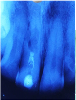
Figure 2:
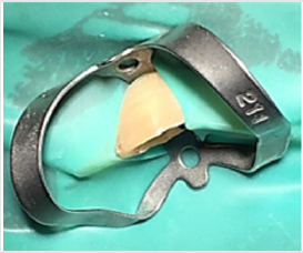
Figure 3:
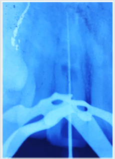
Figure 4:
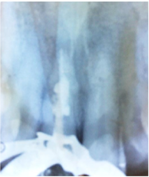
Figure 5: .
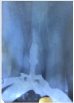
After removing the medication, obturation was done with Hyflex corresponding Gutta Percha and the remaining pulp chamber was obturated with Guttaflow Bio seal sealer (Figure 4). Access cavity restoration was done with light cure composite resin. The patient was recalled after 6 and 12 months (Figure 5, 6) for clinical and radiographic follow up. Clinical examination of tooth was functional without sensitivity to percussion or palpation [11].
Figure 6: .

Discussion
There is always a dilemma of whether to treat a tooth with a questionable prognosis endodontically or extract it and subsequently place an implant. Bell first reported a case on internal resorption in 1830. Since then there have been numerous reports in the literature [14]. Two types of internal root resorption are generally described: the internal root canal inflammatory resorption and the internal root canal replacement resorption. In the inflammatory resorption, the resorptive process of the intraradicular dentin progresses without adjunctive deposition of hard tissues adjacent to the resorptive sites. The phenomenon is associated with the presence of granulation tissues in the resorbed area and identifiable with routine radiographs as are radiolucent zone centered on the root canal. In the replacement resorption, the resorptive activity cause defects in the dentin adjacent to the root canal, with concomitant deposition of bone like tissue in some regions of the defect. It results in an irregular enlargement of the pulp space with partially or fully obliterated area of the pulp chamber. Internal resorption is the result of an inflamed pulp and the clastic precursor cells recruiting through the blood vessels. Treatment of internal resorption is quite predictable as it is easy to control the process of internal root resorption via severing the blood supply to the resorbing tissues with conventional root canal therapy. Intraoral X-ray of IRR is characterized by the radiographic appearance of an oval shape enlargement within the pulp chamber or the root canal. However, the early diagnosis of the IRR is difficult by examination of a conventional X-ray. If IRR is suspected, several shots under different angles of incidence are recommended. In the treatment of internal resorption, the use of calcium hydroxide also has two other important goals: to control bleeding, and to necrotize residual pulp tissue and to make the necrotic tissue more soluble to sodium hypochlorite. Because of the limited access by instruments to all areas of the resorption cavity, chemical means are needed to completely clean the canal. Studies on the effectiveness of sodium hypochlorite and calcium hydroxide to remove the resorptive and other tissues from the root canal indicate that they have an additive or even synergistic effect [15]. In cases where the resorption has not perforated, it is usually enough to use calcium hydroxide paste in the canal once from 1 to 2 weeks. This allows removal of the residual tissue at the next appointment by irrigation and instrumentation. In our treatment protocol, we choose Guttaflow Bio seal (COLTENE) sealer due to its versatile property of Bioceramic component & gutta-percha particles. Upon contact with fluids, this material provides natural repair constituents, such as silicates and calcium, which contribute to the activation of biochemical processes, providing additional support to the root canal regeneration. A novel material for root canal filling that combines gutta-percha in a powder form with a particle size of less than 30 μm and a sealer. The sealer has also showed least cytotoxicity as well as inflammatory reaction [15].
Conclusion
It is puzzling in diagnosing and treating a root resorption case, therefore a suitable management is perilous. Thorough investigations and discussion are required for the management especially when the prognosis of the tooth is poor upon consultation. Absence of periapical lesion and no signs and symptoms at the 12-months review provided a favorable outcome to once a tooth of hopeless prognosis.
To Know More About Lupine Publishers Pinterest Click on Below Link: https://www.pinterest.com/lupineonlinegroup/
Friday, September 4, 2020
Lupine Publishers | Management of Mesiodens In Mixed Dentition- Molariform and Tuberculate: A Case Report
Lupine Publishers | Journal of Pediatric Dentistry

Abstract
Timely intervention is the key to any setback in the mixed dentition. Teeth which are supplemental to the normal dentition are supernumeraries, the most common being mesiodens, present in the premaxillary region. Certain pathological consequences may arise due to mesiodens like unaesthetic midline diastema, rotation, displacement, root resorption and cyst formation. The current case report presents the management of developing malocclusion in the anterior region due to the presence of mesiodentes- a molariform and a tuberculate. The molariform mesiodens was impeding the eruption of the maxillary right central incisor, thus the surgical removal of both mesiodentes was planned and executed. In addition to this soft tissue uncovering was done for the unerupted maxillary central incisor. On follow up, uneventful healing was observed successfully.
Keywords:Mesiodens; Mesiodentes; Mixed Dentition; Molariform; Tuberculate
Introduction
Supernumerary teeth, or hyperdontia, is a term that describes teeth that are surplus in number when compared to the normal complement of teeth [1]. The etiology of supernumerary teeth remains unclear and not yet completely understood [2]. Among the various proposed theories that have attempted to explain the causes behind the development of supernumerary teeth, current literature favors the ‘lamina hyperactivity theory’ that states hyperdontia results from independent, locally conditioned hyperactivity of the dental lamina [1]. The most commonly occurring supernumerary tooth is the ‘mesiodens’, a term that was initially coined by Balk in 1917. As the name suggests, the mesiodens is usually located mesial to the central incisors in the premaxillary region [3]. With a frequency of prevalence between 0.15-3% in the permanent dentition and 0.02-1.9% in the primary dentition, it may occur as single or multiple, unilateral or bilateral, may be erupted or impacted and frequently found in conjection with cleft lip and palate and syndromes like Cleidocranial dysostosis, Gardner’s syndrome, Ellis-Van Creveld syndrome, Ehlers- Danlos syndrome, Incontinentia Pigmenti, and Tricia-Rhino- Phalangeal syndrome [4,5]. Also, mesiodentes may vary in shape from simple conical form to a larger, more complicated crown shape with several tubercles [6]. The dysfunctional nature of mesiodens is known to cause a variety of clinical complications such as being unaesthetic, pathological disturbances in the normal eruption and positions of adjacent teeth, altered growth and development in the area, retention of primary teeth, odontogenic cysts, caries, pulp necrosis of the adjacent teeth, dilaceration of developing tooth, nasal teeth, gingival and periodontal problems [2,7]. Accordingly, their early diagnosis and management is vital to waive off complications of such kind.
Case Report
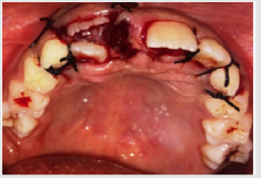
A 9-year old female patient reported to the department of Pedodontics and Preventive Dentistry with the complaint of irregularly placed upper front teeth and wanted it to be corrected. The patient was normal and healthy with non-contributory medical and dental histories. The extra oral examination did not reveal any abnormalities. Intraoral examination revealed a Class I mixed dentition with a missing upper right central incisor and an erupted molariform mesiodens in its place. In addition, there was a firm bulge palpable in the upper right central incisor area which suggests the impediment in the path of eruption of the central incisor by the erupted mesiodens (Figure 1). An occlusal radiograph was taken to rule out the possibility of multiple supernumerary teeth and surprisingly another unerupted and impacted inverted mesiodens with an incomplete root was found mesial to the upper left central incisor (Figure 2). The SLOB technique confirmed that the impacted mesiodens was present palatially. Both informed and written consent was obtained from the parents before initiating the treatment. We decided to extract both mesiodentes under local anesthesia. The erupted mesiodens was extracted by intra- alveolar extraction. The impacted mesiodens was surgically removed by raising a mucoperiosteal flap from maxillary first premolar to contralateral first premolar (Figure 3). Rotary cutting instruments with simultaneous irrigation were used for removing enough bone around the impacted mesiodens for its easy retrieval. Soft tissue uncovering was done for the unerupted right central incisor by placing an elliptical incision over the incisal portion of the palpable bulge (Figure 4). The extraction socket was checked for any pathological tissue and the flap was relocated and sutured with interrupted sutures (Figure 5). The patient was recalled after a week for suture removal and followed-up after 3 months. Uneventful healing with no associated symptoms was observed.
Figure 1: Preoperative view showing erupted molariform mesiodens along with unerupted maxillary central incisor.
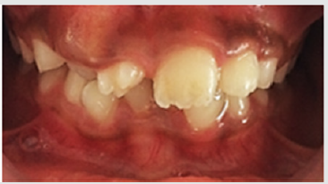
Figure 2: Occlusal radiograph showing unerupted and impacted inverted mesiodens with an incomplete root.
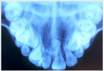
Figure 3: Surgical removal of impacted mesiodens by raising a mucoperiosteal flap from maxillary first premolar to contralateral first premolar.
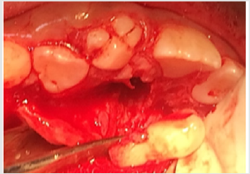
Figure 4: Soft tissue uncovering for the unerupted right central incisor by placing an elliptical incision over the incisal portion of the palpable bulge.
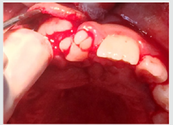
Figure 5: Interrupted sutures placed.
Discussion
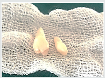
The realm of pediatric dentistry incorporates the practice of interceptive orthodontics, thereby bestowing upon the pediatric dentist, opportunities of providing timely guidance in the development of occlusion. The current case report presents the management of developing malocclusion in the anterior region due to the presence of mesiodens. A mesiodens occurring in the primary dentition is a rarity even though, it being the most common dental abnormality in the permanent dentition [8]. Most cases of mesiodens are discovered during the first decade as maxillary central incisors are erupting and radiographic examinations are performed as an aid to screening for any other malformations and abnormalities [1]. Various theories regarding the etiology of mesiodens have been reported in the literature but the subject remains controversial [3]. Heredity has been suggested to be an etiologic factor based on the observation that supernumeraries are more common in family members; however, it does not follow a simple Mendelian pattern [9]. It was originally postulated that the mesiodens represented a phylogenetic relic of the extinct ancestors who had three central incisors. This is known as phylogenetic theory reversion (atavism) which has now been discarded by the embryologists [10]. The dichotomy theory states that, a mesiodens arises due to the splitting of the tooth bud. On the contrary, Taylor argued that splitting of the tooth bud may either form two equal sized teeth or one normal and one dysmorphic tooth [8]. The hyperactivity theory states that development of mesiodens is due to the hyperactivity of the dental lamina. The ‘field model’, proposes that a tooth bud which is forming at a given location develops according to its position within the field, further determining its shape. The ‘clone model’ postulates that that each tooth class is derived from a clone of ectomesenchymal cells which are programmed by epithelium to produce teeth of a given pattern. Depending upon the specific factors expressed from these ectomesenchymal cells, the shape of the accessory tooth germ forms in the vicinity of the incisors class of teeth becomes evident at the bell stage [11]. This case report presented with an erupted molariform mesiodens and an unerupted tuberculate mesiodens with an undeveloped root (Figure 6). Since the molariform mesiodens was impeding the eruption of the maxillary right central incisor, the surgical removal of both mesiodentes was planned and executed. Timing of interceptive treatment should be as soon as possible following clinical detection of an abnormal eruption pattern. It has been suggested that a tooth delayed in its eruption by more than six months with respect to its antimere should be radiographically investigated. Hogstrum and Andersson [12] suggested two alternatives exist.
The first option involves removal of the supernumerary as soon as it has been diagnosed. This could lead to an unpleasant experience that may have a psychological effect on a very young child and has been said to cause devitalization or deformation of adjacent teeth. Secondly, the supernumerary could be left until root development of the adjacent teeth is complete. The potential disadvantages associated with this deferred surgical plan include; loss of eruptive force of adjacent teeth, loss of space and crowding of the affected arch, and possible midline shifts. In the present case, since the roots of the adjacent teeth were completely formed and the child was at an age where she could sustain a surgical procedure, the surgery was undertaken with utmost attention to detail and caution. Access to mesiodens during surgery must dealt carefully considering the quantity of bone amputation and the possible damage to the adjacent teeth [7]. Follow up is indispensable in such cases since the eruption status should be monitored. The patient revealed satisfactory healing and suitable eruption of the maxillary central incisor. Pediatric dentists are the firsts to usually identify developing malocclusions and thus it is their responsibility to intervene and intercept in an apt manner to prevent future unfavorable sequalae.
Figure 6: Molariform and tuberculate mesiodens.
Acknowledgements
Authors are thankful to their colleagues and faculty.
For More About Lupine Publishers Pinterest Please Click on Below Link:
https://www.pinterest.com/lupineonlinegroup/
Subscribe to:
Posts (Atom)