Friday, March 26, 2021

Lupine Publishers | Association of Mother’s Genetic Sensitivity to the Taste of 6-N-Propylthiouracil (Prop) and Their Children’s Dental Caries Status in Nepal

 Lupine Publishers | Journal of Pediatric Dentistry


Abstract

Introduction: Dental caries is a multifactorial disease and is one of the most prevalent infectious disease that affects mankind. Young children present a unique risk for dental caries as their host-defense systems and bacterial flora are in the process of being developed and also because the newly erupted tooth surfaces are more susceptible to dental caries. Mothers are the persons who generally influence their children via their own food preferences. Hence the mother’s taste perception plays an important role in the development of dental caries in their children. The need of this study is to examine the association of mother’s taste perception to 6-n-propylthiouracil with caries prevalence in their young children as well as with other caries risk determinants such as mothers and their children’s oral hygiene practices.

Material and Method: 180 pair of mothers and their children in the age group of 3 to 6 years of both sexes were selected for the study & 6-n-propylthiouracil testing is done. A trained and calibrated examiner who did not have any knowledge of the mother’s PROP test performed a comprehensive clinical examination of the children to determine the presence or absence of DMFT/dmft.

Results: It is observed that nonstarter mother and children have higher caries prevalence than medium tasters and supertasters. Discussion- Genetic sensitivity to taste is an inherited trait in children from their parents, inheritance from mother being more pronounced.

Conclusion: Dental caries is multifactorial. No significant correlation between susceptibility of mother and child to genetic sensitivity exists, and genetic sensitivity is not the only criteria for severity.

Keywords: 6-n-Propylthiouracil; dental caries; dietary habits

Introduction

Dental caries is a multifactorial disease and is one of the most prevalent infectious disease that affects mankind. The development of dental caries depends on several critical interactions between a susceptible tooth surface, oral bacteria, fermentable carbohydrates and frequency of consumption of sugar [1,2]. Young children present a unique risk for dental caries as their host-defense systems and bacterial flora are in the process of being developed and also because the newly erupted tooth surfaces are more susceptible to dental caries [3]. Parents must also negotiate the dietary transition from bottle feeding to solid food and also take into consideration the child’s tastes. Several risk factors such as salivary counts of mutant’s streptococci, past carious experience and frequency of sugar intake have been evaluated to identify children at high risk of caries [1,4-7]. However, none of the currently available caries screening methods can identify children at high risk of caries quite effectively [1,8].

The role of diet as a direct cause of dental caries has been extensively reported [1,4,9,10]. A high intake of sugar has been correlated with a high dental caries in pre-school and school aged children. A high sugar intake reflects a preference for a sweet substance. Genetic sensitivity to taste may be associated with a preference or no preference of some food by children. Sensitivity to taste is an inherited trait in children [1,11]. In 1991 it was found that there is a genetic variation in the ability to taste the bitterness of the chemical 6-n-propylthiouracil (PROP) [1,12]. This variation was found to be associated with food preferences in children. Fox in 1931 found that some individuals perceived chemicals related to PROP as bitter (supertaster) while others could hardly perceive them at all (nonstarter). Those who are PROP super taster are typically supertasters in general and tend to dislike sweet while those who are PROP none tasters like sweet food and prefer strong tasting food products. Mothers are the persons who generally influence their children via their own food preferences. In many households’ mothers generally reward their children using sugary snacks which might increase their children’s preference for that kind of food. Hence the mother’s taste perception plays an important role in the development of dental caries in their children. This study was carries out with following aims and objectives:

Aim

To find the association between mother’s sensitivity to 6-n-propylthiouracil (PROP) and their children’s dental caries status

Objectives

To compare mother’s and child’s oral hygiene practices with their dental caries status. To determine the taste quality and taste preferences of food products among these study groups.

Materials and Methods

220 pairs of mother and children were randomly selected from consecutive children who were accompanied by their mothers visiting the Department of Pedodontics and Preventive Dentistry in the institution. Out of these 220 pairs of mother and children 180 pairs fulfilled the inclusion criteria. A single trained and calibrated examiner who did not have any knowledge of the mother’s PROP test performed a comprehensive clinical examination of the children to determine the presence or absence of DMFT/dmft. All examinations were performed with a mouth mirror and explorer in a lighted operatory after taking consent from the mother. Demographic information of mothers, their oral hygiene practices, their oral health status and the presence of grandparents in the household was collected by an open-ended questionnaire. A second questionnaire was used to collect data regarding the oral hygiene and feeding practices of the child (e.g. frequency of tooth brushing, intake of sugary food and frequency of intake of sugary food).

Inclusion Criteria

For children

a) Age groups of 3 to 6 years.
b) ASA physical status I/II and stable mental condition
c) Should be accompanied by mother.

For mother

a) Participant cooperation and acceptance of the study programmer.
b) Should be healthy and mentally stable.
c) Should be literate.

Exclusion Criteria

For children

a) Should not be less than three years and not over six years
b) Should not have any medical / hereditary conditions.
c) No acute dental diseases.

For mothers

a) Should not be pregnant
b) Should not suffer from systemic diseases
c) Should not be allergic to 6-n-propylthiouracil (PROP)
d) Should not be under any medications that could alter the taste sensation and affect salivary flow.

Prop testing

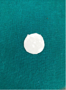
Figure 1: Filter paper with PROP solution.
Lupinepublishers-openaccess-pediatric-dentistry-journal

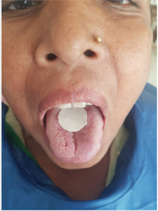
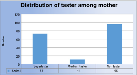
single examiner who did not have any knowledge of the mother’s and children’s oral health status had conducted the PROP test on the mothers. A small piece of filter paper (2 cm circle of Whatman’s grade 1 filter paper) containing approximately 1.6 mg (measured by calibrated dropper) of 6-n-propylthiouracil (PROP) was used to determine each mother’s taste type (Figure 1). The mother was asked to put the piece of filter paper in the mouth and moisten it with saliva for 30 seconds (Figure 2). After removing the filter paper, the mother was asked to quantify the intensity of bitter taste on the modified Green’s scale and was classified of supertasters (>60), medium tasters (12-60) and no tasters (<12). All examinations and PROP testing were carried out at the same time of the day (mid-morning) throughout the study. All the data gathered was tabulated and statistically analyzed. Student’s paired t test and chi square test was done.

Figure 2: PROP test on mother.
Lupinepublishers-openaccess-pediatric-dentistry-journal

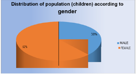
Figure 3: Distribution of population (children) according to gender.
Lupinepublishers-openaccess-pediatric-dentistry-journal

Figure 4: Distribution of taster among mother.
Lupinepublishers-openaccess-pediatric-dentistry-journal

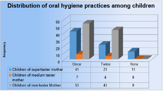
Figure 5: Distribution of oral hygiene practices among children.
Lupinepublishers-openaccess-pediatric-dentistry-journal

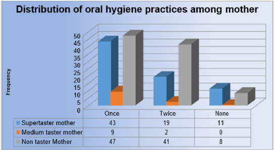
Figure 6: Distribution of oral hygiene practices among mother.
Lupinepublishers-openaccess-pediatric-dentistry-journal

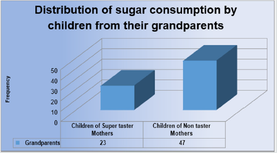
Figure 7: Distribution of sugar consumption by children from their grandparents.
Lupinepublishers-openaccess-pediatric-dentistry-journal

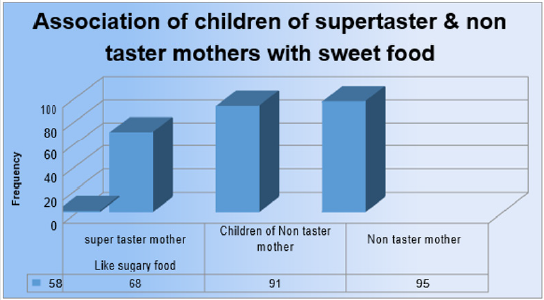
Figure 8: Association of children of supertaster & non taster mothers with sweet food.
Lupinepublishers-openaccess-pediatric-dentistry-journal

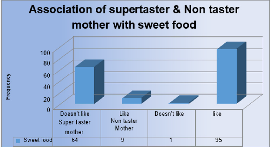
Figure 9: Association of supertaster & Non taster mother with sweet food.
Lupinepublishers-openaccess-pediatric-dentistry-journal

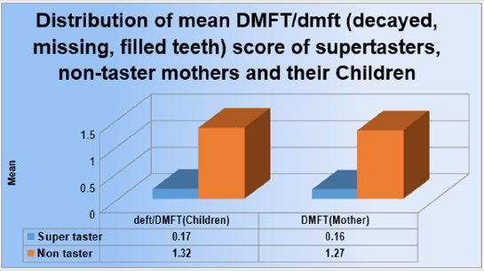
Figure 10: Distribution of mean DMFT/dmft score of supertasters, non-taster mothers and their Children.
Lupinepublishers-openaccess-pediatric-dentistry-journal

Table 1: Association of supertaster & non taster mothers and their children with sweet food.

Lupinepublishers-openaccess-pediatric-dentistry-journal

Table 2: Distribution of mean DMFT/dmft score of supertasters, non-taster mothers and their Children.

Lupinepublishers-openaccess-pediatric-dentistry-journal

Discussion

The present study comprised of 180 pairs of mother and child. The mothers were subjected to PROP testing and classified into super tasters, medium tasters and non-tasters according to the Green’s labeled magnitude scale. The mothers who were super tasters found the taste of PROP to be extremely bitter and the non-taster mothers described the taste of PROP to be tasteless while the medium taster mothers failed to categories the taste of PROP. The super taster mothers perceived the taste of PROP in lower concentration (1.6 mg per ml). This may be due to the presence of high-density fungiform papilla and taste receptors in the tongue compared to medium tasters and non-tasters. This may make the super taster mothers dislike sugary food. Mothers being the primary care givers to children influences the food habit of children. Mothers who prefers sugary foods are more likely to prepare and feed their children the same then those who do no prefer sugary food. In our study it was found that the mean dmft/ DMFT score among non-taster mothers (1.32) and their children (1.27) was higher than the mean dmft/ DMFT of super taster mothers (0.17) and their children (0.16) respectively. The influence of grandparents in the family cannot be neglected. Even in children of super taster mothers, 23 children reported they were given sugary snacks by their grandparents regularly while 47 children of non-taster mothers reported the same.
The pampering of grand parents may also be a causative factor for dental caries in children. The finding of the present study also showed that children who brushed their teeth once or twice per day experienced more dental caries if their mothers were non tasters whereas children of super tasters had less dental caries. This can be explained by the frequent intake of sugary food in case of children of non-taster mothers and also may be influenced by the food prepared by their mothers. Similarly, mothers who were non tasters had a higher dental caries experience than the super taster mothers. This may be due to their increased susceptibility to sugary food. In the present study when the super tasters and non-taster mothers were asked about their liking for sugary food, majority of the super taster mothers (64) stated that they did not prefer sweet food, while 95 non taster mothers stated that they preferred sugary food. In case of the children of super taster and non-taster mothers, the results were significant. The mother’s dietary habits and tastes probably affected the oral health status of the child. When the association between the preference of sugary food in super taster and non-taster mothers and their children was seen, it was found that the children of super taster mothers did not prefer sugary food while it was vice versa for the children of non-taster mothers. This may be due to genetic factors and also might be due to the dietary habits of the mothers which influenced the dental caries status of their children.

Conclusion

In the present study, the results suggested that the children of the mothers who are non-tasters or who prefers sweet food have higher dental caries experience. Factors such as the presence of grandparents in the household, frequency of brushing, preference of food and dietary habits may play an important role in the development of dental caries in children. Thus, mother’s genetic sensitivity to the bitter taste of prop can be used as a useful adjunct to identify children who are at risk of developing dental caries.

Read More Lupine Publishers Pediatric Dentistry Journal Articles:
https://lupine-publishers-pediatric-dentistry.blogspot.com/

Friday, March 19, 2021

Lupine Publishers | The Time Has Come for Pediatric Dentistry and Medicine to Develop A New Thought Process, Rather Than Just Thinking Outside the Box When Caring for Newborn Babies and Infants, We Need A Totally New Box

 Lupine Publishers | Journal of Pediatric Dentistry


Introduction

The concept of thinking outside the box is no longer valid, we now need to create a new box. The volumes of research showing the merging of medicine and dentistry is growing rapidly and those who fail to grasp its importance will be left behind. Our primary goal in caring for newborn infants and babies should be to allow them to grow and develop to their maximum potential. It’s time for the old, outdated conservative ideas, to be shed and have the Dental and Medical community understand that the separation of medicine and dentistry is rapidly disappearing.

Collaboration does not turf concerns

The primary example of this is in the diagnosis and treatment of tethered oral tissues also known as TOTS. In the early 1970’s less than 22 % of US women were breastfeeding their newborn infants, many who were, did so because they could not afford the cost of infant milk formulas [1]. The formula industry has always advertised the benefits of their product over mother’s milk and since so few mothers were nursing, difficulties of nursing due to tongue or lip ties were dismissed or just plain ignored by the medical providers. Today, we have over 80% of mothers breastfeeding , we also are aware of the many long-term benefits for both the infant and mother. We also know that mothers who wish to breastfeed include all socioeconomic levels of society. It is not a fad as many in the medical community refer to treatment of TOTs.
Yet, the medical community still all too often rejects the idea that TOTS do prevent a baby from achieving a good secure attachment to a mother’s breast, resulting in a plethora of symptoms for both mother and infant, often resulting in the mother giving up and using a bottle. Some explain this failure of the Medical community to embrace the release of these TOTS as overstepping our scope of practice and consider it their turf, yet they do nothing. The effects of TOTS begin in the oral cavity, where we as pediatric dentists, are the experts. Improving the health care of mothers and infants and should not involve turf wars and protectionism for physicians and dentists. We need to effectively join forces to create a successful breastfeeding outcome. Dentistry and medicine treat the same entity, where often one or many health problems can overlap into both professions’ territory. This is a key reason why Medicine needs to develop a cooperative team approach to breastfeeding issues with Dentists [2,3]. The understanding in treating infant oral health is growing and changing, this presents an excellent opportunity for physicians and dentists to find ways in to effectively join forces to create a successful breastfeeding outcome.

Infant- Mother Bonding

It is time to look beyond just the infant’s latch, but the actual potential complication that a poor latch creates beyond the act of breastfeeding comfortable for the Mother. Medicine recognizes the term attachment theory, which states that the babies initial bonding with the mother lasts a lifetime, if it is short circuited, the mother may develop signs of post-partum depression, which rather than treating symptoms pharmacologically, in many cases this may be resolved if we , as pediatric dentists can fix the inability of the infant to latch onto the breast by a simple in office surgical release, not just a simple snip, known as a frenectomy [4]. Breastfeeding plays an integral role in forming the deep attachment between mother and baby.

Failure to thrive

Infants are far too often placed in hospitals for failure to thrive, parents are occasionally subjected to Child abuse accusations because their child appears malnourished, and in severe cases have had nasal-gastric or tubes inserted directly into the stomach surgically to literally force feed infants [5]. All this has happened because the physician, pediatrician, ENT or GI doctors never even consider the ankylosed tongue as the primary cause.

Brain growth and development

The window of opportunity to help many babies may close with-in the first 90 days after the infant’s birth [6]. Studies indicate that a baby’s brain grows by 1 percent each day beginning right after birth. A newborn brain grows extraordinarily fast right after birth but slows down to a growth rate of 0.4 percent per day by the end of three months. An infants’ brains grow by 64 percent in the first 90 days, according to the study. The average brain size was 20 cubic inches (341 cubic centimeters) at birth, and 34 cubic inches (558 cubic cm) at 90 days. Simply stated, the brains of newborns grew from about 33 percent of the average adult brain doubles in size in the first year, and by age three it has reached 80 % of its adult volume.

Air induced reflux

It was those hellish hours between darkness and dawn when my husband and I would take turns walking around the house with our baby inn our arms, praying and, mostly in my case, sobbing because we couldn’t console our baby. Infants suffering from reflux are still being treated with adult anti-reflux drugs rather than looking a t the most likely source of gas build up, the swallowing of air during the infant’s latch due to a poor latch and seal onto the mother’s breast. Adult pharmaceuticals such as Peracid and Nexium are not approved drugs for children under age one [7-10]. Studies show they are not effective. Yet the use is skyrocketed in recent years. Simply releasing TOTS will allow a good secure latch to occur in most infants, quickly resolving the issue. Medications like ranitidine (Zantac) or omeprazole (Prilosec) can prevent absorption of calcium and iron and increase the risk of certain intestinal and respiratory infections. Children who used PPIs had a 22% increased likelihood of fracture, while children who used both PPIs and H2-blockers had a 31% increased likelihood of fracture.

Sleep disordered breathing

Tethered oral tissues (TOTS) is also a common undiagnosed source of obstructive airway problems, obstructive sleep apnea and reflux in infants and newborns [11-16]. The American Academy of Pediatric Dentistry (AAPD) recognizes that obstructive sleep apnea (OSA) occurs in the pediatric population. In order to reduce such complications, AAPD encourages healthcare professionals to routinely screen their patients for increased risk for OSA and to facilitate medical referral when indicated The most common form of Pediatric OSA is a disorder of breathing characterized by prolonged, partial upper airway obstruction and or intermittent/ complete obstruction (obstructive apnea) that disrupts normal ventilation during sleep and normal sleep patterns. These cycles of awakening prohibit the infant as well as toddlers having apnea from reaching deep, restful sleep. For this reason, children with untreated OSA may be inappropriately diagnosed as having ADHD. Often the ankylosed tongue is again ignored and not considered as a part of a differential diagnosis. Findings from studies that used complementary research methods have converged to strongly suggest that inadequate sleep quality and quantity are causally linked to sleepiness, inattention, and probably other cognitive and behavioral deficits that impact daytime functioning, with potential implications for long-term development.

Conclusion

It has been stated that it can take a 17-year lag to change and understand traditional research. This lack of knowledge puts those responsible for enabling new research at a disadvantage [17]. A staggering 36,000 randomized controlled trials (RCTs) are published each year, on average, and it typically takes about 17 years for findings to reach clinical practice [18]. We do not have this time to waste in treating our infants and newborns. We need to establish a good collaboration and trust between our two professions and develop a good understanding of how tethered oral tissues can have a significant impact on the overall growth and development of newborns and infants [19].

Read More Lupine Publishers Pediatric Dentistry Journal Articles:
https://lupine-publishers-pediatric-dentistry.blogspot.com/


Friday, March 12, 2021

Lupine Publishers | Anterior Open Bite Using Simões Network in Growing Patient: A Case Report

 Lupine Publishers | Journal of Pediatric Dentistry


Abstract

The anterior open bite is characterized by the negative vertical overhang occurring in the anterior region. It consists of a discrepancy in the vertical direction and is one of the malocclusions with greater aesthetic-functional impairment, besides dental and skeletal alterations. It has a high prevalence in the deciduous and mixed dentition and its etiology is multifactorial, highlighting the deleterious oral habits as the most prevalent. The objective of this study was to present the clinical case of a growing female patient presenting an anterior open bite associated with thumb sucking, by means of treatment with the functional orthopedic device Simões Network (SN3). During the first 12 months follow-up, we observed facial and intraoral oral changes and forward, the improvement of functional and craniofacial relationships, observed through complementary tests.

Keywords: Open Bite; dentition, mixed; orthopedics

Introduction

The balance of soft tissue growth and facial changes are important for the craniofacial development. Mineralized bone is formed through a process known as ossification by the membrane activity providing the function of remodeling and displacement. Breath, chewing, phonation and swallowing functions are prior to the regular growth development [1]. Thumb or pacifier sucking, and tongue thrusting may cause a disorder knowing as an anterior open bite. The severity of the malocclusion will be according to the magnitude, frequency and time of the habit [2]. Prolonged breastfeeding will be recommended to avoid nonnutritive sucking habits, as the sucking of fingers, pacifiers and bottle feeding [3,4]. Anterior open bite (AOB) is defined as the lack of incisal contact between anterior teeth in centric relation. AOB creates aesthetics problems, speech disorders and tongue thrusting habit [5]. This malocclusion requires early treatment due to all the etiological factors mentioned before. The stability will be achieved in a long term; thus, the pediatric dentist must be alert and minimize the problem as soon as possible in attempt to decrease the time of the treatment and to maintain the stability 5. The auto correction index is low when the correct habits are achieved [6,7]. The prevalence in the population ranges from 1,5% to 11%. Some authors also describe that 17% to 36% of those seeking orthodontic treatments feature AOB [8-11]. This malocclusion may also occurs due to a skeletal component classified open bite into dental and skeletal, associated with excessive molar height, divergent upper and lower occlusal planes, steep mandibular plane angle, increased gonial angle, short mandibular ramus, downward rotation of posterior part of the maxilla or palatal plane tipped up anteriorly, increased lower anterior facial height and decreased upper anterior facial height. According to severity, modalities of treatment are required: growth modulation; orthodontic mechanotherapy and the combination with orthognathic surgery [12-14]. Orthopedics devices is a therapy to readapt the muscular system which is very efficient in growing patients resuming the facial balance [15]. This article presents a clinical case of growing female patient, with anterior open bite treated with the functional orthopedic device Simões Network (SN3) [16].

Case Report

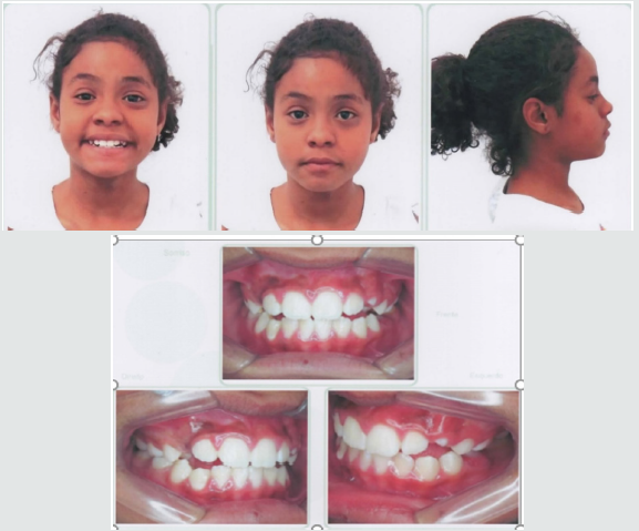
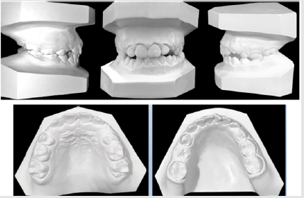
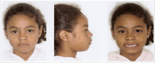
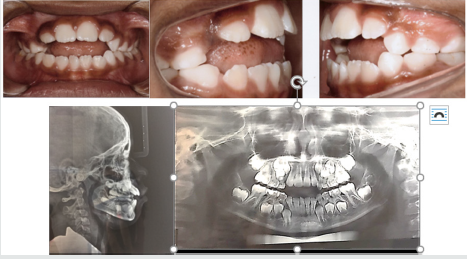
A female patient, 8 years and 4 months of age, melanoderma, came for treatment at the Postgraduate Course in Orthodontics of Brazil University (São Paulo, SP, Brazil). A facial analysis detected convex facial profile, lack of lip closure, and a decrease in nasolabial angle (Figure 1). The patient exhibited thumb sucking habit, mixed breathing, atypical swallowing and speech. shows angle class II malocclusion, 6 millimeters of an anterior open bite, mild crowding and a supernumerary Figures 2&3 tooth in the anterior lower jaw with mandibular midline deviation to the right. Cephalometric Rx shows proclined upper incisors due to the thumb sucking the objetives

Figure 1: Convex facial profile, a decrease in nasolabial angle and the upper lip covering the incisor.

Lupinepublishers-openaccess-pediatric-dentistry-journal

Figure 2: Anterior open bite, mild crowding and a supernumerary tooth in the anterior lower jaw and proclined upper incisors.

Lupinepublishers-openaccess-pediatric-dentistry-journal

Figure 3: Angle class II malocclusion, supernumerary tooth in the anterior lower jaw and an 6mm open bite.

Lupinepublishers-openaccess-pediatric-dentistry-journal

for the first phase of the treatment were to eliminate the thumb sucking, the open bite, dental deviations, provide arch expansion and the extraction of the supernumerary tooth.

Treatment Progress

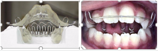
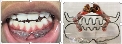
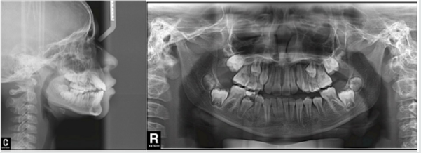
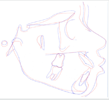
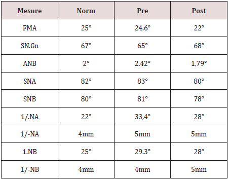
A removable appliance known by Simões Network (SN3) composed with a stainless steel bimaxillary grid (“lower winglets model”) that simulates the incisors occlusion and provides the correct tongue position [15] (Figure 4). The screw was expanded with one-quarter turn biweekly. After 3 months of the treatment beginning o, we added a lip bumper to improve lip seal (Figure 5). We recommended the use for 10 or 12 hours a day. The supernumerary extraction was performed 7 months of the treatment beginning. Figure 6 shows the final of the first stage. Figure 7 shows Angle class I malocclusion and Figure 8 shows the Cephalometric and panoramic Rx after 23 months with the orthopedic appliance. Cephalometric superimposition (Figure 9) and analysis (Table 1) indicated dentoalveolar open bite pretreatment and the correction posttreatment.

Figure 4: SN3 appliance, bimaxillary anchorage with “lower winglets model”.

Lupinepublishers-openaccess-pediatric-dentistry-journal

Figure 5: A lip bumper was added to improve lip seal.

Lupinepublishers-openaccess-pediatric-dentistry-journal

Figure 6: The correction of open bite and the improvement of the alignment.

Lupinepublishers-openaccess-pediatric-dentistry-journal

Figure 7: The arch expansion and molar Class I.

Lupinepublishers-openaccess-pediatric-dentistry-journal

Figure 8: Cephalometric and panoramic Rx after 23 months of treatment.

Lupinepublishers-openaccess-pediatric-dentistry-journal

Figure 9: Cephalometric superimposition revealed maxillary incisor retrusion and mandibular incisor in normal bite.

Lupinepublishers-openaccess-pediatric-dentistry-journal

Table 1: Summary of cephalometric measures.

Lupinepublishers-openaccess-pediatric-dentistry-journal

Discussion and Conclusion

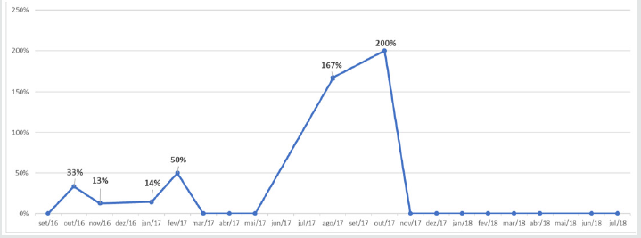
According to the authors, AOB is a challenge daily faced because and it can result speech and swallows’ problems, tongue posture and imbalance between jaw postures [17-19]. Bonna 2016 alerts that the orthodontics or orthopedic devices are fundamental but without family support the habit suppression will not be achieved [20]. The objective for the first phase of treatment were to eliminate thumb sucking, open bite and arch expansion with orthopedic appliance Simões Network SN3 and after a lip bumper was included to improve seal lip. These goals were achieved during the first stage. Graphic 1 shows best fit reduction open bite from May to November 2017. Even pubertal increments offer best time for orthopedic treatment helping determine the predictability, growth direction, patient management and total treatment time, we did not wait to treat because the disadvantages of the open bite [21]. This reported case was successfully treated with SN3 remained stable after the AOB correction. For the second phase with fixed orthodontic treatment will be necessary [22-29].

Graphic 1: Closure open bite variation during time.

Lupinepublishers-openaccess-pediatric-dentistry-journal


Friday, March 5, 2021

Lupine Publishers | The Effects of Breastfeeding on the Process of Tooth and Jaw’s Development

 Lupine Publishers | Journal of Pediatric Dentistry


Abstract

From the nutritional point of view, it has been proven that breast milk has many benefits for the baby, and it is advisable for all mothers to give their baby milk, and if possible do not replace that with the bottle. In other words, we can say that the sucking mechanism used during bottle-feeding is markedly different from that used during breast-feeding. The Federation of Orthodontists of France announced in a report that breastfeeding not only prevents allergies and gastrointestinal infections and overweight, it also promotes the regular growth of the baby’s face. Some of the researches prove this hypothesis.

Keywords: Bottle Feeding; Breast Feeding; Sucking Mechanism; Growth of Baby Face; Allergies; Gastro Intestinal Infections; Overweight

Introduction

After birth, the baby learns how to suck on her mother’s breast. She instinctively brings forward her lower jaw and tongue; then starts sucking with full power so that all the muscles of her tongue, cheeks, lips, and jaw are involved. In all infants, since jaws are not fully developed at birth, sucking breast milk helps the jaw to grow as well as the teeth in the future [1-3]. Breast-feeding has been indicated as one of the main factors which are responsible for the correct growth and formation of dentofacial structures during the infancy [1-3]. Breastfeeding is a useful action for developing and growing teeth and jaws of infants [1-4]. The mechanism used in for the time of bottle-feeding is markedly different from that used during breast-feeding [5-7]. In the course of sucking mother’s milk, more muscles are activated to get milk than to drink milk from the bottles. During this action, the baby inserts more of the nipple into his mouth, consequently, moves the jaw up and down, and sucks the breast with all force to release the milk. To achieve this, the facial and oral muscles of the baby are involved in milking activities. This improves the shape of the jaws, and healthy teeth are expected to be in the correct eruption direction without any deviation and abnormalities [1-4].

The Main Cause of Abnormal Tooth Formation During Infancy

One of the factors leading to abnormal teeth and also leading children to orthodontic or speech therapies [8] is the abnormal orofacial muscular imbalance pattern of the tongue [9-11] known as tongue thrust. This problem is more common among children who are fed through the bottle and is often not seen among those who are breastfed. In other words, the breast-fed baby has more forceful gums and mandibles to extract the milk from the mother’s breast while a baby who is fed with a bottle, does not have to use extra jaws force because by a simple sucking a rapid flow of milk will be obtained. Of course, it should not be taken for granted that all children who use the milk bottle suffer from jaw problems, but it should be remembered that breastfeeding give better evolution to the jaws and teeth than the nourishment from the bottle.

Overview of Some Researches

The early transition from breastfeeding to bottle-feeding may contribute to inadequate mandibular development which can be a dominant and deleterious factor in the development of occolusofacial problems. In this part, we look at some research which may point out this strong hypothesis. Some studies have cited that breastfeeding is a protective factor against malocclusion: Labbok and Hendershot have suggested that increased bottlefeeding duration may contribute to the prevalence of malocclusions [12].

Viggiano et al. and Karjalainen et al. have indicated that breastfeeding can be a positive factor to prevent the development of posterior cross bite in the primary dentition [6,13]. Warren et al. reported that breastfeeding promotes normal palate development and weakens the formation of a deep and high-arched palate [14]. Several studies agree that bottle-feeding may be responsible for the development of sucking habits which may lead to some forms of malocclusion [6,14,15].

Conclusion

Breastfeeding acts on the process of sucking which are influencing the development of facial bones and muscles. Infants who are breastfed have greater facial muscle activities compare to those who are bottle-fed. In other words, breast feeders present an excellent orofacial muscle work out which helps to develop good their bony jaw structures. Moreover, breastfeeding prevents against orthodontic problems and malocclusions (for instance: overbite, posterior crossbite, tongue thrust, oral habits and etc.) that are cited in some researches.

Read More Lupine Publishers Pediatric Dentistry Journal Articles:
https://lupine-publishers-pediatric-dentistry.blogspot.com/
Friday, February 26, 2021

Lupine Publishers | Treatment of Infected Primary Teeth using Modified Antibiotic Paste

 Lupine Publishers | Journal of Pediatric Dentistry


Abstract

Objectives: Treatment of pulpectomized primary molars with chronic infection using a mixture of three antibiotics: Metronidazole, Ciprofloxacin, and Doxycycline mixed with Macrogol or Propylene Glycol (modified 3MIX-MP) as an intracanal medicament before the complete cleaning and shaping and obturation.

Study design: A 7 years old child with infected primary molar came to our clinic for treatment. A detailed medical history and drug allergy were taken. Ciprofloxacin (500mg), Metronidazole (500mg) and Doxycycline (100mg) tablets divided in the proportion of 1:3:3 (one part of Ciprofloxacin, three parts of Metronidazole, and three parts of Doxycycline) and mixed with propylene glycol to form an ointment. Biomechanical preparation was done. The modified 3MIX-MP paste placed in the pulp chamber then temporary filling. The patient was recalled after 2 weeks. The tooth was obturated and restored then a stainless-steel crown placed. Then reevaluated at 3rd, 6th, and 12th months.

Results: Excellent clinical and radiographic success when compared to conventional pulpectomy and non-instrumentational lesion sterilization tissue repair therapy.

Conclusion: Treatment of Primary molar with modified 3MIX-MP, followed by instrumentation and obturation provided excellent clinical and radiographic success when compared to non-instrumentational lesion sterilization tissue repair therapy.

Keywords: Pulp infection; Pulpectomy; Modified antibiotic paste; Primary molars; Chronic, infected pulp; Modified 3 MIX-MP; Pulpectomy; Triple antibiotic paste; Primary teeth

Introduction

The first topical antibiotic introduced to endodontics was Grossman’s polyantibiotic paste in 1951, later many topical antibiotics have been introduced with varying combinations, few of those include Septomixine forte; PBSC (Combination of Penicillin, Bacitracin, Streptomycin and Caprylate sodium), and Clindamycin. However, none of these combinations has proven to be 100% successful in eliminating all the bacterial strains from the root canal system [1-5].

Materials and Methods

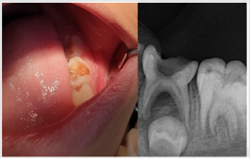
A child aged 7 years old with chronic infection related to the lower left primary molar came to our clinic for treatment of the infected molar (Figure 1). Treatment was explained to the parents and written informed consent was taken from parents before start of the study. A detailed medical history and previous illness with a history of drug allergy were taken from the parents, then the mentioned primary molar was diagnosed clinically, the molar was badly decayed with signs of chronic infection such as: gingival swelling and tenderness to percussion. A radiographic examination was done and a per radicular radiolucency was found, with no excessive root resorption. Commercially available chemotherapeutic agents such as Ciprofloxacin (500mg) (Omacip, NPI Pharma, Oman), Metronidazole (500mg) (Anazol, JPI, Saudi Arabia), and Doxycycline (100mg) (Tabocine, TPMC, Tabuk) tablets were obtained [6,7], then these tablets were crushed into fine powder using sterile porcelain mortar and pestle. These powdered drugs were transferred into three separate sterile glass containers, capped tightly and stored in the refrigerator until its use. Just before use, each powdered drug was divided in the proportion of 1:3:3 (one part of Ciprofloxacin, three parts of Metronidazole, and three parts of Doxycycline) and were mixed with propylene glycol and polyethylene glycol to form an ointment. Reddy GA et al. Trairatvorakul and Detsomboonrat, Jaya et al., Cruz et al. also followed the similar protocol of preparation of 3MIX antibiotic paste [8-11].

Figure 1: Preoperative illustration.

Lupinepublishers-openaccess-pediatric-dentistry-journal

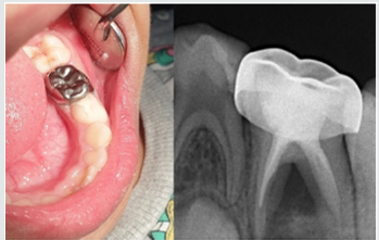
Figure 2: Postoperative illustration.

Lupinepublishers-openaccess-pediatric-dentistry-journal

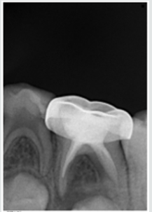
Preoperative clinical and radiographical signs and symptoms were recorded. The tooth was anesthetized using 2% Xylocaine with 1:80,000 adrenalin and isolated with rubber dam. Access opening was performed using round bur, Biomechanical preparation was done using k files from size 10–25. The root canals were chemically cleaned with 1% sodium hypochlorite solution and dried with paper points. The 3MIX-MP paste placed in the pulp chamber and pressed with dampened cotton pellet and temporized with Cavit. The patient was recalled after 2 weeks for evaluation. The tooth was obturated with reinforced zinc oxide eugenol (IRM, Dentsply) using lentulo spirals. Then restored with glass ionomer restorative material (Riva self-cure, SDI) and reinforced by placing stainless steel crowns (Figure 2). Further, the treated tooth was reevaluated both clinically and radiographically at 3rd, 6th, and 12th months intervals postoperatively (Figure 3). At the time of revisits, the tooth was examined clinically for any signs of failure that includes a report of spontaneous pain, presence of swelling, sinus tract and mobility. Radiographic evaluation was done to check the radiolucency and signs of resorption. The tooth was asymptomatic without pain, swelling, sinus tract and mobility also there was no increase in furcation radiolucency or development of root resorption which is abnormal for the age of the child.

Figure 3: 12 months Follow up.

Lupinepublishers-openaccess-pediatric-dentistry-journal

Results

Excellent clinical and radiographic success when compared to conventional pulpectomy and non-instrumentational lesion sterilization tissue repair therapy.

Discussion

This study was approved by “Research Ethics Committee, Taibah University, College of Dentistry, TU CD-REC”. The concept of Non-Instrumentation Endodontic Therapy introduced by Niigata university school of dentistry; Japan has gained reputation as it proved to attain 100% sterility in the root canal system [12- 15]. They recommended a technique similar to pulpotomy where debriding only the pulp chamber of chronically infected primary teeth and placing medicament (ciprofloxacin, metronidazole, and minocycline) near the root orifice without preparing the radicular portion. Cruz et al. suggested vehicles such as macrogol and propylene glycol (3MIX–MP) and demonstrated that these vehicles will carry the medicament deep into the dentinal tubules, thus aid in effective eradication of bacteria [11]. Metronidazole (Nitroimidazole compound) due to its wide spectrum of antibacterial action against anaerobes (Ingham et al. 1975) gained importance as the 1st choice drug for triple antibiotic paste preparation [16,17]. Metronidazole binds to the DNA and disrupts its helical structure and thus leads to rapid cell death. However, metronidazole even at higher concentrations could not eradicate all the bacteria thus indicating the necessity of some additional drugs to sterilize these lesions [15]. The two other antibacterial drugs, i.e. ciprofloxacin, and minocycline, in addition to metronidazole (3MIX) were added in an effort to eliminate all bacteria [8,10,15,18]. The 2nd choice of drug ciprofloxacin is a synthetic fluoroquinolone with rapid bactericidal action. It inhibits the enzyme DNA gyrase of bacteria. It exhibits very potent activity against Gram-negative bacteria but very limited activity against Gram-positive bacteria. Most of the anaerobic bacteria are resistant to ciprofloxacin. Hence, it is often combined with metronidazole in treating mixed infections. The 3rd choice of drug was minocycline. It is a semisynthetic derivative of tetracycline, primarily bacteriostatic, inhibiting protein synthesis by binding to 30S ribosomes in susceptible organisms and exhibits broad spectrum of activity against Gram-positive and Gramnegative microorganisms [3].

In our present study we replaced Minocycline with Doxycycline due to the difficulty in obtaining Minocycline, and before using the Doxycycline as a replacement we have done further searches for previous studies to ensure that both medications have the same effect and this replacement will not affect the efficacy of the mentioned mix. The already done studies concerning the difference between both Doxycycline and Minocycline revealed that still no statistically significant differences had been demonstrated in clinical trials when comparing Minocycline with Doxycycline, and investigators had concluded that both are equally effective. And they differ in their adverse event profile [19]. Considerably fewer adverse effects have been reported for Doxycycline than Minocycline; the adverse effects for Minocycline are 5 times more common than for Doxycycline [19]. We have followed the same protocol of Reddy GA et al. of extirpation of both necrotic coronal as well as all accessible radicular pulp tissue and then complete obturation, which is reported successful clinically over 16th month follow-up [9]. Although the previous studies have demonstrated that the LSTR (Lesion Sterilization Tissue Repair) technique as one of the successful techniques for management of chronically infected primary teeth, the controversies aroused about the duration of therapeutic activity of the medicament and leaving the infected material in the radicular region. So that the present study planned where in treated tooth were revisited after 2 weeks for medicament removal and obturation.

Conclusion

All the primary teeth with chronic infection which were treated using modified 3MIX-MP, followed by the instrumentation and obturation provided excellent clinical and radiographic success when compared to conventional pulpectomy and noninstrumentational lesion sterilization tissue repair therapy.

Read More Lupine Publishers Pediatric Dentistry Journal Articles:
https://lupine-publishers-pediatric-dentistry.blogspot.com/
Friday, February 19, 2021

Friday, February 12, 2021

Lupine Publishers | Varied clinical and Oral Presentation of Beckwith – Wiedemann Syndrome - Report of a Case from Saudi Arabia

 Lupine Publishers | Journal of Pediatric Dentistry

Abstract

Beckwith – Wiedemann syndrome is congenital, genetic and epigenetic pathologies with low prevalence and diverse clinical presentations. It is characterized by triad of omphalocele, macroglossia and gigantism. This syndrome has been widely studied with a current emphasis on improvement of prenatal diagnostic techniques and a multidisciplinary approach towards treatment. We report a case of BWS from Saudi Arabia, with unique presentations and misleading history which delayed diagnosis, due to cultural and religion constraints.

Keywords:Congenital; Epigenetic; Genetic; Prenatal

Introduction

Genetic and epigenetic changes or a human genomic imprinting disorder is characterized by phenotypic variability which might shows its occurrence either as sporadic or inherited. The pathology presents wide range of effect on psychological and social wellbeing of patients and families. One such congenital, multigenic, multisystem human genomic imprinting disorder with complex molecular etiology and variable complex phenotype is Beckwith – Wiedemann Syndrome (BWS). Beckwith-Wiedemann Syndrome is most common overgrowth syndrome described by Beckwith in 1963 and Wiedemann in 1964 with similar findings. It is rare congenital deformity with low prevalence but at same time have high prevalence within genetic abnormalities of overgrowth [1]. The presentation of triad features of omphalocele (exomphalos), macroglossia and gigantism was described earlier as EMG syndrome which now is referred as Beckwith – Widemann Syndrome. The incidence of BWS reported is approximately 1:13700 births and the major cause is thought till date is genetic and epigenetic defects within the chromosome 11p15.5 regions [2].

BWS presents wide array of clinical manifestations such as congenital abdominal wall defects as hernia (exomphalos), gigantism, macroglossia, nevus flammeus, ear pits/hearing loss, midface hypoplasia, cardiac anomalies, hemihypertrophy, genitourinary anomalies and musculoskeletal abnormalities. To standardize the diagnostic criteria various attempts have been made to classify the major and minor criteria. Elliot et al described the diagnosis of BWS with the presence of either three major features (abdominal wall defect, macroglossia, gigantism) or two major and three minor features (ear pits, nevus flammeus, hemi hyperplasia, nephromegaly, neonatal hypoglycemia) [3]. In spite of diverse clinical presentations of BWS, most of the cases do not show characteristic features at birth but develop later in life. Also, children with BWS have significantly increased risk of cancer during early childhood which need strict follow up and monitoring. Here, we present a case of BWS with unique dental and medical presentation and its differential diagnosis with literature review.

Case Report

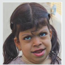
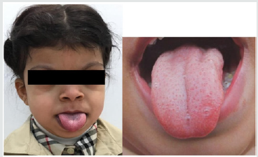
A 5-year-old female patient, accompanied by her mother, presented to the dental unit with complaint of decay tooth in upper front region of mouth. Extra oral examination revealed dysmorphic features, coarse facies and developmental problems (Figure 1). Intra oral examination of hard tissue showed high arched palate, decayed teeth in relation to 51, 52, 55, 61, 62, 74, 75, 84,85. Oral soft tissue examination revealed macroglossia, enlargement of fungiform papillae and mild loss of filiform papillae (Figure 2). Speech and feeding difficulty were noticed due to macroglossia. History revealed she is the youngest 7th child born out of consanguineous marriage in 30th week by cesarian section. She has a chronic history of constipation for 9 months of age. She passes hard stool once in every 8 to 10 days, by spending long time in washroom. It is associated with decrease in appetite and abdominal pain. She was given Movicol (half the adult dose) twice a day for constipation without any medical prescription. She was also tried with lactulose, glycerin suppository and mineral oil. Under medical supervision fleet enema and contrast enema were performed to relieve constipation and to rule out Hirschsprung disease.

Figure 1: Photograph showing dysmorphic features and hypertelorism.

Lupinepublishers-openaccess-pediatric-dentistry-journal

Figure 2: Macroglossia with enlarged fungiform papillae and loss of filiform papillae.

Lupinepublishers-openaccess-pediatric-dentistry-journal

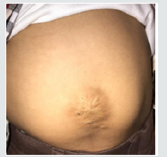
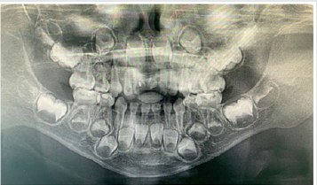
Other medical findings noticed omphalocele, ear pits, large child at 90th centiles, large rounded eyes with hypertelorism, abdominal soft lax, enlargement of kidney, distention of left renal pelvis with significantly distended urinary bladder, abnormal anatomy of the colon located in left abdomen and partial colonic non – rotation with no evidence of obstruction (Figure 3). Based on the clinical and past medical history a diagnosis of Beckwith – Wiedemann Syndrome (BWS) was made. Series of laboratory investigation were reviewed which presented negative urine examination, alpha – fetoprotein, karyotype, microarray and methylation analysis for BMS. Patient was advised for gene analysis and targeting testing for parents. The gene analysis of CDKN1C gene showed heterozygous alteration consistent with BWS but targeting gene tests were refused by parents. Panoramic radiograph was advised considering the patient chief complaint, which revealed multiple developing permanent tooth buds, protrusion of anterior teeth, open bite and increase in mandibular dimension (Figure 4). Under preventive measures the patient was treated for the decayed teeth and is under follow up from past 6 months.

Figure 3: Photograph showing abdominal wall defect with surgical scar.

Lupinepublishers-openaccess-pediatric-dentistry-journal

Figure 4: Panaromic radiograph showing multiple developing permanent tooth buds, open bite and increased mandibular dimension.

Lupinepublishers-openaccess-pediatric-dentistry-journal

Discussion

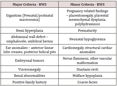
Diagnostic criteria for BWS is still a matter of research due to its varied clinical presentations and overlapping features with other various conditions. The presence of major and minor findings is generally helpful in establishing the clinical diagnosis (Table 1). The oral findings as mentioned in the literature and observed in our case has been tabulated in Table 2 [4,5]. The incidence of BWS is difficult to assess in Saudi Arabia, as most of the cases goes undiagnosed and unnoticed. Also attributed to its diverse clinical presentation and difficulty in diagnosing. In the present case, features of macroglossia, macrosomia, omphalocele, abdominal wall defect (treated immediately after birth and surgical scar observed clinically), Renal involvement, ear crease, high arched palate, open bite and increased mandibular dimension, leads to the diagnosis of BWS. Various molecular mechanisms and alterations have been involved in BWS such as abnormal methylation of H19DMR, loss of imprinting of IGF2, chromosomal rearrangements, loss of imprinting of LIT1, uniparental disomy of 11p15 and CDKN1C mutations [2]. The full gene analysis of CDKN1C gene profile were suggestive of BWS in our case and the alteration is thought to be located in the allele inherited from the mother. Parental testing was advised which was refused by the parents. There are various endocrine and overgrowth syndromes that was considered in the differential diagnosis. These included Simpson-Golabi-Behmel syndrome (mutation in X-linked gene, GPC3), Perlman syndrome (Increased risk of neonatal mortality), Costello syndrome (missense mutation in HRAS), Sotos syndrome (Mutation in NSD1) and Mucopolysaccharidosis type IV (lysosomal storage disorder) [6]. Oral findings like macroglossia of BWS needs differentiation from other lesions like lymphangioma, idiopathic muscular hypertrophy, hemangioma, rabdomyomas, amyloidosis, cretinism and acromegaly.

Table 1: Presenting major and minor features of BWS.

lupinepublishers-openaccess-pediatric-dentistry-journal

Table 2: Oral findings of BWS.

lupinepublishers-openaccess-pediatric-dentistry-journal

The overall risk of BWS for tumor development/malignancies is estimated to range from 4 – 21%. The tumors reported with BWS are mainly embryonal tumors such as Wilms tumor, hepatoblastoma, rabdomyosarcoma, adrenocortical carcinoma and neuroblastoma [7]. The prenatal diagnosis with current technology is increasing representing an important tool to determine some features of BWS before birth. In our case, parents were highly orthodox and refuse to share the detailed prenatal and ultrasonic reports. Few misguided information’s were given by mother which was later clarified with the reports from the subsequent medical hospitals. Patient’s parents were advised for periodic follow up with genetic counselling and the possibility of surgical interventions in the medical units, but they refused to follow and changed the hospitals every time. Hence, an effort was put forward to retrieve the information’s related to the patient while giving her the primary treatment for which she reported to our dental unit. This suggest the need of awareness required in the country like Saudi Arabia, where most of the cases goes unreported/unnoticed or parent’ consent not given or the cultural and religion barriers that prevent reporting such cases. Though the patient was treated with dental fillings, the follow up of the patients is been restricted by the family members.

Conclusion

Beckwith – Wiedemann Syndrome patients usually grow and do well despite being at increased risk of childhood cancer. Hence, strict follow up, awareness of parents and cancer screening is mandatory. Families, physicians and dentists should determine screening schedule including abdominal ultrasound in every three months, blood test to measure alpha-fetoprotein in every six weeks, dental check-up in every six months and other symptomatic treatment schedule as and when required.

Read More Lupine Publishers Pediatric Dentistry Journal Articles:
https://lupine-publishers-pediatric-dentistry.blogspot.com/

980 nm Diode Laser: A Good Choice for the Treatment of Pyogenic Granuloma

Abstract Pyogenic granuloma is a benign non/neo plastic mococutanous lesion . It is a reactional response to constant minor trauma and ca...