
May you celebrate Thanksgiving
Day with love in your heart, prosperous vision in your mind and
gratitude in your being…. Wishing a Happy Thanksgiving from our Interventions
in Pediatric Dentistry Open Access Journal (IPDOAJ)
May you celebrate Thanksgiving
Day with love in your heart, prosperous vision in your mind and
gratitude in your being…. Wishing a Happy Thanksgiving from our Interventions
in Pediatric Dentistry Open Access Journal (IPDOAJ)
Lupine Publishers | Journal of Pediatric Dentistry

Cleft palate is the third most common congenital deformity subsequent to clubfoot and the cleft lip. It may be either unilateral or bilateral and is either complete or incomplete. A multidisciplinary team approach is important to accomplish the various façades pretentiously observed by orofacial cleft. The optimum time of surgical repair is based on the surgeon’s preference, anesthetic risks, co-morbid congenital anomalies, and the apparent psychological impact on the family. Most surgeons repair the cleft lip around 10–12 weeks of age. Cleft palate and lip need early care and attention with a long-term follow-up which requires a team approach involving many specialties.
Cleft palate is the third most common congenital deformity subsequent to clubfoot and the cleft lip [1]. It is seen as anatomical changes which involves the upper lip, nose, and the palate [2]. It represents roughly about 65% of the malformations of the craniofacial section [3]. In this, unilateral cleft lip and palate have been found to have the majority at 46%, followed by isolated cleft palate accounting for 33% of cases. These two being the most common diagnosis among the cleft lip and palate population followed by the others [1].Cleft lips show racial unpredictability, with the highest incidence seen among Asian and Native Americans (1:450 live births) and the lowest incidence seen in the African Americans (1:2,000 live births) [4]. Isolated cleft palate is seen more commonly in females (57%) than in males (43%), with gender differences attributable to the differences in the timing of the embryologic development [4]. About 50% of the isolated cleft palates are found to be in relation with a malformation syndrome, in comparison 15% of combined cleft lip and palate patients without any syndrome [5]. Velocardiofacial syndrome (VCFS) has been found to be the most common syndrome, which is associated with isolated cleft palate [6]. Other etiological factors associated with cleft palate are gestational exposures to alcohol, cigarette smoking, steroids, rubella, anticonvulsants, retinoids, advanced paternal age, folate deficiency and hypoxia [7-9]. An affected parent having a child with a cleft palate is 7%. If one sibling has a palatal cleft with no parental clefts, then future siblings have 2% jeopardy of developing a cleft. That risk upsurges to about 17% if there is one affected sibling and a parent with a cleft [10].
The palate is divided into the primary and the secondary palate. The primary palate comprises of the alveolar arch and secondary palate comprises of the hard and the soft palate. The hard palate is formed by the palatine processes of the maxillae and by the horizontal lamina of the palatine bones. It is covered by oral and nasal mucosa [11].
A cleft palate may be either unilateral or bilateral and is either
complete or incomplete [12]. The Veau System classifies orofacial
clefting into four classes by whether the secondary and/or primary
palates are affected and by laterality [13].
a) Veau Class I: Incomplete cleft, soft palate only (no
unilateral/ bilateral designation)
b) Veau Class II: Hard and soft palate, secondary palate only
(no unilateral/bilateral designation)
c) Veau Class III: Complete unilateral cleft including lip
(primary and secondary palates)
d) Veau Class IV: Complete bilateral cleft
a) Cleft palate is difficulty with feeding [14].
b) Velopharyngeal Insufficiency.
c) Speech distortion especially is the consonant /s/ [15].
d) Hyper-nasality—the perception of excessive nasal
resonance during the production of vowels.16
e) Reduced loudness.
f) Monotone quality.
g) Strangled voice quality - an attempt to be non-nasal in the
presence of VPI.
A multidisciplinary team approach is important to accomplish the various façades pretentiously observed by orofacial clefting. The reconstructive surgeon, otolaryngologists, dentists, orthodontists, speech pathologists, audiologists, geneticists, psychiatrists, maxillofacial surgeons, prosthodontists, and social workers form the most important crucial members of the team treating the patients [17]. The procedure of cleft palate repair has been suggested and gained popularity over many years and is not a one-day ideology. These techniques have undergone numerous modifications that are practiced today. The task of contemporary palatoplasty is not just the closure of the cleft palate but also is to enable optimal speech without compromising the maxillofacial growth [18]. The optimum time of surgical repair is based on the surgeon’s preference, anesthetic risks, co-morbid congenital anomalies, and the apparent psychological impact on the family. Most surgeons repair the cleft lip around 10–12 weeks of age. The rule of 10’s was endorsed by Wilhelmsen and Musgrave that repair of cleft lip should take place when the patient reaches the ensuing cut-offs: weight 10lbs, hemoglobin 10g/dL, and white blood cell count <10,000mm3 [19]. It was Mallard who proposed the commonly used “rule of order 10” for the timing of repair stated as weight over 10lbs, hemoglobin over 10g/dL and over 10 weeks [20].
A recommendation by Dallas operative protocol for treatment of cleft lip and palate cases is that at three months of age, primary unilateral cleft lip and nose should be treated and at six to nine months, palatoplasty should be performed in a single stage; At the age of five years, lower secondary cleft lip and nose; from seven to nine, cancellous bone graft from the iliac to the alveolar cleft; after full growth, orthognathic surgery; from 12 to 18, soft tissue surgery, rhinoplasty or other should be done [21]. However, in developing countries there is a significant risk of neglect of palatoplasty procedure after lip repair. Cleft palate should be initially repaired at the age of 6-9 months of age, as suggested by Agrawal et al, whereas cleft lip should be fixed three to six months after the first operation [22]. Further, all-in-one (AIO) closing procedure includes the repair of the lip, hard and soft palate within a single stage. De Mey et al. in his study has shown the AIO surgical protocol offers many benefits, such as a single anesthesia, less hospitalization, less snooping during the growth process, lower amount of scar tissue, no secondary operations, being an ideal alternative for countries that do not have a health system able to afford the costs of a multistage, multidisciplinary treatment [23]. The operative schedule plays an important role in the prognosis of patients with cleft lip and palate, so that the timing of the primary palate closure can influence the indication for orthognathic surgery. According to Broome et al. Children who had conventional repair of primary cleft palate during the first year of life need orthognathic surgery more than those who underwent repair of the soft palate at three months of age, and hard palate at six [24]. To ensure the proper development of speech, the Protocol of Marburg (Germany) establishes the closure of the soft palate at six months of age and hard palate not before 13 years of age. Likewise, the Malek operating protocol anticipates the closure of the soft palate to allow speech without compensatory mechanisms and delays the closure of the hard palate to minimize iatrogenic effects in the face [25].
Nonsurgical treatment of the cleft palate is attempted with prosthodontic devices designed to correct velopharyngeal incompetence. Indications for use of prosthodontic devices are rare and largely of historical interest. Candidates who may benefit from prosthodontic devices are those who do not want or are too high risk for surgery, those in whom surgery has failed, or patients who would benefit from better alignment of the maxillary segments prior to definitive surgery [26]. The major drawback is that the prosthesis must be realigned every 2 weeks until growth is finished. Additionally, the device may be irritating to the fragile mucosal surface, difficult to clean, and require cooperation on the child’s behalf. Obturation is practical beginning at ages 3 to 4 years. The principle advantage is achieving as high or a higher rate of velopharyngeal competence than with surgery while avoiding potential surgical complications, such as restricted maxillary growth. Cleft patients are capable of normal facial skeleton development, but corrective surgical procedures are known to impair maxillary growth and may lead to midface retrusion [27]. Optimal timing of cleft palate repair must consider other medical conditions and speech development. Early repair has been shown to benefit speech development but may inhibit facial growth to a greater extent as transverse facial growth is not complete until 5 years of age. In the past, surgery on the palate was often delayed until maxillary growth was completed or when deciduous molars achieved proper occlusion. Recently, speech outcomes have taken precedence and as a result, most experts repair cleft palates beginning at 10 months of age [25-28]. Some advocate waiting until 2 years of age to operate on large cleft palates. Surgery on isolated soft palate clefts has been advocated as early as 3 months of age. The most common surgical techniques for repair of the soft palate are the Furlow doubleopposing Z-plasty and the intralveolar veloplasty. The bony palate is often repaired using the Von Langenbeck palatoplasty, the Veau- Wardill- Kilner palatoplasty, or a Bardach two-flap palatoplasty. Vomer flaps are used in conjunction with the above hard palate repairs to repair the nasal mucosa [29,30]. Rhinoplasty in cleft lip and nose patients can lead to the formation of inclusion cysts, which can be either epidermoid or the mucous cysts, which is based on the type of the epithelium seen [31].
In case of repair involving the patients with bilateral cleft lip and palate, a deformity in the central portion of the upper lip is seen which usually requires multiple revisions throughout childhood. The other deformities seen are the lack of redness in the midline, with relative excess laterally (whistle deformity); incompatibility of the lip vermilion color when compared with the hypoplastic prolabium vermilion; enlargement and absence of depression in the philtrum; straight lateral columns; absence or deformation of the cupid’s bow and philtrum landmarks [32]. In case of secondary deformity associated with bilateral clefts, Abbe flap technique is the surgical method with the initial phase of the technique being the evaluation of bilateral cleft lip and palate and rotation of a pedicle flap of the lower lip in a defect created in the center of the upper lip. The pedicle should remain in place for two to three weeks to neovascularization and posterior section with safely. To ensure proper extension, this procedure should be performed with the premaxilla in the correct position. The cases in which the premaxilla is deficient should undergo Le Fort I surgery before the Abbe flap. In the Texas Children’s Hospital, Koshy concluded that this flap is effective for replacing structural components and functional defects of the total thickness of the upper and lower lips resulting from the resection of cancer, birth defects or traumatic amputation [32]. Vertical excess of the premaxilla has been a challenge for orthodontists and surgeons according to Meazzini et al. constituting a serious cosmetic problem that does not improve spontaneously with growth [33]. Orthodontic intrusion during the growth phase offers advantages such as the use of low-intensity forces, improves the relationship between the front teeth and upper lip due to control of the premaxilla, and allows further growth of the impaired maxilla in patients with bilateral cleft. In severe cases, such as 7-8 mm superior bulge, orthodontics alone is not enough to achieve the intended objectives and should not be indicated because of the risk of root resorption and, considering the expected mandibular growth, the good positioning of the premaxilla is important [33]. The two most common abnormalities after primary lip repair according to Koh are the loss of the philtrum setting and the obliteration of the cupid’s bow [34]. Cleft lip and palate patients are at higher risk for chronic middle ear disease, hearing loss, hypochromic anemia, and hypoproteinemia, [35] particularly individuals from lower social classes. Patients undergoing Millard lip repair are at increased risk for development of cholesteatoma and tympanic membrane perforation. However, the type of palatoplasty and age did not influence the otologic and audiologic indicators of children of 5-6 years with unilateral cleft [36].
Cleft palate and lip need early care and attention with a longterm follow-up which requires a team approach involving many specialties. However, there are many complications associated with the cleft palate and lip, therefore a systematic approach is, was and always will be the need of the hour.
Read
More Lupine
Publishers Pediatric Dentistry Journal Articles:
https://lupine-publishers-pediatric-dentistry.blogspot.com/
Lupine Publishers | Journal of Pediatric Dentistry

Periodontal infections are very common in children and should be properly diagnosed and treated after obtaining proper history. They can arise due to multiple factors, of which an important role is played by various bacteria. Familial and genetic factors are also important. Timely treatment is essential for best outcome. In this review article authors have tried to summaries available information about the condition.
Keywords:Periodontal disease; infection; gingivitis; plaque
In medical dictionaries, the word periodontium is said to be derived from the Greek term’s peri-, which means “around,” and -odons, which means “tooth.” Hence, literally, it means that which is around the tooth. Periodontium includes the tissues that surround and support the teeth. Those tissues are gingiva, cementum, periodontal ligaments, and alveolar bone [1]. Periodontal diseases consist of a group of conditions that are considered nowadays ubiquitous among children, adolescents, and adults [2] . The term “periodontal diseases” includes any inherited or acquired disorders of the tissues that are investing and supporting the teeth (gingiva, cementum, PDL, and alveolar bone) [2]. Another researcher defined periodontal diseases as chronic infectious disorders caused primarily by bacteria . Also, destructive forms or type of lesions are lesser in younger children than older children or adults [3]. Children and adolescents can have any of the several forms of periodontitis as discussed in the proceedings of the 1999 International Workshop for a Classification of Periodontal Diseases and Conditions (like aggressive periodontitis, chronic periodontitis, and periodontitis as a manifestation of systemic diseases) [3]. However, aggressive periodontitis is more commonly seen in younger children and chronic lesions in younger children [3].
An up-to-date classification of the periodontal diseases allows
the clinician to consider the full range of periodontal disorders that
can affect the patient and provides a basis for their diagnosis and
subsequent management [4]. Hence, nowadays researchers classify
the following types of periodontitis in children:
a) Gingival diseases, Plaque-induced gingivitis, Gingival
diseases associated with dental plaque only (without/with
local factors which predispose to plaque retention), Gingival
diseases modified by systemic factors, Gingival diseases
modified by medications, Gingival diseases modified by
malnutrition, Non-plaque induced gingival lesions, including:
those of specific bacterial, viral, fungal or genetic origin;
lesions of systemic conditions; traumatic lesions or foreign
body reactions
b) Chronic periodontitis.
c) Aggressive periodontitis.
d) Periodontitis as a manifestation of systemic diseases,
associated with hematological disorders, Associated with
genetic disorders.
e) Necrotising periodontal diseases, Necrotising ulcerative
gingivitis, Necrotising ulcerative periodontitis.
f) Abscesses of the periodontium, Gingival abscess,
Periodontal abscess, Peri coronal abscess
g) Periodontitis associated with endodontic lesions.
h) Developmental or acquired deformities and conditions,
Localised tooth-related factors that modify or predispose to
plaque-induced gingival diseases/periodontitis, Mucogingival
deformities and conditions around teeth, including gingival
recession and gingival overgrowth, Mucogingival deformities
and conditions on edentulous ridges
i) Occlusal trauma
Gingivitis of varying severity is found very commonly in children and adolescents [3]. Gingivitis was added as a new category of periodontal infections in 1999. Plaque-induced gingivitis is common in young as well as older age groups and modifying factors can be identified from the history and examination that can influence the natural course and management [4]. Plaque displays typical properties of biofilms or microbial communities ; the plaque ecology is a very important determinant in disease development [5]. People now opine that gingivitis may be more important than previously thought. From the Fifth European Workshop in Periodontology in 2005, it was concluded that gingivitis and periodontitis are an extension of the same broad inflammatory disease [6]. However, there is a wide range in individual susceptibility and not all individuals with gingivitis will progress to destructive periodontitis. It is also significant that the terminology has changed from ‘adult periodontitis’ to ‘chronic periodontitis’ because this highlights the increased international awareness that periodontitis is not just confined to adults over the age of 35 years, but can begin in early teenage years and progress slowly throughout the teens [6,7]. A subgingival calculus, smoking and Diabetes mellitus are plausible predisposing factors (6). A diverse microbial flora can be found in the subgingival plaques in this condition, and a bacterium, Tannerella forsythensis has been significantly associated with loss of attachment and the conversion of periodontally healthy sites to diseased sites over a three year period in adolescents, in a study [8].
The term ‘ Early onset periodontitis’ is now replaced with ‘Aggressive periodontitis’. It is a rapidly developing disease with family history, and mainly occurs in people lesser than 30 years [6]. Aggressive periodontitis can be localized or generalized. Localized aggressive periodontitis (LAgP) patients have interproximal attachment loss on at least two permanent first molars and incisors, with attachment loss on no more than two teeth other than first molars and incisors. Generalized aggressive periodontitis (GAgP) patients show generalized interproximal attachment loss including at least three teeth that are not first molars and incisors [3]. LAgP occurs in children and adolescents without clinical evidence of systemic disease and is characterized by the severe loss of alveolar bone around permanent teeth. Linkage studies of the Brandywine population (a segregated group of people in Maryland that represents a relatively closed gene pool) have found a gene conferring increased risk for LAgP on chromosome 4 [3]. A number of functional defects have been reported in neutrophils from patients with LAgP. These include anomalies of chemotaxis, phagocytosis, bactericidal activity, superoxide production, FcgIIIB (CD16) expression, leukotriene B4 generation, and Ca2+–channel and second messenger activation [3]. The defect in chemotaxis is considered to be an intrinsic defect by some investigators.
In children, the most common form of infection belonging to the classification of periodontal disease, is plaque-associated gingivitis , which is usually reversible when the plaque is removed but, in a few cases, leads to a more serious teeth destruction with bone loss [9]. Chronic gingivitis is common in children. It usually causes gum tissue to swell, turn red and bleed easily. Gingivitis is both preventable and treatable with a usual routine of brushing, flossing and professional dental care in children. However, if left untreated, it can finally progress to more serious forms of periodontal disease [10].
Microbial dental plaque and the presence of periodontal pathogens are required for the beginning of periodontal disease, but the host defense mechanism is also an important factor that affects the progression and severity of periodontitis in individuals affected by systemic diseases [6]. Some of the periodontal pathogens present in periodontal pockets in children are Actinobacillus actinomycetemcomitans, Prevotella intermedia, Eikenella corrodens and Capnocytophaga sputigena [11]. Aggressive form of periodontal infection with tooth loss is uncommon in children, and generally indicates a systemic illness [11]. Such diseases are usually hematological and genetic disorders. The types of hematological disorders are:
a) Acquired neutropenia
b) Leukemias
c) Other
The types of genetic disorders are:
a) Familial and cyclic neutropenia
b) Down syndrome
c) Leukocyte adhesion deficiency syndrome
d) Papillon-Lefèvre syndrome
e) Chédiak-Higashi syndrome
f) Histiocytosis syndromes
g) Glycogen storage disease
h) Infantile genetic agranulocytosis
i) Cohen syndrome
j) Ehlers-Danlos syndrome (Types IV and VIII)
k) Hypophosphatasia
l) Others.
Dental plaque in LgAP has predominantly A actinomycetemcomitans ,releasing virulence factors like leukotoxin, lymphocyte suppression factor, fibroblast and keratinocyte inhibitor, collagenase, and lipopolysaccharides [12]. P gingivalis also has host specific proteases called gingipains that causes its virulence [12]. In CP (Chronic periodontitis) and GAgP, the plaque bacteria are more in variety and diversity, and consists of P gingivalis, P intermedia, T forsythia, Eubacterium sp and F nucleatum [12]. Generalized AP shows equal number of A actinomycetemcomitans, Campylobacter rectus, and Spirochetes [13]. The serum level of antibody IgG2 reactive against A. actinomycetemcomitans is generally higher in LAP than in GAP and CP patients [12]. Hence the bacterium definitely plays a role. Gingival problems that are commonly seen in children and adolescents, and their pathogenesis are as follows:
Eruption Gingivitis: Some gingival inflammation normally accompanies eruption process . Poor oral hygiene by neglect or as a consequence of malalignment of the erupting teeth will aggravate gingival inflammation . Usually, the condition will subside as the oral hygiene improves and the tooth reaches normal occlusion . Plaque control regimen is the treatment of eruption gingivitis [14].
Pubertal Gingivitis: Pubertal gingivitis , which is also called steroid hormone-related gingivitis is defined as exacerbation of gingivitis by fluctuation in gonadotrophic hormone levels during puberty . A similar condition is seen during pregnancy and in females taking contraceptives. The phenomenon of this condition can be explained as any increase in the levels of oestrogen and progesterone in the gingival tissues resulting in vasodilatation and proliferation, increase in gingival vascularity, and increase in susceptibility of inflammation in the presence of local factors [14].
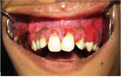
Acute necrotising ulcerative gingivitis is somewhat rare in children. It is illustrated in Figure 2. Generalized Aggressive periodontitis is highlighted in Figure 3.
Diagnosis of periodontitis in children is mainly clinical based on appearance of lesions and family illness. However, radiographic investigation should be performed when it is clinically required iand affects patient prognosis and management [12]. Although panoramic radiography gives important information on unerupted permanent and missing teeth, the alveolar bone levels are best assessed and viewed by intraoral periapical and Bitewing radiography [12]. Figure 1 illustrates Bitewing radiograph.
Figure 1: Bitewing radiography.
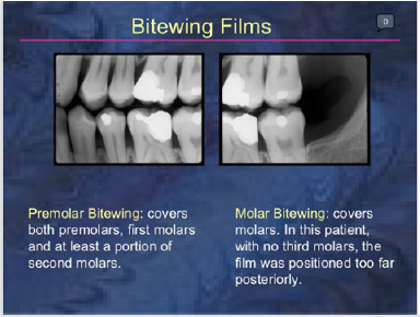
Figure 2: Acute Necrotising Ulcerative gingivitis.
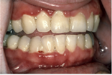
Figure 3: Generalized Aggressive periodontitis.
Plaque control program can be planned based on the chronological age and manual dexterity of the child and can be done by caregiver if necessary [12]. Scrub technique of tooth-brushing technique has been found to be useful in children, but the modified Bass technique can be taught to adolescents with expertise [15]. Periodontal lesions predominantly are interdental, and older individuals can be advised use of interdental cleaning aids [16]. Nowadays, Triclosan with copolymer and dentifrices containing Fluoride have been documented to significantly reduce attachment loss in adolescents aged 11–13 years [12]. Keyes’s technique involving a paste of baking soda, salt, and hydrogen peroxide as a dentifrice and subsequent irrigation with saturated salt solution carried out with a regimen of tetracycline hydrochloride orally, is old and has been prescribed to patients having AgP ;it has shown good results also [17]. Scaling and root planning can definitely reduce microbial load and bring down Cytokine levels, but not in Aggressive periodontitis where motile bacteria are involved and recolonization with reinfection is very common [12,18]. Systemic antibiotics are indicated where A. actinomycetemcomitans is involved and recolonization occurs very frequently. The dosage should be adjusted as per the child’s weight and age. As per adult doses and individuals weighing 40 kg and more, Tetracycline in 250 mg qid and doxycycline in 100 mg od doses have the ability to concentrate in the gingival crevice fluid and inhibit the growth of A. actinomycetemcomitans. It exhibits anticollagenase effect also, which can thereby inhibit tissue destruction and aid in bone regeneration.
Periodic review is essential to prevent recurrences in such cases, especially AgP (20). Qualitative chair side diagnostic aids for guessing the risk for future attachment loss for severe cases of CP and AP consists of the use of chair-side assays such as ELISA for detecting the presence of A. Actinomycetemcomitans, P. gingivalis and P. intermedia in plaque samples, and Benzoyl-DLarginine- napthylamide assay for trypsin and other end products of proteolytic enzymes in plaque samples containing P. gingivalis, T. forsythia, and Treponema denticola . X ray can also be required for assessment and review.
CP usually has a slower progression rate and a better prognosis . It responds to therapy better ,as compared to AgP [12]. Considering the multifactorial aetiology and predisposing factors that lead to the development of AgP in the susceptible host, special care needs to be taken that all facets affecting the diagnosis and therapeutic outcomes are addressed well [12]. AgP affects young individuals with early loss of teeth which influences quality of life. Early diagnosis with meticulous therapeutic intervention and monitoring of the child and adolescent is therefore advised with thorough periodontal examination and early intervention [12].
Periodontitis is very common in children. Knowledge of the different types of periodontitis is optimal for correct diagnosis and therapy.
Read More Lupine Publishers
Pediatric Dentistry Journal Articles:
https://lupine-publishers-pediatric-dentistry.blogspot.com/
Lupine Publishers | Journal of Pediatric Dentistry

The American Association for Dental Education (ADEA) is working on several initiatives to foster an interest in academic careers. One of these strategies is a student fellowship aimed at allowing the student at the School of Dental Medicine to explore early experiences in academia. In 2016, the School of Dental Medicine of the University of Puerto Rico (SDMUPR), Medical Sciences Campus, began this fellowship. A pipeline of SDMUPR students with interest in pediatric dentistry and academics has evolved from this initiative.
Keywords:The pipeline of future pediatric dentists; mentoring in pediatric dentistry
Abbreviations: ADEA:American Association for Dental Education; SDMUPR: School of Dental Medicine of the University of Puerto Rico
The School of Dental Medicine of the University of Puerto Rico (SDMUPR), Medical Sciences Campus, began in 2016 an ADEA Fellowship Program in Dental Academic Careers (ADEA/ADCFP) to encourage students to discover and experience the options available in the academic work setting[1]. This initiative began a parallel pipeline. Students with interest in academics and pediatric dentistry applied continuously to the ADEA/ADCFP and achieved successful educational projects in pediatric dentistry with their mentor assistance. This article will relate the SDMUPR4 years’ experience with this pipeline project of future pediatric dentists with interest in academics.
ADEA’s Dental Academic Careers Fellowship Program was founded in 2006 to provide students and residents of the USA and Canadian Dental Schools with the opportunity to venture into academia, providing a structured mentoring to be able to observe and reflect on what an academic career requires[1]. The student is required to complete eight components during the ADEA Fellowship Program in Dental Academic Careers. These components include building an action plan, bi-monthly meetings with a mentor, reflective essays before and after an academic experience, interviews with faculty, academic practice, research practice, presenting a poster, and submitting a portfolio with the evidence of all activities made[1]. In 2016, the SDMUPR began with the ADEA/ADCFP with SDM/UPR students. Since then, every year, at least one student applies to the pediatric dentistry track (Table 1). The mentor is a pediatric dentistry faculty at the SDMUPR, and they work together for eight months on a pediatric educational dentistry track project (Figures 1-3). The number of Applications and First-year Enrollment for Advanced Dental Education Programs continues to rise in the USA[2]. With proper guidance and early experiences, the students can learn and explore their areas of interest in dentistry.
Table 1: SDMUPR ADEA/ADCFP Fellows.
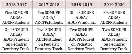
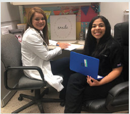
Figure 1: Faculty and student participating in academic activities.

Figure 2: Student presenting a poster at ADEA’s Annual Meeting.
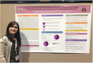
Figure 3: Faculty and student in by-monthly meetings.
Twenty-six students have completed the ADEA Academic Dental Careers Fellowship Program at the UPRMSC School of Dental Medicine and one of the students who participated in the Program in 2016, currently is a pediatric dentist, from the 2017-2018 cohort, two are pediatric dentistry residents at the SDMUPR, and from the 2019-2020 cohort, one applied to pediatric dentistry programs and is waiting for interviews invitations.
We want to thank Dr. Jocelyn Medina Paneto, professor and co-liaison of the SDMUPR ADEA/ADCFP, Dr. Carla Rodríguez, main mentor of the pediatric dentistry track, and all the faculty of the School of Dental Medicine of the University of Puerto Rico, Medical Sciences Campus, for their collaboration in this Program.
The author of this article has no conflict of interest to disclose.
Read
More Lupine
Publishers Pediatric Dentistry Journal Articles:
https://lupine-publishers-pediatric-dentistry.blogspot.com/
Lupine Publishers | Journal of Pediatric Dentistry

The concept of dental dam or rubber dam was first developed by Barnum
in 1864. He reported struggling with saliva contamination during
treatment for a long time. On one occasion, while he was treating a
mandibular molar and saliva was flowing all over the oral cavity, he
came up with a new idea, making a hole in his protective napkin and
putting it around the tooth. This idea resulted in developing rubber
dam, the main problem of which was not being fixed around the tooth. To
address this problem, rubber dam punches, a set of metal clamps, and
other equipment of rubber dam were introduced in the following years
[1]. Rubber dam is a thin, 15 cm square disposable rubber sheet, which
is of two types: latex and non-latex (nitrile). In dentistry, rubber dam
is used for the isolation of the operative site from the oral cavity in
order to increase the safety and quality of dental procedures. Rubber
dam prevents the patient from aspiration or swallowing dental
instruments as well as cross infection and contaminated aerosols [2].
This brief letter focused on the role of rubber dam against transmission
of contaminated aerosols, particularly COVID-19.
Dental aerosol or splatter is produced from dental instruments, such as
ultrasonic scalers or dental handpieces. Aerosol is a particle less than
50 μm in diameter and has the potential to stay airborne for a long
period before it settles on surrounding surfaces or gets into the
respiratory tract. Aerosol droplets which are >0.5-10 μm have an
improved ability to transmit severe acute respiratory syndrome.
Particles larger than 50 μm are defined as splatter and are airborne
only for a short period. With that being said, almost 99% of airborne
particles can be eliminated through the use of a high-volume evacuator
during treatment [3].
According to the World Health Organization (WHO), viral epidemics are a
serious threat to public health. Severe acute respiratory syndrome
coronavirus 2 (SARS-CoV-2) first appeared
in the city of Wuhan, in December 2019. On February 11, 2020,
WHO named the virus, coronavirus diseases 2019 (COVID-19). COVID-19
leads to severe respiratory problems and in some cases even death. It is
transmitted through contaminated aerosol droplets >5-10 μm from
coughing or sneezing. Thus, close contact (within 1 m) with an infected
person causes the mucosa, oral or nasal cavity, and conjunctiva to be
exposed to respiratory droplets containing coronavirus infection [4].
Since dental health care providers are constantly in close proximity to
patients, they are at risk of microbial or viral infection that can be
transmitted through atmospheric aerosols.
In a study conducted by Samaranayake et al. [5] the effectiveness of the
rubber dam in preventing contaminated aerosols during therapy was
examined. The result demonstrated that the rubber dam significantly
reduces the contaminated aerosol particles from the operational site up
to 3-foot distance (91,44 cm) by 70%. Moreover, the use of rubber dam
decreases the production of saliva and blood contamination during dental
treatment as well as the potential airborne particles between the
clinician and patient [3]. In addition, in the case in which the
gingival is exposed, the split-dam method is useful. According to the
evidence provided, it is possible that the use of rubber dam contributes
to the prevention of COVID-19. However, conducting more clinical or
laboratory studies investigating the role of rubber dam against
respiratory diseases, particularly COVID-19 is recommended in order to
obtain more accurate and valid data.\
Read More Lupine Publishers
Pediatric Dentistry Journal Articles:
https://lupine-publishers-pediatric-dentistry.blogspot.com/
Lupine Publishers | Journal of Pediatric Dentistry

Pain sensation is evolutionarily the primary basis of sensitive human
activity, and it is quite stable. The stability of electrodontometry
(EOM) indicators is demonstrated by a narrow zone of the pain threshold
of 2-6 mkA of healthy front teeth, which was first evaluated by Rubin L.
R. in 1953. This value was confirmed by many domestic researchers
[1,2]. Rubin tried to extend it to all teeth, but the indicators of the
lateral teeth, although stable, are 5-15 mkA higher [3-5]. The most
important feature of the sensitivity of the dental pulp is that it is
represented by an exceptional pain [6,7]. Its sensitivity is very high
-2-6 mkA- and sharply differs from the sensitivity of the skin and all
surrounding tissues, including the gums and periapical tissues. It is
located at the level of 100-200 mkA. The pulp, perceiving tactile,
thermal and electrical irritation, translates it into a painful
sensation. In the human tooth, the circulatory and nervous systems of
the pulp are separated as a thin soft string and are protected from
external influences by a thick tube of enamel and dentin. The nerves in
the pulp are 20.5% by weight.The pulp is represented by two main pain
points that determine pain sensitivity. In the crown pulp, these are
odontoblasts and dentine tubes with 1-2 mm of nerve branches of A-Delta
fibers entering them. At the root it is a narrow thin apical part of the
canal, where the pulpar nerves of the pulp core from C- fibers are
tightly placed together with the blood vessels. Secondary cement is also
involved in creating the apical opening, preventing myelination of the
nerve tissue there[8]. The presence of myelin A-Delta fibers in the apex
has not been proven. Pain fibers A-Delta are slightly myelinated,
located in the crown, have a higher rate of conduction (dentin pain).
They respond to the pulse current as an acute pain sensation. C-fibers
transmit late, dull and not always clear pulp pain. It is interesting
that all foreign authors use A-Delta fibers [6], to consider
themechanisms of electrodontometry (EOM) from the standpoint of the
theory of Branstrom, with an emphasis
on EPT (Electrical pulpal test) as dentin stimulation. Foreign
researchers exclude the reaction of pain by C-fibers, which make up the
bulk of the nerve structures of the tooth.
Interestingly, EPT is rated as a low-value test for primary or permanent
teeth during apex formation [9].Many children with normal teeth do not
respond even to high stimulation currents [9].We believe that for the
reaction of all neuroreceptors in single teeth, a more universal AC
stimulator with a broad total effect, mainly on C-fibers, is needed.The
aim of our investigation was: to evaluate tooth maturation in children
aged 6.5 - 15 years in dynamics in the study of pain caused by
electrotesting in three directions:
a) Reduction of the pain threshold of the central incisors,
b) x-ray width of the apex, and
c) The nature of the pain: pain or not pain.
280 teeth were examined in 280 children from 6 to 15 years (central incisors). Patients were divided by age into 5 groups:1 - 6,5 – 7 years; 2 – 7-8 years; 3 – 9-10 years; 4 11-12 years; 5 – 13-15 years.After receiving informed consent, the oral cavity was examined, the child was instructed, and the central incisors were isolated from saliva. The threshold of pain sensitivity of the dental pulp was determined using the IVN-01 Pulpotest-Pro electrodontodiagnostic device. Measurements were made on the central upper incisor. The pulp tester used created a series of alternating current pulses, devoid of polarization, with a frequency of 50 Hz with a gradually increasing current strength on a scale from 0 to 200 mkA until the first sensation: pain/non-pain[10-15]. The active electrode was located in a PVC tube into which the contact gel was inserted. The diameter of the active electrode was 4 mm. The passive metal electrode was in the hand of the subject. When pain occurred, the subject released the button, thus stopping the current supply, and the value of the EOM was recorded on the display. Indications of the pain threshold after a two-time (one- time) measurement were recorded.In addition to the digital indicator of EOM, the response to electrical stimulation was determined. The main criterion for pain was to divide it into pain / non-pain. All patients according to indications, as a rule, aesthetic, carried out radiography. Intraoral and x-ray images were used to measure the diameter of the tooth apex in mm[16-18].
The results are shown in Table 1. Figure 1 shows a descending curve similar for all three parameters under study: EOM, apex, and pain.The highest EOM index was observed in the first age group (140.86 mkA), exceeding that in the second age group by more than 1.5 times (91.95 mkA). At the same time, the average diameter of the apex decreased from 2.55 to 1.96 mm. For both parameters, the differences between the first and second age groups were statistically significant (t-criteria 5.70 and 6.8, respectively). Between the second and third age groups, the difference in EOM indicators was sharper— 91.95 mkA to 39.75 mkA-with almost the same smooth decrease in the diameter of the apical opening (1.96 to 1.39 mm). For both parameters, the differences between the second and third age groups were statistically significant (criterion t = 7.48 and 8.90). Indicators of EOM in the third and fourth age groups differed a little, although the average value of EOM in the fourth group is slightly lower: 35.83 mkA compared to 39.75 mkA (the differences were not statistically significant, the criterion t = 0.70, and the diameter of the apex was significantly different: 0.82 mm compared to 1.39 mm (criterion t = 11.40). In the fourth and fifth age groups, the EOM as well as the apex diameter differed significantly, but without a sharp decrease in these indicators: from 35.83 to 24.35 mkA and from 0.82 to 0.72 mm (criterion t = 2.21 and 2.36, respectively). A sharp drop in EOM indicators occurred at the age of 6.5 to 11 years with a furthermore gradual decline (Figure 1).In the first age group, 13 subjects had no pain at all, and 6 (38%) subjects had a pre-pain sensation that was close to tactile. In the second group, 4 children had no pain, and 5 had afeeling of pre-pain in the form of cold or heat with spreading in the gum above the tooth (14.5%). In the group of 9-10 years, 4 subjects had a feeling of pre-pain (7.14%), in 11-12 years in 5%, in 13-15 years in 1.92%(Figure 2).
Table 1: Electrodontometry of teeth, apex diameter and sensations (pain, non-pain) in 280 children of different ages.
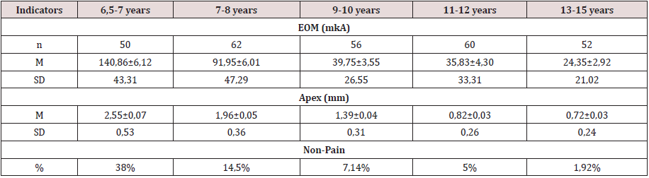
Figure 1: The Value of the pain threshold (A) and the value of the apical opening (B) of the central incisors in children of different ages:1 - 6,5 – 7 years; 2–7-8 years; 3– 9-10 years; 4 11-12years; 5–13-15 years.
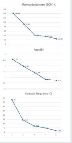
Figure 2: Central incisors of a 6.5-year-old girl. EOM 199 and 195 mkA (apex 3 mm; there is no pain when measuring).
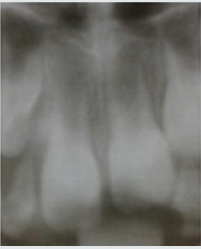
Figure 3: Central incisors of a 12-year-old girl, EOM 13 and 15 mkA (apex 0.8 mm; pain felt during measurement).
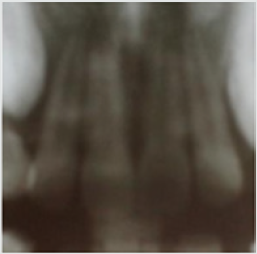
Pain is one of the main feelings. With age, the senses improve. The difference in pain assessment is visible on the graph (Figure 1). The youngest ones have the greatest feeling of “non- pain”. Children replaced real pain with feelings of cold, heat, etc. We have combined these feelings into the concept of pre-pain.Pain sensitivity of a permanent tooth is the main pain criterion of a person, in which, unlike animals, there is contact with the researcher. The phenomenon of a sharp decrease in the sensitivity of permanent teeth that are in the process of eruption has been established. A close relationship has been established between the reaction of A-Delta fibers and pulsed diagnostic current. The role of C-fibers – the basis of the tooth’s nervous system-is underestimated. The electric current due to its strength and the large electrode covers the entire pulp, both coronal and apical (Figure 3). In our literature, there is a tendency to prohibit devices that operate from alternating sinusoidal current with a frequency of 50 Hz [1,2]. The regularities between the degree of tooth eruption and the reduction of its pain threshold to a minimum level, while reducing the apical narrowing, and clarifying the pain response in a young subject were determined. The formation of the apex consists in its gradual narrowing, including ingrowth of cement into the apical part of the root canal. It is dominated by C-fibers. Pulse pulp testers do not “feel” the pulp in the erupting teeth.It should be noted that when the final apex was formed, the threshold value did not always correspond to the previously accepted norm of 2-6 mkA. In one 15-year-old patient, the initial pain threshold was 125 mkA.
The phenomenon of reducing the pain sensitivity of permanent teeth during eruption due to the reaction of C-fibers has been established. Pulse pulp testers do not work in teeth during apex formation.
Read More Lupine Publishers
Pediatric Dentistry Journal Articles:
https://lupine-publishers-pediatric-dentistry.blogspot.com/
Lupine Publishers | Journal of Pediatric Dentistry

The dental care of children is an area that requires special
attention. The dental visit, even in the early years of life, allows the
child to have, early on, greater contact and familiarity with the dental
environment, thus having the possibility to learn new habits
in addition to positive experiences with regard to oral health. Thus, it
is extremely important to know the view of children about the
dental care provided by the institution CEUPL-ULBRA. We randomly
selected children aged 3 to 11 years, 13 males and 8 females.
Data collection was performed through interviews and story-design after
the service, as well as analysis of the medical records to
record the procedures performed. For the analysis of the drawings and
for the interview, four categories were considered:
a) Dental environment
b) Dental treatment
c) Dentist image and
d) Behavioral manifestation
The most frequent categories in storytelling were the environment and
dental treatment, with the most cited curative
procedures. The operator / dentist’s image according to the drawing was
considered technical. According to the interview, the
clinical procedure itself was considered a positive point of care,
especially when it was associated with pain relief. The most negative
point was evidenced at times that led to some kind of discomfort in the
child such as anesthesia, taste of the prophylactic paste and
noise of high rotation. The perception of the operator’s image was
considered humanized in all responses. Most children showed
satisfaction with their smile and some reported the need to return to
the dental clinic for new procedures. Only a small portion
was free of oral problems. It is concluded that: the need for dental
follow-up is not consistent with the oral health condition of the
children evaluated and that the care process follows the curative model.
Keywords:Pediatric Dentistry;child psychology;health evaluation
Pediatric dentistry is the dental specialty that takes care of children’s oral health. It is known that the great fear presented by adult patients in the dentist’s chair originates from the negative experiences of dental treatments that occurred in childhood. For this reason, the role of pediatric dentists is of great relevance in dentistry. Pediatric dentists are responsible for the care of children from infants to adolescence, and their exercise is comprehensive, as it is not limited only to the prevention and solution of oral problems, it also plays an important role with regard to the psychological and educational aspects of the patient [1].The practice of children’s dental clinic shows that children have some peculiarities, such as growth and development, biodynamics, tissue and organic responses, behavior, and personality structure. These peculiarities cause the sociological methods and the techniques of physical examination to have a different approach from that performed in adults, despite having the same diagnostic and therapeutic purpose [2]. According to Melo et al. [3], the dentist’s approach must be in accordance with the child’s age and psychological development, and can be used from a more playful language in early childhood to logical explanations in early adolescence, so that in many situations children are driven to overcome fears and phobias. During clinical practice, it can be observed that lessinvasive procedures do not generate major behavioral reactions, whereas more invasive procedures are directly related to rejection and fear in the face of treatment [3].The negative attitude towards dental treatment is a process that begins in childhood, and the origin and cause must be investigated by the Pediatric Dentist before any behavior control technique is applied [4]. According to Gomes et al. [5], the child may manifest fear and anxiety in several ways, the most frequent symptoms being tachycardia, sweating, palpitations, tremor, flushing and gastrointestinal complications.
Behind a behavior, positive or negative, there are a multitude of characters that exert marked influences on children, such as: age; the socioeconomic class of the parents; temperament; psychological development; the environment in which you live. Through such knowledge, the operator / pediatric dentist will have more baggage for the application of certain measures, and for a better understanding of the types of behavior presented by the children[5]. According to the literature, several control techniques can be used, such as: orders; compliments; reward; suggestions; containment; distraction; restriction; dominance by voice and sayshow- do. The pediatric dentist must, therefore, play an active role in the psychological and educational sectors, allowing the avoidance of possible trauma that almost always determines incompatible relationships with the dentist or with the clinical environment or even with the surgical procedures [6]. The control of fear and anxiety during dental treatment must be performed throughout the service. Thus, it is essential to use basic conducts to control the situation, such as verbalization, associated with pharmacological techniques for muscle relaxation or psychological conditioning, reducing the wear of the professional in relation to the patient. The proper use of these behavioral control techniques is fundamental for the success of the planned treatment and consequent restoration of the child’s oral health. The choice of behavioral approach techniques may vary according to the professional’s criteria, being influenced by factors observed during anamnesis, such as age, child’s behavior, and parental acceptance. In this sense, it is of great relevance to establish which procedures generate more behavioral disorders through specific control protocols and techniques for the care of pediatric patients, because regardless of the procedure, it is clear that they present some type of discomfort such as fear and / or anxiety, proving to be of great importance for professionals to update themselves in offering treatment options and techniques so that this moment becomes more dynamic and comfortable, recognizing, first of all, each child with their particularities.Therefore, knowing the child’s perception about the dental experience is extremely important for understanding the dental practice developed within the different environments that offer this service. Such knowledge allows the dental surgeon to identify possible failures committed and can develop new forms of interaction during care, thus modifying negative behaviors and / or reinforcing positive ones. This will allow the use of more effective methods so that they accept and understand the need for the procedure[7].Based on this principle, this study evaluated the perception of children between 3 and 11 years of age regarding dental treatment, the figure of the dentist and their own oral health condition, through analysis of information obtained by interview, drawing on the topic and analysis of medical records.
The present study is characterized in a cross-sectional analytical and descriptive design, where the bibliographic research took place through the consultation of online publications such as: LILACS, SCIELO, BVS, PubMed. The search strategy occurred through Health Sciences Descriptors (DECs) registered in Portuguese as: Pediatric Dentistry, Child Psychology and Health Assessment, which are terminologies that make up electronic articles and made possible their search, in addition to the terms in English: Pediatric Dentistry, Psychology Child and Health Evaluation. 100 publications were found on the proposed theme, including subjects related to fear, anxiety and behavior management, with 33 articles selected, 3 master’s dissertations, 3 conclusion papers of a specialization course and 2 doctoral theses. For inclusion criteria, articles in Portuguese or English were selected, complete and published from 2009 to 2019, in addition to national books that addressed methods of controlling behavior in pediatric dentistry, considering the reliability of the selected material. As exclusion criteria, articles, monographs, and dissertations that do not fit the research objectives, in addition to those that are not available in full.The research was carried out at the Pediatric Dentistry’s School Clinic of the Lutheran University Center of Palmas, during the second semester of 2019.The object of the study was children assisted by the children’s clinic (I and II) of the institution, and the data were collected in the second semester of 2019. A random sample of 21 children aged 3 to 11 years participated in the research. The variables used in this study relate to those observed by the child, in relation to the procedures and the dental environment, and were adapted using a questionnaire already validated, and a drawingstory about their care.
As inclusion criteria, children in need of care participated in the research, whether for the first time or not at the school clinic. Children with motor difficulties or mental disabilities were excluded. Children who refused to do the drawing and answer the questions even with the parent’s permission, were also excluded. Children who failed to answer just one question were not excluded from the sample. Children who stopped making the drawing, but answered the questions, were not excluded from the sample. Based on previous studies and according to several authors [8- 13]the application of the instrument was carried out through a questionnaire already validated, where the child’s perceptions regarding the situation of care were recorded. All data were collected in the clinic environment, each child was approached individually. This collection was made after the service, with the application of a questionnaire containing 8 questions of the type:
a) What is a dentist?
b) Are you happy with your teeth? why?
c) Do you think you need to take more care of your teeth?
why?
d) While you were with the dentist, how did he treat you?
e) How was your reaction during the consultation?
f) What did you like most about the consultation?
g) What did you like least?
h) Finally, how do you feel now?
After the questionnaire was completed, the child was invited to carry out a drawing-story about his care (each child was provided with crayons and a blank sheet to carry out the drawing.), And afterwards, describe the meaning of the researcher to the researcher. In addition to the interview, the researcher was responsible for collecting data regarding the patient’s clinical history at the school clinic. The answers and the description of the drawing were faithfully transcribed and evaluated. Those responsible were informed of the research, and those who wished toparticipate signed the free and informed consent form. The level of invasion of the procedure to which the children were subjected was also subject to classification where they were divided into groups 1 and 2, being classified as invasive procedures (extraction; endodontics; restorations, which require absolute isolation) and not invasive (prophylaxis; topical application of fluoride and use of sealants that are carried out without absolute isolation) respectively. Only those drawings that met the following requirements were included in the study:
focus on the proposed theme
j) be completed
k) be clear for interpretation
l) in isolation or with the help of the child’s oral description.
A fluctuating reading was carried out for the initial knowledge of the material produced. Subsequently, the drawings were systematically observed, and the texts obtained for each of them were read, as well as their responses. Thus, the quantification allows to define the shared thought collectively among the researched group. Twenty-one children aged 3 to 11 years participated in the study, with the frequency of each age as follows: 3 years (1), 4 years (3), 5 years (1), 6 years(1), 7 years (3 ) and 8 years (5), 9 years (4) 10 years (2) and 11 years (1). The male gender had the highest frequency (13).
It was found that the majority of respondents reported to more than one subcategory during the interview. The perception of the operator’s image was considered humanized in all reports and the design of the treatment model was described by most children as a curative, with preventive treatment being little mentioned. In the positive view, objects from the dental environment were highlighted, such as, for example, a dental chair, a Robinson brush, and a dental sucker. In the negative view, the most frequent responses were in relation to the dental procedure itselfwhen the treatment generated pain or discomfort: “I did not like the needle” and “I thought that noise was bad”. In the negative view, discomfort related to other objects was also highlighted, such as a needle and explorer probe.
It was identified that most of the interviewees reported to more than one subcategory during the elaboration of the drawingstory. The dental environment category was most frequently addressed, being the subcategories, operator, and equipment. The other categories demonstrated that, according to the child’s view, the institution’s dental treatment presents itself as a curative and technical model; but humanized, where the interviewees reported having been treated with great empathy. The moment of consultation in the days of the survey was reported by the interviewees as a pleasant moment and the environment was referred to as peaceful. The children’s behavior during the observation made by the researcher was satisfactory (positive). The operator was sometimes mentioned in phrases such as: “nice, nice and polite” as well as “he treated me very well”, “she explained what she was going to do”.The appreciation of the dental treatment received by each one was evaluated by means of a positive view (what he liked best) and a negative view (what he liked least). In the positive view, the dental procedure was the most mentioned, when related to the child’s pain relief, as identified in the excerpts: “I liked it when she passed the ointment, and when she removed the tooth, I didn’tfeel anything”; “I liked to pull the tooth out it because it hurt.” In the negative view, the dental procedure was also the most cited when it caused a sensation of pain or discomfort, as identified in the excerpts: “When she put the needle it hurt” and “I didn’t like that thing to brush my teeth”.
In the analysis of the medical record, despite the innumerable invasive procedures, most of the subjects showed positive behaviors, as the drawings and the speeches that showed tranquility, empathy towards the dentist, establishing dialogue were expressive; it was evident in the studied group that there is a relationship of trust and good communication between professionals and patients.The self-perception of the oral condition was evaluated as positive by most children, and the most frequent reasons were demonstration of health (8), absence of pain (6), self-care (4) as observed in the report “I’m happy because they they are beautiful ”. For those who negatively assessed their oral health condition, the presence of pain related to the carious process was the main related reason (5). Regarding the need for dental care, most children believe that they should attend other dental appointments (12), for one or more reasons, and the condition “To treat decayed tooth” was mentioned (9) times and procedures related to prevention were cited (3) times. Children who do not intend to return or only want to return in case of pain.It was found that the child’s behavior in the dental consultation can be determined by a series of factors, such as maturity, relationship with parents, approach to the dentist, past experiences, office environment, this because their handling, in some circumstances, becomes a great challenge for the professional. The professional’s positive interaction with the child brings out the image of a humanized professional. In both cases, the most apparent reaction in the child during and after the interview was joyful and calm. Behaviors related to anxiety, fear and scared were mentioned a few times (3), (1) and (2).In the present study, children who claimed to be happy with their oral health condition, even though they were compromised, demonstrate the inability to recognize oral health as an integral part of systemic health, which demonstrates a deficiency in health actions aimed at this evaluated population.
To demonstrate the view that the child has on the care that receives the drawing technique, it proves to be efficient because it is a pleasant activity and easy to perform. This can be seen in the course of this research. All children, after explaining the research, readily accepted the invitation and expressed satisfaction in drawing and reporting their drawings.The analysis of the story-drawings about the dental care provided by the Clinic of Pediatric Dentistry of CEULP-ULBRA showed very positive aspects regarding the actions developed by academics and teachers. The view of the infant patient that was part of this study reflects the effectiveness of the work performed by the teams of the Pediatric Dentistry Clinic of CEULP-ULBRA. The description of the drawings, through the children’s speeches, denoted a scenario of tranquility and empathy. It was very evident that there is a relationship of trust, good communication between academics and children. Communication between the dentist and the child, aimed at a friendly and friendly relationship during care, is essential for the success of dental treatment and, therefore, for the establishment of healthy behaviors. This condition was perceived in the storydesigns carried out by the patients as being from a cordial setting, with good communication and goodwill.Therefore, it is suggested that further studies be carried out in order to identify the best way of working with health professionals in order to encourage the practice of prevention as a health promotion strategy in addition to raising the awareness of children and their guardians. about the importance of periodic dental consultations for the benefit of your children’s health.
For most of the subjects participating in the research, the context of the dental consultation is revealed to be a pleasant situation, characterized by an educational-curative practice, and permeated by a humanized view of the dental professional, configuring itself in a pleasant situation. It was found that even in the midst of invasive procedures, in the days of the research, the children had behaviors of collaboration and demonstration of interest in the care and tranquility during the consultation. It was found that the evaluation process, through the technique of drawing-story, is rich and authentic. Therefore, the technique can be considered an excellent methodological alternative when compared to the use of questionnaires, which can induce the respondents’ answers, limiting the quality and depth of the evaluation process. According to the interview and the medical record findings, the interviewees’ oral health is not consistent, requiring dental follow-up for both new procedures and for hygiene and oral health care instructions.
Read
More Lupine
Publishers Pediatric Dentistry Journal Articles:
https://lupine-publishers-pediatric-dentistry.blogspot.com/
