Friday, June 25, 2021

Lupine Publishers | Effectiveness of a School Oral Health Education Program in Yangon, Myanmar

 Lupine Publishers | Journal of Pediatric Dentistry


Abstract

Aim: We evaluated the effectiveness of oral health education in terms of oral health literacy, knowledge, behaviors, and oral health status among middle school students in Yangon, Myanmar.

Materials and Methodss: This 1-year study enrolled 10- to 11-year-old students. At baseline, a dentist provided health education to group A (n=247), and peer group leaders reinforced it. Group B (n=195) received no education program. After 6 months, the dentist provided education to both groups, and classroom teachers reinforced it. Both groups received questionnaire surveys and oral examinations at baseline, 6 months, and 1 year.

Results: There were no between-group differences at baseline. After 6 months, group A showed significant improvements in oral health literacy, knowledge, and behavior as well as reduction in dental plaque, gingivitis, and plaque bacterial levels. Group B only showed improved brushing and mouth rinsing behaviors. After 1 year, all items were significantly improved in both groups, although sweet snack/drink consumption behaviors remained unchanged and dental caries increased. At 1 year, group differences were not significant.

Conclusion: Oral health education is effective in improving oral hygiene, decreasing gingivitis, and reducing plaque bacteria levels by improving oral health literacy, knowledge, and behaviors. However, eating behaviors and dental caries were difficult to change within 1 year

Keywords:School oral health; Oral health education; Middle school

Abbreviations: HPS: Health-Promoting School; OHLI: Oral Health Literacy Instrument; CMOHK: Comprehensive Measurement of Oral Health Knowledge; OHBs: Oral Health Behaviors; DT: Decayed Teeth; FT: Filled Teeth;

Introduction

Dental caries and periodontal diseases are the most common oral health burdens in industrialized countries and in developing countries [1,2]. In some developing countries, dental caries affect a substantial proportion of children and are a public health problem [3,4]. Dental caries are increased among school children in developing countries where limited oral health services [1] and school-based oral care programs are not established [5,6]. Compared to children without poor oral health, children with poor oral health are 12 times more likely to have restricted activity days when missing school [7]. More than 50 million school hours are lost annually because of oral diseases [8]. A health-promoting school (HPS) constantly aims to strengthen its capacity to promote healthy living, learning, and working condition [9]. An HPS has the following key features: [10]

a) Healthy school policies,
b) A physical school environment,
c) A social school environment,
d) Health skills and education,
e) Links with parents and the community, and
f) Access to school health services.

An effective school health program uses cost-effective investments, and each country can improve education and health simultaneously through a health program [11]. Much evidence indicates that an oral health promotion program in school is needed and that it can easily be incorporated into general health promotion, school curricula, and activities [8].

Schools provide an ideal setting for oral health promotion and are one of the best places to give oral health information to children to achieve the goals of a health education program [2,8]. Oral health education messages can be reinforced regularly throughout the school years. A single session of health education cannot achieve the set goals, particularly among young children, and health education programs with reinforcement have shown some effectiveness [12,13]. In schools, peers may strongly influence other students’ behaviors and share experiences and feelings, and peers represent an important role in an adolescent’s socialization process [14]. Teachers have a positive influence on society and are guides and role models for students [11]. Trained teachers and peers can have a key role in the reinforcement and enhancement of the success of school-based oral health education programs [13]. Low oral health literacy is associated with poor oral health knowledge, oral behaviors, and oral health status [15]. School-based oral health education interventions can have positive impacts on improving oral health literacy to develop behavioral outcomes and clinical oral health status among students [16,17].The basic education system in Myanmar consists of primary education (kindergarten to grade 4), lower secondary education (middle grade years 5–8), and upper secondary education (high grade years 9–10). The basic education curriculum is taught from age 5 to 16 years in Myanmar. Secondary schools are usually combined and contain middle and high schools [18]. In Myanmar, 6224 lower secondary (i.e., middle) schools exist and contain 129,945 teachers with approximately 2.79 million students [18].

In 2015, 4539 dentists practiced in Myanmar and the dentist per population ratio was 1:16,000 [19]. A wide gap exists between dental healthcare services and the dental health condition of the population. Not all citizens in Myanmar are covered by health insurance [20]. A comprehensive evaluation and research helps to strengthen a school’s health program and determine the current outcomes of ongoing activities and the program’s cost-effectiveness or benefits [8,21]. In Myanmar, very limited data are available regarding the impact of health education on the clinical oral health status of students [22]. Moreover, the lack of annual school oral health data is a problem when planning a school-based oral health program. Therefore, implementing a cost-effective school-based oral health promotion program is necessary in a country where the number of dental professionals are limited. A previous study [23] in Myanmar reported that students with a mixed dentition had a high prevalence of dental caries and gingivitis, as well as poor oral hygiene and inadequate oral health care habits. Therefore, the present study was targeted at middle school students and aimed to evaluate the effects of oral health education on oral health literacy, knowledge, and behaviors and oral health status among this student population in Myanmar.

Materials and Methods

Sampling methods and sample size

This 1-year follow-up study was conducted in Yangon, Myanmar from December 2017 to December 2018. Two townships with a similar sociodemographic status in Yangon region were selected. Two public schools in each township were randomly chosen; in total, four schools participated in this study. All were middle schools with the same academic performance level, curriculum, and school environment, and none of the schools had organized an oral health education program. In each township, one school constituted group A and the second school constituted group B. After obtaining consent from the school authorities, a written informed consent form was sent to the students and their guardians. The exclusion criteria in this study were students who had systemic diseases or neuromuscular dysfunction, students with fixed orthodontic treatment, and students without their parents’ agreement to participate. The required sample size for this study was 362 students (181 per group), based on a power test of 0.80 and compensating for a 10% loss of response rate. A previous study by Ueno et al. [24], which was also part of a school oral health promotion program, was used as the parameter for sample size calculation.

Study procedure

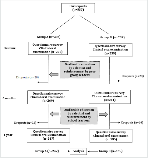
Because the school authorities wanted all students to receive the same benefit, we invited all grade 5 students in the four schools to participate. Therefore, the number of students in each group exceeded the upper limit of the estimated sample size. From 569 eligible students, 32 students were excluded and 537 students (group A: 298 students; group B: 239 students) completed the questionnaire surveys and clinical oral examinations at baseline. Among these, 442 students (group A: 125 boys and 122 girls; group B: 87 boys and 108 girls) completed the 1-year follow-up examination; thus, the follow-up rate was 82.3% (Figure 1). The dropout rate was 18%. The common reasons for dropout included transfer to another school or absence on the examination day. The questionnaire survey and oral examinations were conducted at baseline, 6 months, and 1 year. The study design was a comparative study of two groups; one group received health education at the baseline and at 6 months while the second group received health education only at 6 months. This study received ethical approval from the ethics committees of the Department of Medical Research in Myanmar (No. Ethics/DMR/2017/064), University of Dental Medicine (Yangon), and Tokyo Medical and Dental University (no. D2017-018). The study was conducted in full accordance with the Helsinki Declaration of the World Medical Association. Information regarding approval can be provided on request.

Figure 1: Study flowchart.

Lupinepublishers-openaccess-pediatric-dentistry-journal

Questionnaire survey

To evaluate the students’ skill in reading and understanding information about oral health and to evaluate their knowledge about oral health, 10 items from the reading comprehension section of the Oral Health Literacy Instrument (OHLI) [15] and nine questions from the Comprehensive Measurement of Oral Health Knowledge (CMOHK) questionnaire [25] were used. In the modified OHLI and CMOHK, a “correct” answer was scored with 1 point and an “incorrect,” “do not know,” or “not answered” response was scored with 0 points. Thus, the modified oral health literacy score ranged from 0 to 10 points, and the modified CMOHK score ranged from 0 to 9 points. The original English versions of these questionnaires were translated into the Burmese language and then back-translated. Expert panels confirmed the content and consistency. In a pilot study, the translated Burmese versions were tested among 25% of the participants. Based on the feedback, minor corrections were made so that the questionnaire would be suitable for students. Test–retest reliability was conducted 2 weeks later for reliability testing. The interclass correlation coefficient for oral health literacy and CMOHK were 0.78 and 0.82, respectively. Internal consistency, assessed using Cronbach’s alpha, was 0.70 and 0.73 for the OHLI and CMOHK, respectively. Questions regarding oral health behaviors asked the students about daily tooth-brushing frequency (“less than once a day,” “once a day,” “twice a day,” and “more than twice a day”), daily mouth-rinsing habit (“ once a day,” “twice a day,” “three time a day,” and “never”), having a dental visit within 6 months, and daily consumption of sweet snacks and/or drinks from snack shops at schools and near their home (“yes” or “no”). The validity of these questionnaires was reported in a previous paper [23].

Clinical oral examination

One dentist conducted the oral examinations of the students by using a dental mirror and a community periodontal index probe under an artificial light. In the oral examination, the dentist assessed the students’ dental caries status (using the decayed, missing, filled teeth [DMFT] index), based on the World Health Organization guidelines [26]; oral hygiene status, based on the debris index (i.e., DI-S) [27]; and the gingival status of 12 anterior teeth, based on the papillary, marginal, attached gingiva (PMA) index [23,28]. Intra examiner agreement value was defined as “excellent” for dental caries (0.90) and as “good” for DI-S (0.76), PMA (0.77). After the clinical examination, the dentist advised the students in both groups to receive dental treatment, if necessary.

Bacterial tests

A bacteria counter machine (Panasonic, Tokyo, Japan) was used to measure the bacteria in dental plaque from the buccal surface of the cervical portion of the upper left permanent first molar [23]. After the clinical examination, the same investigator conducted the bacterial tests.

Oral health education

At baseline, the dentist provided health education to group A and specially trained peer group leaders. From baseline to 6 months, the peers repeated the dentist’s information by using posters to their classmates in a small group. This activity required only 5–10 minutes per week. By contrast, group B students did not receive education program. After 6 months, the dentist provided both groups health education. From 6 months to 1 year, specially trained classroom teachers reinforced the health education with the students by using posters. They reinforced it once weekly for 5–10 minutes. The dentist used visual modes (e.g., slide projector, photo albums, posters, dental models) for education, followed by self-checkup training by using a hand mirror and dental mirror to improve the students’ self-evaluation of their oral health status. These mirrors were then given to students who received education so they could continue daily checkups at home. The 30-minute health education focused on healthy teeth and gingiva, signs and symptoms of dental caries and gingivitis, treatment, and the prevention of dental diseases. The health education messages were written on posters and displayed on a wall in their classrooms. The topics, instructions, and training program presented the same content to both groups. Burmese language was used for delivering health education. Toothbrush and toothpaste were provided to all participants after the clinical examination.

Statistical analysis

The questions regarding oral health behaviors were categorized under the following domains: tooth-brushing frequency (≤1 time daily [0] or ≥2 times daily [1]); mouth-rinsing behavior (“never” [0] or “ ≥ once a day” [1]); dental visit within 6 months (“no” [0] or “yes” [1]); and daily sweet snack/drink consumption (“no” [0] or “yes” [1]). The clinical outcome of the bacterial test results was categorized as level 1–4 (i.e., “low”; 0) or level 5–7 (i.e., “high”; 1). The association of categorical variables was analyzed by using the Chi-square test and McNemar’s test. Mean differences in continuous values for the within group comparisons were analyzed by using paired t-tests from baseline to 6 months (i.e., the first 6 months) and from 6 months to 1 year (i.e., the second 6 months). The betweengroup comparisons were analyzed at baseline, at 6 months, and at 1 year by using the Student t-test. Statistical Program of Social Science (SPSS, version 21; IBM, Tokyo, Japan) was used for data analysis. The significance level for all results was set at P < 0.05.

Results

Oral health literacy and knowledge

At baseline, no significant difference existed between the two groups for any item. Table 1 shows the Oral health literacy and CMOHK scores of the students at baseline, 6 months, and 1 year. The mean literacy score significantly increased in both groups from baseline to 6 months and from 6 months to 1 year. With regard to the CMOHK, a significant improvement occurred in group A in the first 6 months and in the second 6 months. However, in group B the mean CMOHK score increased significantly only in the second 6 months. In the comparison between the two groups, oral health literacy and CMOHK scores were significantly improved in group A than in group B at 6 months. However, no significant differences existed between the two groups at 1 year.

Table 1: Comparison of the Oral Health Literacy and CMOHK scores in Groups A and B at baseline, 6 months, and 1 year.

Lupinepublishers-openaccess-pediatric-dentistry-journal

aThe P value is based on the intragroup comparison between baseline and 6 months and between 6 months and 1 year. bThe P value is based on the intergroup comparison at baseline, 6 months, and 1 year.

Oral health behaviors

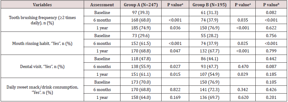
The comparisons of the percentage of students’ oral health behaviors (OHBs) are in Table 2. Students in both groups reported a significant increase in tooth-brushing frequency (≥2 times per day) and in adopting a daily mouth-rinsing habit from baseline to 6 months and from 6 months to 1 year. Dental visit experience was significantly increased in group A for both 6-month periods, whereas it was significantly improved in group B for only the second 6-month period. However, daily sweet snack/drink consumption behaviors did not significantly change after 6 months or at 1 year in either group. In addition, at 6 months, OHBs significantly improved in group A, compared to group B, except for dental visit experience and dietary habit. After 1 year, no significant difference existed between the two groups in OHBs.

Table 2: Comparison of oral hygiene behaviors at baseline, 6 months, and 1 year.
Lupinepublishers-openaccess-pediatric-dentistry-journal

aThe P value is based on the intragroup comparison between baseline and 6 months and between 6 months and 1 year. bThe P value is based on the intergroup comparison at baseline, 6 months, and 1 year.

Oral health status

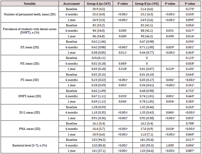
Table 3 shows the oral health status of the students at baseline, 6 months, and 1 year.

Number of permanent teeth: The number of permanent teeth in both groups significantly increased until 1 year. This parameter was not significantly different between the two groups at baseline, 6 months, and 1 year.

Dental caries: The percentage of students with dental caries in the permanent teeth (DMFT index) was significantly increased in group B from baseline to 6 months; however, no significant changes occurred in group A in the first 6 months and in the second 6 months. At 6 months and at 1 year, group B had a significantly higher percentage of students with DMFT than did group A. The number of untreated decayed teeth (DT) was significantly reduced in group A in both 6-month periods. By contrast, the number of untreated DT was significantly reduced in group B in the latter 6 months. A comparison between the two groups at 6 months revealed a significant reduction in group A. No significant changes occurred in the mean number of missing teeth within each group or in the between-group comparison within 1 year. A significant increase in filled teeth (FT) was detected in both groups in the first 6 months and in the second 6 months. At 6 months, group A had a significantly higher number of FT than did group B. After 1 year, no significant differences were detected between the two groups. Dental caries (DMFT index) was increased in both groups from baseline to 6 months and from 6 months to 1 year. No difference in the DMFT values existed between the groups at baseline, 6 months, and 1 year.

Oral hygiene condition: The mean DI-S was significantly reduced in group A in the first 6-month and second 6-month periods. However, in group B, the DI-S did not show a significant reduction until 1 year. At 6 months, the mean DI-S score was significantly reduced in group A, compared to group B. The intergroup comparison of the mean DI-S revealed no significant changes at 1 year. Gingival condition. In group A, gingivitis (PMA index) was significantly reduced in the first 6-month and second 6-month periods. However, gingivitis in group B significantly worsened from baseline to the 6-month follow up. A significant reduction in gingivitis only occurred in group B from 6 months to 1 year. Gingivitis was significantly different between the two groups only at 6 months.

Oral bacteria level: In the intragroup comparison, the percentage of group A students with a high level of bacteria (i.e., level 5–7) significantly decreased from baseline to 6 months and from 6 months to 1 year. However, the percentage of group B students with a high bacteria level (i.e., level 5–7) significantly reduced from 6 months to 1 year. The reduction in the bacterial level was significantly different between group A and group B only at 6 months.

Discussion

In this study, we evaluated the effectiveness of oral health education on oral health literacy, knowledge, behaviors, and oral health status among middle school students in Yangon, Myanmar. Our findings indicated that an oral health education program had significant positive effects on the oral health of middle school students in Myanmar, and it may be an economically cost-effective education method. Group A students had a significant improvement in oral health literacy, knowledge, behaviors, increased number of FT, and reduction in plaque, gingivitis, and plaque bacteria levels from baseline to 6 months. This outcome may be completely attributed to the impact of health education with self-checkup training by the dentist. Daily self-checkup training could help children observe and judge their oral health condition correctly. Moreover, peer group leaders could lead their peers and effectively deliver information. Several studies [6,29-31] report that health education with a peer group approach effectively improved the oral health related knowledge, behaviors and oral hygiene status of students.

Table 3: Comparison of the oral health status at baseline, 6 months, and 1 year.
Lupinepublishers-openaccess-pediatric-dentistry-journal

aThe value is based on the intragroup comparison between baseline and 6 months and between 6 months and 1 year.
bThe value is based on the intergroup comparison at baseline, 6 months, and 1 year.
cThe value is based on the intragroup comparison between baseline and 6 months and between 6 months and 1 year.
dThe value is based on the intergroup comparison at baseline, 6 months, and 1 year.

However, group B students also had significant improvements in oral health literacy, tooth-brushing and mouth-rinsing habits, and FT during the first 6 months. This finding indicated that, even in the absence of health education, the impact of school dental checkups improved oral health related knowledge, behaviors, and number of filled teeth among the group B students. Dental screening in the school setting, followed by informing their guardians about the oral condition of their child as detailed recommended to received dental treatment improved the children’s oral health [32]. Moreover, some children may have tried to search and obtain correct answers and gain knowledge about oral health through various sources.
In this study, a control group without health education was not used because of ethical reasons. All students have the right to receive the benefits of health education; therefore, we divided the students into two groups—the early intervention group (group A) and the late intervention group (group B)—and compared their results.
During the second 6 months, significant improvements occurred in both groups in oral health literacy, knowledge, and behaviors (except sweet snack/drink consumption) with a reduction in plaque accumulation, gingivitis, and plaque bacterial level. No significant differences occurred between the two groups at the 1-year examination. This finding may be because, compared to group A, the students in group B surprisingly improved their knowledge, behaviors, and clinical oral health status from 6 months to 1 year. These results suggested that health education by a dentist could be effectively augmented and reinforced by school teachers. Previous papers [33-35] also report the same outcomes as in this study and indicate that reinforcement of health education addressed to students by school teachers helped the students to improve their knowledge, their behaviors related to oral health, and their oral hygiene status (based on the DI-S). Another reason for this finding is that the students in group B had previous exposure to an oral examination by the same dentist and they were therefore willing to follow the dentist’s instructions.
A previous paper [23] suggested that a bacterial count using a bacteria counter machine requires only 1 minute to obtain and the device is easy to use. Moreover, students or children can easily recognize their oral hygiene status by checking the face icon. In addition, the machine can be used as a motivating factor for students. Education messages and instructions were the same in the first 6 months and the second 6 months in group A. Therefore, the progress in group A in the second 6 months was not substantial, compared to the progress in the first 6 months. However, they maintained good oral health knowledge, behaviors, and oral health status until the 1-year follow up.
The findings of this 1-year study were in contrast with those of a previous study [36], which reported that students who received health education earlier had better improvements in tooth-brushing behaviors and the bacteria plaque score than did students who received an education program later. However, the reinforcement used in the aforementioned study was different from the reinforcement used in the current study. The DMFT was significantly increased in both groups within 1 year. Several studies [37-40] have been conducted in Bangladesh, Tanzania, Greece, and Indonesia to evaluate the impact of health education on dental caries. The investigators in these studies had a similar conclusion as that of this study. In the current study, the dental caries status was recorded by using the DMFT index, which thus permitted the recording of carious cavities.
One year may be a short period for detecting changes in caries. A significant number of students visiting the dental clinic might have had a positive effect on the increased number of FT after the education program. A previous study [37] conducted in Bangladesh also reported that an education program significantly reduced untreated dental caries among adolescents. The comparison of the two groups revealed that students who received dental screening with oral health education had a significant increase in the number of dental visits and FT and reduction in untreated dental caries.
This study’s findings indicated that school oral health education programs are a potential solution for the shortage of dentists and for poor oral health behaviors and poor oral health status of students such as those in Myanmar. Investigators in a previous 2-year study in Pakistan [6] concluded that dentist-led, teacher-led, and peer-led strategies of education methods are equally effective for improving oral health knowledge and oral hygiene status among students. In the current study, sweet snack/drink consumption behaviors did not change in either group during the 1-year period. Some previous studies [6, 35,40] demonstrated a significant reduction in sweet snacks consumption behavior after implementing health education, which contrasts with the findings of the present study. In Myanmar, every school has a school canteen and most snacks and drinks available for students are sweet and unhealthy foods. The temptation to ingest sugary foods and drinks is difficult for students to refuse during their snack time at schools. To change this dietary habit, school officials need to modify unhealthy snacks to healthy snacks in school snack shops. Moreover, the tooth-brushing environment is unsatisfactory in schools, and environmental changes are definitely needed in schools. School oral health promotion programs in Myanmar require effective cooperation between school authorities, teachers, peer groups, families, professionals, and support from the government to improve the oral health status of students.
One limitation of the present study was that the four selected schools were not representative of the whole Myanmar population. Moreover, the duration was only 1 year. Further long-term studies with larger samples should be conducted for the generalization of the results. Another limitation is that some questions used categorical responses to record sugar consumption practices at the school snack shops. In addition, oral hygiene behavior outcomes were based on information derived from self-reported surveys (i.e., information and response bias exist). The improvement in the oral health status of the participants, as well as a decrease in the risk indicators of dental caries and gingivitis [23], indicated that the students well improved their oral health literacy and knowledge, and they adopted good oral hygiene behaviors long after the education programs. We believe that these favorable outcomes of a school oral health education program could be a good model for expanding health-promoting schools in Myanmar and in other developing countries.

Conclusion

Oral health education in schools improved the students’ oral health knowledge, oral hygiene behavior, and clinical oral health status of Myanmar middle school students. The impact of peer and school teachers as health educators is an economical cost-effective method and could be used as a model for future school oral health education programs in Myanmar.

Acknowledgements

We would like to express our deepest gratitude to the school authorities and students who participated in the study.

Competing Interests

The authors declare that they have no competing interests.

Read More Lupine Publishers Pediatric Dentistry Journal Articles:
https://lupine-publishers-pediatric-dentistry.blogspot.com/


Friday, June 18, 2021

Lupine Publishers | Hemoglobin Genotype Polymorphism in Gravidas Women Attending Federal Medical Center Yola, Nigeria

 Lupine Publishers | Journal of Pediatric Dentistry


Abstract

Background/Objective: Hemoglobin is the iron-containing oxygen-transport metalloprotein in our red blood cells, hemoglobin is made up of globin chains which are encoded by their respective genes located on chromosome 11 and16 with several alleles. Many of these alleles suffer point mutations in the DNA sequence that lead to single amino acid substitutions in the globin moiety, resulting in the production of abnormal hemoglobin polymorphism and which are associated with a wide range of moderate to severe hemolytic anemia in pregnant women. Therefore, this study aims to examine hemoglobin genotype polymorphism among gravidas women in Yola.

Materials/Methods: 904 pregnant (i.e. gravida) women with age range of 18 to 41years in the antenatal ward of the hospital participated in this study and 2mls of venous blood were aseptically collected from each participant into EDTA vacutainer. The hemoglobin genotype was determined within 5hours of blood collection using Helena electrophoresis tank.

Results: 750.3(83%) of women had hemoglobin genotype of AA while 144.6(16%) women had hemoglobin genotype of AS. In addition, hemoglobin genotype AC was seen in 9(1%) of the gravida’s women while hemoglobin genotype SS and SC was not seen in this group of women within the study period.

Conclusion: Gene frequencies with regard to the hemoglobin genotype polymorphism in gravidas women has shown a general formula of AA > AS > AC indicating high prevalence of AA over AS and AC in Federal Medical Center Yola Nigeria.

Keywords:Hemoglobin polymorphism; gravidas women

Introduction

Hemoglobin is the iron-containing oxygen-transport metalloprotein in red blood cells of humans and most vertebrates. Hemoglobin in our blood carries oxygen from respiratory organs to the rest of the body, where it releases the oxygen to metabolize nutrients to generate energy that powers our body’s physiology and collects the resultant carbon dioxide back to respiratory organs to be expelled from the body. Hemoglobin is made up of heme, which is the iron-containing portion, and globin chains, which are proteins and the globin protein consists of chains of amino acids. There are several different types of globin chains, such as: alpha, beta, delta, and gamma. Hemoglobin Polymorphism in this study refers to the occurrence of variety of hemoglobin types in pregnant (i.e. gravidas) women. Hemoglobin types include:
a) Hemoglobin A (Hb A) which makes up about 95%-98% of hemoglobin found in adults; it contains two alpha (α) chains and two beta (β) protein chains[1].
b) Hemoglobin S (HbS) which is an abnormal hemoglobin with a single nucleotide substitution (GTG for GAG) in the gene for beta globin on short arm of chromosome 11, resulting in the replacement of a glutamic acid residue with valine at the sixth position of both (i.e. homozygous state) or single (i.e. heterozygous state) globin chain[2].
Hemoglobin C (HbC) is also an abnormal hemoglobin similar to HbS but in HbC, lysine replaces glutamic in the globin chain. Deoxygenation of either HbS or HbC exposes valine or lysine residue on the surface of the molecule, which forms hydrophobic interactions with adjacent chains, the resulting polymers align into bundles, causing distortion of the RBC into a crescent or sickle shape, consequently, reduces flexibility and increase deformability, which hinders passage of the cell through narrow blood vessels[3] resulting in sickle cell episodes. Sickle cell disorders include the homozygous state for Hemoglobin S, or sickle cell anemia (SS), the heterozygous state for Hemoglobin S or the sickle cell trait (AS), and the compound heterozygote state of Hemoglobin S together with other hemoglobin variants such as C or D can result in hemoglobin AC[3].The globin chains are encoded by their respective genes located on chromosome 11 and chromosome 16 and are both known to have several alleles[4].Many of these alleles suffer point mutations in the DNA sequence that lead to single amino acid substitutions in the globin moiety, resulting in the production of hemoglobin polymorphism. The abnormal hemoglobin genotype occurs when an affected individual inherits mutated globin gene(s) such as hemoglobin S, C, D, and E from both parents. Abnormal hemoglobin genotypes are inherited in an autosomal codominant fashion and occur by different combinations[5]. Several abnormal hemoglobin genotypes have been discovered but the most commonly encountered abnormal hemoglobin genotypes among Nigerians include AS, AC, SC, and SS[5].It has been reported that abnormal hemoglobin genotypes have been associated with a wide range of moderate to severe hemolytic anemia, leading to a high degree of morbidity and mortality among affected individuals as well as susceptibility to renal medullary carcinoma[6]in addition, World Health Organization ranked Nigeria as first in terms of high prevalence of infants born with abnormal hemoglobin genotype[7]. Therefore, this study aims to examine hemoglobin genotype polymorphism among gravidas women in Yola in other to elucidate the risk of giving birth to children with abnormal hemoglobin as well as risk of developing hemolytic anemia during gestation period in this locality.

Materials and Method

This retrospective and descriptive study was carried out at the hematology department of Federal Medical Center Yola in Adamawa State, Northeastern Nigeria. 904 pregnant (i.e. gravida) women with age range of 18 to 41years in the antenatal ward of the hospital participated in this study.

Statistical analysis

Statistical analysis was performed using SPSS computer software version 20.0 (IBM Chicago, IL, USA). Descriptive values were given as mean and standard error of mean. Categorical variables were expressed as the number of cases and the percentage value.

Sample collection and analysis

2mls of venous blood were aseptically collected from each participant into a tripotassium Ethylenediaminetetraacetic acid (K3 EDTA) anticoagulant vacutainer. The hemoglobin genotype was determined within 5hours of blood collection as followsa portion of the blood was put in a clean khan tube and washed 3 times with normal saline (0.85% sodium chloride). Distilled water was added to the washed red cell in ratio of 1:4 to lyse the blood sample. The lysed samples were applied on Helena cellulose acetate paper using the Helena plate and applicator, and the paper was placed in the Helena electrophoresis tank (Consort) containing a commercially prepared Tris-EDTA-Borate buffer, the pH of the buffer is 8.6. The electrophoretic separation was allowed at room temperature for 3minutes at 220V. A commercially prepared Helena known hemoglobin were run as controls along with the test, and the results were read immediately after the end of the test time.

Results

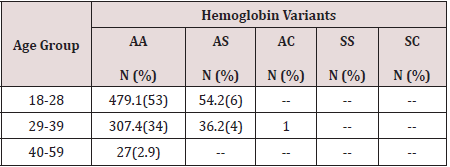
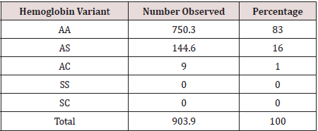
Hemoglobin genotype polymorphism in gravidas women have been analyzed and 750.3(83%) of the women had hemoglobin genotype of AA while 144.6(16%) women had hemoglobin genotype of AS. In addition, hemoglobin genotype AC was seen in 9(1%) of the gravidas women attending the antenatal clinic of federal medical center Yola as shown in Table 1.Age distribution shows that, 479(53%) and 54.2(6%) of hemoglobin genotype AA and AS respectively was seen in women within the age of 18 to 28years while 307.4(34%) of the hemoglobin genotype AA and 36.2(4%) of hemoglobin AS was observed in women within the age range of 29 to 39years. 1% of hemoglobin AC occurred in women within the age of 29 to 39years. In addition, among women at the age of 40 to 59years, 27(2.9%) was observed at P<0.05 as shown in Table 2. None of the gravidas women had hemoglobin SS or SC.

Table 1: Hemoglobin variants of gravidas women.

Lupinepublishers-openaccess-pediatric-dentistry-journal

Table 2: Hemoglobin variants per age group of gravidas women.

Lupinepublishers-openaccess-pediatric-dentistry-journal

Discussion

The analysis of hemoglobin polymorphism among pregnant women in Federal Center Yola revealed that gene frequencies with respect to the hemoglobin genotype polymorphism in gravidas women has shown a general formula of AA > AS > AC indicating high prevalence of AA over AS while AC genotype was the least of the hemoglobin genotype observed in this study. This result is in agreement with the earlier report by Medugu et al.[8] In addition, 83% of the women had hemoglobin genotype AA indicating that these women had normal allele of hemoglobin A in a homozygous state while 16% of women had hemoglobin genotype AS to reflect the presence of hemoglobin A (HbA) and abnormal hemoglobin S (HbS) in a heterozygous state and inherits one normal allele and one abnormal allele encoding hemoglobin S (hemoglobin genotype AS). Hemoglobin genotype AS is also called sickle cell trait which is generally regarded as benign condition but this condition have been reported to cause medical complications in exercise, muscle contraction or dehydrated state[9] and by consequence, 16% of pregnant women in this center may be at risk of anemia hence women with hemoglobin genotype of AS may require additional medical attention during vaginal child birth which usually involves levels of muscular contractions. Furthermore, 1% of women had hemoglobin genotype AC and none of the women had genotype SS or SC and this low level of homozygous state of abnormal hemoglobin may be due to high level of medical education among couples or/ and that women with homozygous state of abnormal hemoglobin may be unable to keep pregnancy hence their absence in this study

Conclusion

Gene frequencies with respect to the hemoglobin genotype polymorphism in gravidas women has shown a general formula of AA > AS > AC indicating high prevalence of AA over AS and AC in Federal Medical Center Yola Nigeria.

Read More Lupine Publishers Pediatric Dentistry Journal Articles:
https://lupine-publishers-pediatric-dentistry.blogspot.com/
Friday, June 11, 2021

Lupine Publishers | Management of Neonatal Teeth: Two Case Reports

 Lupine Publishers | Journal of Pediatric Dentistry


Abstract

Neonatal teeth are abnormalities in the chronology of eruption in which one or more teeth, which erupt during the first 30 days following birth and cause problems such as trauma-induced ulcers of the tongue, damage to the mother’s breasts, a reduction in the quality of breastfeeding, weight loss and even aspiration of the neonatal tooth. The removal or not of these teeth should be assessed on a case-by-case basis. The aim this article was to report the clinical management of two cases of neonatal teeth. The two female babies were attended in a pediatric dental clinic. The first patient was a 2-month-old baby that had difficulty during breastfeeding due to the mobility of one of the neonatal teeth. The clinical and radiographic exam showed that the three teeth were part of the normal dentition. The two mandibular teeth had approximately one third of the crown erupted and moderate mobility and opted for follow-up. The maxillary tooth was fully erupted and had severe mobility and we opted for its removal. The clinical and radiographic exam confirmed that the two teeth were part of the natural dentition. The teeth presented the clinical crowns fully erupted and both mobile, the left tooth having more mobility than that of the right tooth and opted for removal of more mobile left central incisor. In both cases, the extractions of teeth with high mobility were performed by greater risk of aspiration and caused more difficulty during breastfeeding. The follow-up showed a quick recovery, easier breastfeeding, including weight gain.

Keywords: Neonatal teeth; newborn; pediatric dentistry

Introduction

It is important that pediatricians and pediatric dentists are knowledgeable about the normal characteristics and anomalies in a newborn baby’s oral cavity, as a foundation for early diagnosis [1,2]. One of the dental anomalies found in a newborn’s oral cavity is natal and/or neonatal teeth, whose prevalence is one in every 3,000 live births [1]. Natal teeth are present at birth, while neonatal teeth appear in the oral cavity during the first 30 days of life [3]. The etiology of these teeth can range from hereditary factors, or a superficial position of the tooth germ, infections or even as a sequela of congenital syphilis [3-7]. There is evidence of a relationship between these anomalies and Pierre Robin [7] syndrome and also with premature and low birthweight children [8]. It is important to determine if the tooth is part of the normal dentition or if it is a supernumerary. If a radiograph confirms the tooth is a supernumerary, extraction is indicated, as well as in cases of excessive mobility, which can present a risk of aspiration [4-12]. If an extraction is indicated, it should be performed after the first week of life. Prior to this, the baby is incapable of absorbing vitamin K and the breast milk is lacking in this vitamin. To avoid bleeding, surgical procedures before this age should be preceded by vitamin K supplementation [6]. These teeth can also be associated with ulcers forming on the tongue surface in newborns, known as Riga-Fede [3-5,13,14], which can lead to nutritional deficiencies in the baby [2,5,11,15]. Therefore, clinical management should be evaluated with discretion and on a case-by-case basis. In this sense, the aim this article was to report the clinical management of two cases of neonatal teeth.

First Case Report

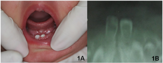
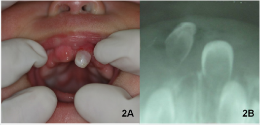
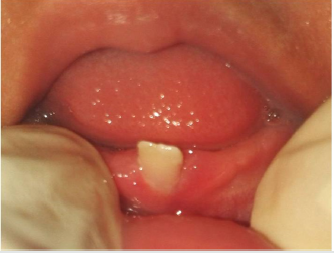
A female patient, dark skinned, full-term birth, two months old, was attended to the pediatric dental clinic of the Brazilian Association of North Rio Grande. Mother’s main complaint was the presence of three teeth that appeared when the child was less than one month old and the difficulty, they caused during breastfeeding due to the excessive mobility of one of the teeth. At the time of the consult, the child weighed less than expected for her age. According to the mother, the child was born with bumps on both the maxilla and mandible, in the area of the future central incisors. After 15 days, two lower teeth erupted, right first, then the left. After 20 days, an upper tooth erupted. The mother denied any family history of neonatal teeth. During the intraoral clinical exam, two mandibular incisors and one maxillary incisor were observed (Figure 1A). The two mandibular teeth had approximately one third of the crown erupted and moderate mobility. The maxillary tooth was fully erupted and had severe mobility (Figures 2A & 2B). No lingual ulcer was found during the exam. Two radiographs, upper and lower anterior regions, were taken using a #2 film and a modified occlusal technique. The exposure time was reduced by one half (0.2 seconds) and the baby was placed in a supine position, in the mother’s lap. Both used a lead apron for radiation shielding.

Figure 1: A) Neonatal teeth in the region of lower central incisors and lack of ulcer on the ventral surface of the tongue; B) Lower occlusal radiograph showing neonatal lower incisor teeth.

Lupinepublishers-openaccess-pediatric-dentistry-journal

Figure 2: A) Neonatal tooth in the region of upper left central incisor and bump in the region of upper right central incisor; B) Upper occlusal radiograph showing the neonatal teeth in the region of upper left central incisor.

Lupinepublishers-openaccess-pediatric-dentistry-journal

Figure 3: A) Topical anesthetic EMLA (Lidocaine Hydrochloride 2.5%).
B) Topical anesthesia, being careful to avoid that the baby could swallow the anesthetic.
C) Neonatal tooth in the region of upper left central incisor extracted.

Lupinepublishers-openaccess-pediatric-dentistry-journal

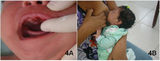
The radiographs suggested that the deciduous teeth were part of the normal dentition. The decision was made to remove the maxillary tooth because, according to the mother, the child had difficulty latching on during breastfeeding due to the high degree of mobility of this tooth, resulting in both irritability and weight loss (Figures 1B & 2B). The mother was informed about the recommendations for treatment, along with risks and benefits after which she gave written authorization to proceed with treatment, photographic documentation and scientific publishing. The procedure was performed with the dentist and mother in the knee-to-knee position, and the baby reclined in the dentist’s lap. No vitamin K prescription prophylaxis was performed because the baby had already achieved the normal safe levels. Initially, the mucosa was dried and a topical anesthetic (EMLA - lidocaine hydrochloride 5%) (Figure 3A) was applied with cotton three times, being cautious to prevent its ingestion by the baby. The extraction was performed with sterile gauze (Figure 3B & 3C). Cotton gauze was applied with pressure for hemostasis and the mother was told to immediately begin breastfeeding because the breast milk contains immunoglobulins which promote healing and the intimate contact with the mother provides security to the baby [4] (Figure 4A & 4B).

Figure 4: A) Clinical appearance immediately after the extraction.
B) Child being breastfed, just after the extraction. The breastmilk has a hemostatic effect and helps to calm the baby.

Lupinepublishers-openaccess-pediatric-dentistry-journal

The day after the procedure, the mother was contacted by phone and she reported that there was a small clot in the area of the extraction. She also indicated that the child latched on more easily during breastfeeding and the absence of any irritability. The next day, the mother was again contacted by phone and she reported that the clot was gone. After seven days the healing was complete, and within thirty days the child had gained weight.

Second Case Report

Figure 5: A) Neonatal teeth in the region of lower central incisors and lack of ulcer on the ventral surface of the tongue.
B) Taking the radiograph using the modified occlusal technique.
C) Radiograph of two neonatal teeth in the region of lower central incisors.

Lupinepublishers-openaccess-pediatric-dentistry-journal

A 30-day-old white female patient was seen at the same dental center as the previous case. The guardian reported the presence of neonatal teeth as the primary concern, along with difficulty breastfeeding due to the mobility they presented. According to the mother’s report, the child was born with a bump in the region of the lower incisors and two days later the bump disappeared, replaced by two teeth. These teeth initially presented with slight mobility, which gradually worsened over time. The patient had a family history of neonatal teeth with the aunt having the same condition when born. Mother also reported the child was gaining less weight than was expected at that age. Upon performing an intraoral clinical examination, two teeth were seen in the mandibular region corresponding to the area of the lower central incisors (Figure 5A). The teeth presented with the clinical crowns fully erupted and both mobile, the left tooth having more mobility than that of the right tooth. The soft tissue surrounding the teeth appeared normal and the tongue and sublingual region demonstrated no ulceration. Two radiographic films were taken using the same protocol as in the first described case (Figure 5B). The radiographs confirmed that the two teeth were part of the natural dentition (Figure 5C). The mother was informed of the recommendations for the treatment, along with risks and benefits after which she gave written authorization to proceed with treatment, photographic documentation and scientific publishing. The mother opted for removal of more mobile left central incisor, which had a greater risk of aspiration and caused more difficulty during breastfeeding. The extraction was performed following the exact same protocol as the first case (Figure 6). Additionally, the same healing process occurred as in the first case. We followed-up by phone the day following and one week following. Then one month after the exam, the patient returned to the clinic where the right mandibular central incisor presented with no increase in mobility (Figure 7).

Figure 6: A: Topical anesthesia, being careful to avoid that the baby could swallow the anesthetic; B: Lower left central neonatal tooth being extracted. C: Child being breastfed, just after the extraction.

Lupinepublishers-openaccess-pediatric-dentistry-journal

Figure 7: Healing one-month post-extraction.

Lupinepublishers-openaccess-pediatric-dentistry-journal

Discussion

The included clinical cases reference the occurrence of upper and lower central incisor neonatal teeth. The epidemiological data shows that these eruption anomalies are most common in lower central incisors, around 85% of the cases, and upper central incisors in around 11% [6,7,16]. According to Cunha et al. [15], natal and neonatal teeth are often seen in pairs, which is corroborated in the second case presented. The two patients were female, according to the literature this is the more affected sex [3,6,7,16,17]. The etiology of these teeth is still unknown [18]. Nevertheless, some authors report that it may or may not be a genetic condition [6,12,17]. The increased gain in body weight that was observed after the surgical extractions was similarly reported by other authors [4,5,7,13,14,19]. These cases and the benefits seen by both the mother and patient reiterate the importance of early management by a dentist. Neonatal teeth are often associated with excessive mobility and weight loss, affecting the baby’s health and quality of life.

Read More Lupine Publishers Pediatric Dentistry Journal Articles: https://lupine-publishers-pediatric-dentistry.blogspot.com/

Friday, June 4, 2021

Lupine Publishers | Scoping Review of Dental Anxiety among Children and Adolescents in Saudi Arabia

 Lupine Publishers | Journal of Pediatric Dentistry


Abstract

Background: Dental anxiety (DA) has shown to be a complex concept associated with multiple factors including parents and children previous episodes of DA, poor oral health, avoidance of appointments, and unstable overall health. Thus, exploring this issue among Saudi children is highly important.

Aim: This scoping study aims to review the nature of research conducted on dental anxiety among Saudi children and adolescents.

Methods: Arksey and O’Malley’s scoping review methodology was used. Relevant cross-sectional studies, case reports, cohort studies, randomized and non-randomized control trials, systematic and scoping reviews published between 1900 and the end of January 2020 were extracted from MEDLINE via PubMed, MEDLINE via Voids, Web of Knowledge, Cochrane, CINAHL, Embase, and Scopus, and were assessed for their eligibility.

Results: A total of 18 eligible studies were included. Six articles evaluated validity of different DA measures; four assessed effectiveness of different distraction techniques in reducing DA; four examined association of previous dental experiences with DA; and four explored causative factors associated with DA.

Conclusion: To advocate reducing the level of children’s DA, dentists must not consider only common causative factors such as anaesthesia, dental extraction, and numbness; but also socio-dental aspects related to DA including children’s previous painful experiences, gender, age, and oral health related quality of life.

Key Message

Dental practitioners must consider the common causative factors and socio-dental aspects associated with dental anxiety as this could help in reducing dental anxiety level among children.

Keywords: Dental anxiety; dental fear; dental phobia; children; scoping review

Abbreviations: DA: Dental Anxiety; OHRQoL: Oral Health Related Quality of Life; ACDAS: A beer Children Dental Anxiety Scale; CFSS-DS: Children’s Fear Survey Schedule-Dental Subscale

Introduction

Dental anxiety (DA) is described as a sensation of apprehension about dental therapy that is not essentially associated with a specific external stimulus [1]. A recent review has demonstrated that evaluation of DA is important for not only identifying and managing patients with high levels of DA, but also managing pain in dental patients [2]. DA has been shown to be a complex decision making process in dental settings that is associated with multiple factors such as age, gender, parents and children previous episodes of DA, poor oral health, a history of excessive dental caries, avoidance of appointments, and poor oral health related quality of life (OHRQoL) [3-8]. DA is considered as a public health issue that commonly affects more children and adolescents than adults [9]. DA is reportedly caused by fear-evoking sensations, sights, and sounds, and fear of pain from dental drills and needles. It has uncovered that the reported levels of DA in childhood worldwide ranged from 10% to 29.3% [10]. This wide variation in DA prevalence may be related to the use of different measures of DA and different cut-off points to differentiate between those who are dentally anxious and those who are not. It is vital to reduce the disparities in the evaluation of DA among children and adolescents, as this will help in determining the most effective psychological techniques for alleviating DA as well as pain management strategies [11]. Nevertheless, dentists find it stressful and time-consuming to deal with children who experience DA [8,12]. Interestingly, dentists’ appearance has been shown to play a key role in reducing DA in children [13]. Further, dentists who have completed postgraduate courses in DA and undergone training for managing DA demonstrate better attitudes and higher use of behavioral management techniques with anxious children [14]. A scoping review aims to detect gaps in the literature, identify the main concepts reported, explore a wide territory of research, and highlight evidences that impact and can potentially improve practice in the field [15]. The present study seeks to identify gaps in the dental literature based on the nature and types of DA research conducted among Saudi children and adolescents, to explore the territory of DA in Saudi children and adolescents, and lastly, to report the results of research conducted on DA among Saudi children and adolescents. Scoping studies can also be used to identify the range of existing studies about a specific topic and to understand how those studies have been carried out [16]. This review determines the range of research conducted on DA among Saudi children and adolescents and how the research in this area has been conducted. The complexity and multifactorial of DA, as mentioned previously, [3-8] mean that it is highly important to investigate this issue among Saudi children and adolescents. This scoping study, therefore, aims to review and evaluate the nature of research conducted on DA among Saudi children and adolescents.

Materials and Methods

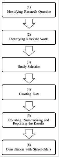
Figure 1: Arksey and OMalley’s Strategy of Conducting a Scoping Review in Healthcare.

Lupinepublishers-openaccess-pediatric-dentistry-journal

The scoping review strategy of Arksey and O’Malley was adopted in the current study [17]. This strategy consists of six key stages, as shown in Figure 1, and is described in detail below.

Stage: Identifying the research question

DA is considered as a key barrier in accessing dental care and individuals’ OHRQoL [5,18,19]. Impact of dental treatment on relationships and irritability were more common among patients with DA [20]. This highlights the significance of studying the nature of research on DA. This scoping review, consequently, emphasizes on exploring research on DA among Saudi children and adolescents.

Stage: Identifying relevant work

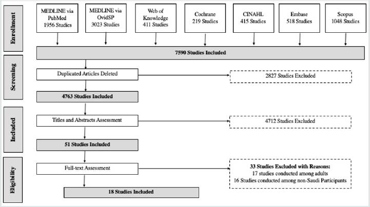
For this review, seven different resources were searched for relevant published material: MEDLINE via PubMED, MEDLINE via OvidSP, Web of Knowledge, Cochrane, CINAHL, Embase and Scopus. Figure 2 illustrates the process of searching and excluding articles. A combination of free-text search terms and controlled vocabulary were used: ‘dental anxiety*’.mp., ‘dental fear’.mp., ‘dental phobia’. mp., and (‘children’ or ‘adolescent’).mp. or ‘children*’.mp. or ‘adolescent*’.mp. All studies were considered, with the exception of studies that were conducted among adults and/or non-Saudi participants. This scoping review had no gender restrictions and included studies with all types of settings, including dental clinics, schools, and private or governmental clinics in Saudi Arabia. An Endnote library was established for organizing, classifying and systematizing relevant studies. A total of 7590 eligible studies were identified. All duplicated articles were deleted (n = 2827). As a result, 4763 articles were evaluated based on the inclusion and exclusion criteria in the next stage.

Stage: Selecting relevant studies

Based on the inclusion criteria for this scoping review, the following publications were included:
a. Articles published between 1900 and the end of January 2020.
b. Studies conducted among Saudi children and/or adolescents.
c. Studies on humans that were in the form of cross-sectional research, case reports, case-control studies, retrospective and prospective cohort studies, clinical randomized and nonrandomized control trials, systematic reviews, literature reviews, and scoping reviews. Based on the exclusion criteria, the following were excluded.
a) Studies that were not written in English.
b) Research conducted on animals.
c) Studies conducted among non-Saudi children and/or adolescents and
d) Studies carried out among adult participants.

The two reviewers independently searched all the sources. They initially screened the titles and abstracts of all the identified studies through the search to assess their applicability. The reviewers then conferred and discussed the eligibility of the included studies. There were no disagreements among them about the included articles. In total, 4712 studies were excluded based on the assessment of their titles and abstracts (see Figure 2). This left 51 articles for the fulltext assessment. At the full-text evaluation stage, both reviewers excluded 33 articles because they did not meet the inclusion criteria. This left 18 articles for the comprehensive review in the next step.

Figure 2: The Process of Searching the Literature and Defining Relevant Studies.

Lupinepublishers-openaccess-pediatric-dentistry-journal

Stage: Charting data

In this stage, the two examiners individually read all 18 included articles and then conferred to examine their applicability to this study. The main author then assessed the eligibility of all the included studies based on the determined inclusion and exclusion criteria. The other examiner independently evaluated and confirmed the eligibility of the studies.

Stage: Collating, summarizing and reporting the results

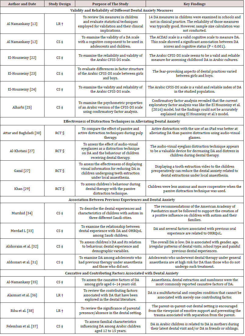
This study uses qualitative content analysis to condense and report the findings of the included studies. The findings were categorized under four headings to illustrate differences between the results based on the likeness of their aims and nature (see Table 1).

Table 1: Studies on Dental Anxiety among Saudi Children and Adolescents and their Key Purposes and Findings.

Lupinepublishers-openaccess-pediatric-dentistry-journal

Stage: Consulting with stakeholders

This phase of the scoping study is not applicable to the current review.

Results

In total, 18 articles published between 2011 and 2019 were included in this study. The process of searching the literature and identifying relevant articles is depicted in the flowchart in Figure 2. Six of the articles were focused on evaluating the validity and reliability of different DA measures, such as A beer Children Dental Anxiety Scale (ACDAS) and Children’s Fear Survey Schedule-Dental Subscale (CFSS-DS) [21-26]. Four of the selected studies assessed the effects of different distraction techniques on reducing the level of DA among Saudi children [27-30], and four other studies investigated the association of previous dental experiences and DA among Saudi children [31-34]. The last four studies explored the causative and contributing factors associated with DA, with one of the studies focusing specifically on the presence of a parent in the dental clinic [35-38]. Table 1 presents the included studies and their key aims and results. Most of the articles included were cross-sectional (11 studies) in nature, and the remaining studies were categorized as randomized controlled trails (four studies) and literature reviews (three articles). The key findings of these 18 included studies are described and discussed in detail in the following section.

Discussion

The findings of this review are categorized into four sections to illustrate and discuss the differences between the results of the included studies: Validity and reliability of different DA measures; Effectiveness of distraction techniques in alleviating DA; Association between previous painful dental experiences and DA; and Causative and contributing factors associated with DA. Following this, some limitations and conclusions based on the review findings are described.

Validity and reliability of different DA measures

Reliable and valid methods for evaluating DA in children could have important benefits for dentists and dental service suppliers [39]. For instance, these methods could be utilized for determining the prevalence of DA, identifying the symptoms and risk factors for DA in a population, and evaluating changes in DA over time [40]. The present study included one review article and one cross-sectional study on DA measures. Al-Namankany, De Souza [21], reviewed DA measures reported between 1960 and 2011 and validated 14 paediatric DA measures, but they were unable to validate 5 scales. Moreover, Al-Namankany, Ashley [26], reported that based on the ACDAS measure, there was a strong association between cognitive status and DA scores. An exploration of the dental literature has uncovered that the CFSS-DS scale is valid and reliable in several countries [41,42]. Further, this review found that the Arabic CFSSDS scale has been assessed four times for its validity and reliability [22-25]. It was concluded that this scale was reliable, consistent and valid [22,24]. However, there were differences in factor structure for this scale between Saudi girls and boys, with regard to factors associated with fear of dental procedures, fear of dentists, fear of strangers and fear of injections [23]. Furthermore, examining the psychometric properties of this scale revealed that the internal consistency of the Hospital Fear Subscale, Dental Fear Subscale and Stranger Fear Subscale corresponded to good and acceptable reliability [25]. However, it might be important to examine the CFSSDS measure across different regions of Saudi Arabia, considering the role of factors such as age, gender, previous DA experiences in parents, poor oral health, and poor oral health related quality of life [3-5]. Despite this, the Arabic version of the CFSS-DS scale is valid and reliable for measuring DA among Saudi children.

Effectiveness of distraction techniques in alleviating DA

Non-pharmacological passive and active distraction techniques could play a major role in alleviating DA among children [43]. The findings of the current review are consistent with this view and reveal that using a passive or active distraction technique could reduce DA among Saudi children and improve their response to their dentists. Passive distraction was not only effective in reducing children’s DA but also effective in helping children co-operate after local anesthesia [27,29]. Moreover, showing tooth extraction videos to children was found to alleviate DA associated with dental extractions under local anesthesia [28]. Yet, Saudi children preferred active distraction with an iPad more than passive distraction using audio-visual glasses [30]. Additionally, the use of music in dental settings is effective in reducing DA [44]. However, no study in the dental literature has directly examined the effectiveness of the music distraction technique on reducing DA among Saudi children.

Association between previous dental experiences and DA

Children’s previous experiences with dental treatment has been found to be associated with increase in their level of DA across several cultures [45,46]. Likewise, this review also found that previous experiences associated with DA was a contributing factor, in addition to gender, age, irregular patterns of dental calls and OHRQoL [32,33]. Though, dentists’ appropriate use of behavior management techniques is strongly associated with them having attained appropriate training and completed postgraduate courses in DA [14]. Yet, our review revealed that no studies so far have examined dentists’ skills and attitudes related to behavior management in Saudi children with DA. The evaluation of DA among special-needs children such as children with Down’s syndrome, autistic children and children with intellectual disabilities has received its fair share of attention in the literature across different countries [47,48]. Focusing on children those children is important because they are at a greater risk of dental diseases due to difficulties associated with expressing their dental needs and interacting with their parents or dentists [49]. Interestingly, there was only one study that evaluated DA among autistic Saudi children; this study concluded that the period of diagnosis was longer in these children and they needed more assistance to advance their oral health [34]. Consequently, evaluating DA in this vulnerable group of children in Saudi may be highlighted.

Causative and contributing factors associated with DA

Several pathways have been associated with DA in children, including informative, cognitive conditioning, verbal threat, and parental aspects [20,50]. Investigating these factors in children in detail may help in alleviating DA. However, only two studies have explored the causative and contributing factors associated with DA among Saudi children. These studies not only reported that the numbness caused by anesthesia and dental extraction were the most common causative factors of DA; but also concluded that DA is a multifactorial and complex occurrence that cannot be explained by merely one contributing factor [35,36]. Moreover, DA in the children was found to be significantly associated with DA in parents, siblings, friends and a parent-in-parent-out dental setting [37,38].Despite these findings, there are some shortcomings in the dental literature on Saudi children with regard to several informative, cognitive conditioning, verbal threat, and parental aspects associated with DA.

Limitations

There are two main limitation in this scoping review. First, there is a possibility that relevant articles were neglected because they were deposited in databases other than those that were searched here or because they were published in a language other than English. Exploring other databases and articles in other languages in the future might prove useful. An additional limitation of scoping studies is the lack of quality assessment of the included articles [16]. However, the emphasis of this study is on delivering a comprehensive review of the nature of the research that has been conducted on DA among Saudi children and adolescents. In this respect, the scope of the included studies with regard to DA was defined, the data charted, and the results collated, summarized, reported and discussed in detail.

Conclusions

In order to reduce the level of children’s DA, dentists must not consider only common causative factors such as anesthesia, dental extraction, and numbness; but also socio-dental aspects related to DA including children’s previous painful experiences, gender, age, and oral health related quality of life.

Acknowledgments

I would like to acknowledge the generous help of my colleague, Dr Eltayeb Mohammed Alhassan (Head of Dental Health Department, Faculty of Applied Medical Sciences, Albaha University, Saudi Arabia), in data extraction and confirmation of the eligibility of the included articles in this scoping review.

Source of Funding

This article did not receive any specific grant from funding agencies in the public, commercial, or not-for-profit sectors.

Conflict of Interest

The author declares no conflicts of interest regarding the authorship and/or publication of this stud Title of the article: Scoping Review of Dental Anxiety among Children and Adolescents in Saudi Arabia.

Read More Lupine Publishers Pediatric Dentistry Journal Articles:
https://lupine-publishers-pediatric-dentistry.blogspot.com/
980 nm Diode Laser: A Good Choice for the Treatment of Pyogenic Granuloma

Abstract Pyogenic granuloma is a benign non/neo plastic mococutanous lesion . It is a reactional response to constant minor trauma and ca...