Lupine Publishers | Journal of Pediatric Dentistry
Abstract
Background/Objective: Hemoglobin is the iron-containing oxygen-transport metalloprotein in our red blood cells, hemoglobin is made up of globin chains which are encoded by their respective genes located on chromosome 11 and16 with several alleles. Many of these alleles suffer point mutations in the DNA sequence that lead to single amino acid substitutions in the globin moiety, resulting in the production of abnormal hemoglobin polymorphism and which are associated with a wide range of moderate to severe hemolytic anemia in pregnant women. Therefore, this study aims to examine hemoglobin genotype polymorphism among gravidas women in Yola.
Materials/Methods: 904 pregnant (i.e. gravida) women with age range of 18 to 41years in the antenatal ward of the hospital participated in this study and 2mls of venous blood were aseptically collected from each participant into EDTA vacutainer. The hemoglobin genotype was determined within 5hours of blood collection using Helena electrophoresis tank.
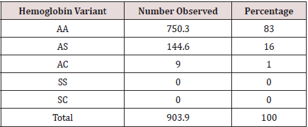
Results: 750.3(83%) of women had hemoglobin genotype of AA while 144.6(16%) women had hemoglobin genotype of AS. In addition, hemoglobin genotype AC was seen in 9(1%) of the gravida’s women while hemoglobin genotype SS and SC was not seen in this group of women within the study period.
Conclusion: Gene frequencies with regard to the hemoglobin genotype polymorphism in gravidas women has shown a general formula of AA > AS > AC indicating high prevalence of AA over AS and AC in Federal Medical Center Yola Nigeria.
Keywords:Hemoglobin polymorphism; gravidas women
Introduction
Hemoglobin is the iron-containing oxygen-transport
metalloprotein in red blood cells of humans and most vertebrates.
Hemoglobin in our blood carries oxygen from respiratory organs
to the rest of the body, where it releases the oxygen to metabolize
nutrients to generate energy that powers our body’s physiology
and collects the resultant carbon dioxide back to respiratory
organs to be expelled from the body. Hemoglobin is made up of
heme, which is the iron-containing portion, and globin chains,
which are proteins and the globin protein consists of chains of
amino acids. There are several different types of globin chains, such
as: alpha, beta, delta, and gamma. Hemoglobin Polymorphism in
this study refers to the occurrence of variety of hemoglobin types
in pregnant (i.e. gravidas) women. Hemoglobin types include:
a) Hemoglobin A (Hb A) which makes up about 95%-98%
of hemoglobin found in adults; it contains two alpha (α) chains and
two beta (β) protein chains[1].
b) Hemoglobin S (HbS) which is an abnormal hemoglobin
with a single nucleotide substitution (GTG for GAG) in the gene
for beta globin on short arm of chromosome 11, resulting in the
replacement of a glutamic acid residue with valine at the sixth
position of both (i.e. homozygous state) or single (i.e. heterozygous
state) globin chain[2].
Hemoglobin C (HbC) is also an abnormal hemoglobin similar
to HbS but in HbC, lysine replaces glutamic in the globin chain.
Deoxygenation of either HbS or HbC exposes valine or lysine
residue on the surface of the molecule, which forms hydrophobic interactions with adjacent chains, the resulting polymers align
into bundles, causing distortion of the RBC into a crescent or sickle
shape, consequently, reduces flexibility and increase deformability,
which hinders passage of the cell through narrow blood vessels[3]
resulting in sickle cell episodes. Sickle cell disorders include the
homozygous state for Hemoglobin S, or sickle cell anemia (SS), the
heterozygous state for Hemoglobin S or the sickle cell trait (AS), and
the compound heterozygote state of Hemoglobin S together with
other hemoglobin variants such as C or D can result in hemoglobin
AC[3].The globin chains are encoded by their respective genes
located on chromosome 11 and chromosome 16 and are both
known to have several alleles[4].Many of these alleles suffer point
mutations in the DNA sequence that lead to single amino acid
substitutions in the globin moiety, resulting in the production of
hemoglobin polymorphism. The abnormal hemoglobin genotype
occurs when an affected individual inherits mutated globin gene(s)
such as hemoglobin S, C, D, and E from both parents. Abnormal
hemoglobin genotypes are inherited in an autosomal codominant
fashion and occur by different combinations[5]. Several abnormal
hemoglobin genotypes have been discovered but the most
commonly encountered abnormal hemoglobin genotypes among
Nigerians include AS, AC, SC, and SS[5].It has been reported that
abnormal hemoglobin genotypes have been associated with a wide
range of moderate to severe hemolytic anemia, leading to a high
degree of morbidity and mortality among affected individuals as
well as susceptibility to renal medullary carcinoma[6]in addition,
World Health Organization ranked Nigeria as first in terms of high
prevalence of infants born with abnormal hemoglobin genotype[7].
Therefore, this study aims to examine hemoglobin genotype
polymorphism among gravidas women in Yola in other to elucidate
the risk of giving birth to children with abnormal hemoglobin as
well as risk of developing hemolytic anemia during gestation period
in this locality.
Materials and Method
This retrospective and descriptive study was carried out at the hematology department of Federal Medical Center Yola in Adamawa State, Northeastern Nigeria. 904 pregnant (i.e. gravida) women with age range of 18 to 41years in the antenatal ward of the hospital participated in this study.
Statistical analysis
Statistical analysis was performed using SPSS computer software version 20.0 (IBM Chicago, IL, USA). Descriptive values were given as mean and standard error of mean. Categorical variables were expressed as the number of cases and the percentage value.
Sample collection and analysis
2mls of venous blood were aseptically collected from each participant into a tripotassium Ethylenediaminetetraacetic acid (K3 EDTA) anticoagulant vacutainer. The hemoglobin genotype was determined within 5hours of blood collection as followsa portion of the blood was put in a clean khan tube and washed 3 times with normal saline (0.85% sodium chloride). Distilled water was added to the washed red cell in ratio of 1:4 to lyse the blood sample. The lysed samples were applied on Helena cellulose acetate paper using the Helena plate and applicator, and the paper was placed in the Helena electrophoresis tank (Consort) containing a commercially prepared Tris-EDTA-Borate buffer, the pH of the buffer is 8.6. The electrophoretic separation was allowed at room temperature for 3minutes at 220V. A commercially prepared Helena known hemoglobin were run as controls along with the test, and the results were read immediately after the end of the test time.
Results
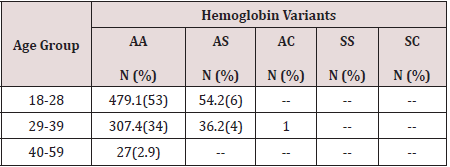
Hemoglobin genotype polymorphism in gravidas women have been analyzed and 750.3(83%) of the women had hemoglobin genotype of AA while 144.6(16%) women had hemoglobin genotype of AS. In addition, hemoglobin genotype AC was seen in 9(1%) of the gravidas women attending the antenatal clinic of federal medical center Yola as shown in Table 1.Age distribution shows that, 479(53%) and 54.2(6%) of hemoglobin genotype AA and AS respectively was seen in women within the age of 18 to 28years while 307.4(34%) of the hemoglobin genotype AA and 36.2(4%) of hemoglobin AS was observed in women within the age range of 29 to 39years. 1% of hemoglobin AC occurred in women within the age of 29 to 39years. In addition, among women at the age of 40 to 59years, 27(2.9%) was observed at P<0.05 as shown in Table 2. None of the gravidas women had hemoglobin SS or SC.
Discussion
The analysis of hemoglobin polymorphism among pregnant women in Federal Center Yola revealed that gene frequencies with respect to the hemoglobin genotype polymorphism in gravidas women has shown a general formula of AA > AS > AC indicating high prevalence of AA over AS while AC genotype was the least of the hemoglobin genotype observed in this study. This result is in agreement with the earlier report by Medugu et al.[8] In addition, 83% of the women had hemoglobin genotype AA indicating that these women had normal allele of hemoglobin A in a homozygous state while 16% of women had hemoglobin genotype AS to reflect the presence of hemoglobin A (HbA) and abnormal hemoglobin S (HbS) in a heterozygous state and inherits one normal allele and one abnormal allele encoding hemoglobin S (hemoglobin genotype AS). Hemoglobin genotype AS is also called sickle cell trait which is generally regarded as benign condition but this condition have been reported to cause medical complications in exercise, muscle contraction or dehydrated state[9] and by consequence, 16% of pregnant women in this center may be at risk of anemia hence women with hemoglobin genotype of AS may require additional medical attention during vaginal child birth which usually involves levels of muscular contractions. Furthermore, 1% of women had hemoglobin genotype AC and none of the women had genotype SS or SC and this low level of homozygous state of abnormal hemoglobin may be due to high level of medical education among couples or/ and that women with homozygous state of abnormal hemoglobin may be unable to keep pregnancy hence their absence in this study
Conclusion
Gene frequencies with respect to the hemoglobin genotype polymorphism in gravidas women has shown a general formula of AA > AS > AC indicating high prevalence of AA over AS and AC in Federal Medical Center Yola Nigeria.
Read
More Lupine
Publishers Pediatric Dentistry Journal Articles:
https://lupine-publishers-pediatric-dentistry.blogspot.com/


No comments:
Post a Comment