Saturday, May 15, 2021

Lupine Publishers | An Assessment of Emergency Care Following Tooth Avulsion Among A Selected Population of Nigerian School Children

 Lupine Publishers | Journal of Pediatric Dentistry


Abstract

Aim: To assess the knowledge of school children on the first aid administered after traumatic tooth avulsion.

Methods: A structured questionnaire was used to obtain information from children of six primary and six secondary schools in Port Harcourt, Nigeria. Information elicited included respondents’ socio-demographics, exposure to dental trauma and knowledge on emergency care for tooth avulsion. Data collected was analysed using SPSS Version 22 and level of significance was set at p <0.05.

Results:There were 411pupils; 194 males and 217 females with a mean age of 12.5 (±2.6) years. One hundred and eighty-three (44.5%) had previous dental injuries. Three hundred and twenty-three (78.6%) would seek treatment in dental clinic after tooth avulsion. Only 16 (3.9%) would replace the tooth in its socket, 160 (38.9%) would take it to the dental clinic and 192 (46.7%) will throw the tooth on a roof top. Only 164 (39.9%) would seek immediate treatment. Twenty-two (5.4%) will use milk as storage/ transport media. There were statistically significant differences between
a) The males and females in their time for seeking treatment (p=0.01).
b) The public and private school pupils on where to go for treatment (p=0.013) and transport media use (p=0.00).
c) The primary and secondary school pupils on replantation of avulsed tooth (p=0.03) and transport media use (p=0.00).

Discussion: Although majority knew who a dentist is, their knowledge of emergency care when avulsion occurs is low; they require oral health education to bridge the gap in their knowledge.

Keywords:Tooth avulsion; traumatic dental injuries; emergency care; Nigerian school children; tooth replantation; transport media

Introduction

The school is an educational institution for learning both within [1] and outside [2] the classrooms under the guidance of teachers [3]. However, recreation and sport activities [2-4] in schools when unsupervised may predispose school children to traumatic dental injuries (TDI). Such injuries occur more among males than females and more commonly in urban children than rural children [5-8]. Sixty percent of TDI have been reported to occur during sporting activities among school aged children [9,10]. Of these, tooth avulsion; the complete displacement of the tooth out of the alveolar socket, is the most severe type of TDI and it occurs in both the primary and permanent dentitions [11]. In the permanent dentition, it occurs commonly among children aged between 6-12 years, [5,8,11,12] during the period in the tooth eruption cycle when root formation is incomplete and the periodontal ligament surrounding erupting teeth are loosely structured and provide very minimal resistance to an extrusive force. Tooth avulsion has an incidence of between 0.5 to 16% [8-14] among children and an incidence of 20.8% was reported among Nigerian adolescents [15].
Tooth loss, as a result of trauma, in addition to being distressing has both functional and psychosocial consequences in the permanent dentition [9,11,16]. First aid carried out minimizes the negative emotional/social consequences and ensuing cost of treatment that may result from premature tooth loss. Timely intervention at the site of the accident and immediate presentation to a dental clinic for professional care would result in a favourable outcome [7,10,16]. Replantation is the treatment option for avulsed permanent teeth and the treatment outcome is dependent on prompt and appropriate intervention at the site of the accident [3,17]. Other factors include short extra oral time, suitable transport media, immaturity of the root apex and patients’ general health [16,18,19]. The first aid carried out at the time of injury includes replacing the tooth within the alveolar socket and immediate referral to the dental clinic [8,13] for replantation procedure. However, if the tooth cannot be replaced into its original position, a suitable storage and transport medium would help maintain the viability of the periodontal ligament cells while seeking prompt professional attention in the dental clinic. The transport and storage media include Hanks Balanced Salt Solution (HBSS), Eagle’s medium, Normal saline, Via span, propolis, milk and coconut water [13,18,19]. The most recommended medium based on maintenance of PDL cell viability, availability, low cost and long shelf life is milk as reported in the reviews by Adnan et al. [18] and Udoye et al. [19]. Parents, teachers and students, especially, are almost always present where these injuries occur and need to be knowledgeable on what to do immediately it occurs. The knowledge of parents, [17,20] school teachers [3,9,10,20-23] and students [24,25] has been assessed in several studies both within and outside Nigeria. Most of these showed a poor awareness and low knowledge of first aid measures following tooth avulsion. Though the knowledge of students has been conducted in a study in Nigeria, it was done over a decade ago in another geopolitical region [24]. The aim of this study was to assess the level of knowledge of first aid administered following tooth avulsion among school children in the South-South geopolitical region in Nigeria.

Materials and Methods

Ethical clearance was obtained from the Research and Ethics Committee of the University of Port Harcourt Teaching Hospital. Consent was sought and obtained from the State Universal Basic Education Board and the proprietors of the private schools. Consent and assent were sought from the head teachers and pupils after being assured of confidentiality, respectively. Children aged 8 to 15 years were selected from six (three public and three private) primary and six (three public and three private) secondary schools using a multistage sampling technique. A structured questionnaire [24] was used to obtain information from each of the children. The information elicited were the socio-demographics such as age as at the last birthday and sex, other information were experience of exposure to dental trauma, response to past traumatic injuries, knowledge of the emergency treatment with particular focus on tooth avulsion and knowledge on the process of replantation. The information collected was entered into data spreadsheet and analyzed using the IBM Statistical Package for Social Sciences (SPSS) software, Version 22.0 (SPSS Inc., Chicago, IL, USA). Descriptive summary statistics was obtained for demographic variables and knowledge of emergency care. Pearson’s chi square was used to assess the differences in knowledge between groups and statistical significance was set at p<0.05.

Results

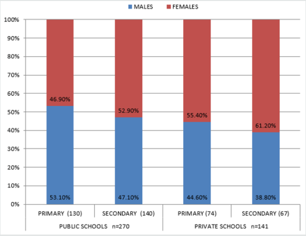
Figure 1: The distribution of the school pupils according to the type of schools, level of education and gender.

Lupinepublishers-openaccess-pediatric-dentistry-journal

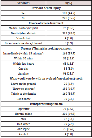
Four hundred and eleven students comprising 194 males and 217 females with a mean age of 12.5 (+2.6) participated in this study. Two hundred and four (49.6%) primary and 207 (50.4%) secondary school pupils participated in the study; 270 (65.7%) attended public schools while 141 (34.3%) attended private schools. Details in Figure 1. One hundred and eighty-three (44.5%) had history of previous dental injuries. Their responses to the questions on their knowledge of avulsion revealed that 323 (78.6%) would go to a dentist if they had tooth avulsion, 76 (18.5%) would see a medical doctor and 4 (1%) would go to the school clinic. Only 16 (3.9%) would replace the tooth in its socket. Although 164 (39.9%) would seek immediate treatment, 160 (38.9%) would take the tooth to the dentist. Two hundred and five (49.9%) would use normal saline as transport medium and only 22 (5.4%) would use milk. When avulsion occurs, 192 (46.7%) would throw it on roof top, while 160 (38.9%) would take the tooth to the dental clinic. Details in Table 1.

Table 1: The knowledge of the school pupils on traumatic dental emergencies (tooth avulsion).

Lupinepublishers-openaccess-pediatric-dentistry-journal

Gender of the subjects

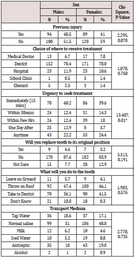
When the gender was considered equal proportion of males and females would seek treatment from a dental clinic (78%) and immediate treatment within 15 minutes (40%). Though 40% of males and females would seek treatment within 15 minutes of the injury, there were statistically significant differences between the males and females (p=0.01) in their timing for seeking treatment (Table 2).

Table 2: The relationship between the Gender and knowledge to the process of tooth replantation.

Lupinepublishers-openaccess-pediatric-dentistry-journal

*p<0.05 is statistically significant

Private and public schools

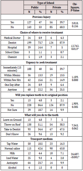
Following avulsion, 4.8%public and 2.1%private school pupils would attempt replacing the tooth within the tooth socket at the site of injury. Three hundred and twenty-three (78.6%) would go to a dental clinic for treatment, however only 164 (39.9%) would seek dental treatment immediately after the injury. More (51.1%) public school pupils compared to 38%private pupils had cultural beliefs on throwing an avulsed tooth on roof tops (p=0.06). Twelve (4.4%) and 10 (7.1%) public and private school pupils, respectively would use milk as a transport medium while 63.8% private as against 42.6% public school pupils preferred normal saline as transport medium. There were statistically significant differences between the public and private school pupils on where to go for treatment (p=0.013) and transport media (p=0.00) following avulsion (Table 3).

Table 3: The relationship between the pupil’s school type and the knowledge of the process of tooth replantation.

Lupinepublishers-openaccess-pediatric-dentistry-journal

*p<0.05 is statistically significant

Level of education

Table 4 shows that 52.9% of primary school pupils and 44.9% secondary school pupils preferred normal saline while 7.4% of primary school pupils preferred milk compared to 3.4% secondary school pupils. There were statistically significant differences between the primary and secondary school pupils on replacing the avulsed tooth back to the sockets (p=0.03), transport media (p=0.00).

Table 4: The association between the type of school, knowledge of emergency dental care and the level of education of the pupils.

Lupinepublishers-openaccess-pediatric-dentistry-journal

*p<0.05 is statistically significant

Discussion

Tooth avulsion is known to commonly occur among children in the mixed dentition phase hence the sample for this study was taken from among the school children. Though school children have teachers in the school premises, sometimes other pupils are the ones present when tooth avulsion occurs. The prognosis of treated avulsed tooth is dependent on prompt treatment which relies greatly on what is done at the site of the accident. Appropriate transport and storage media within the recommended period of storage will help maintain viability of the periodontal ligament cells, thus a favourable treatment outcome [18,19]. In this study 44.5% of the children had experienced dental trauma compared to 6-12.8% reported prevalence of TDI among the school aged children [15]. A good proportion (78.6%) of the participants would choose to receive treatment in a dental clinic. This demonstrates good awareness of whom a dentist is and understanding of the role a dentist plays in the healthcare. Such knowledge may have been fostered by series of outreaches and awareness programmes previously done in most of the schools [22]. However, there was a statistically significant difference in the level of knowledge between the private and public schools (p=0.013), the private school participants seem to know better. This finding is contrary to that reported in South Western Nigeria where the children preferred going to see a medical doctor thereby showing less awareness of the dentists’ role [24].

It was observed that 53.3% would seek professional care within 30 minutes after tooth injury, 23% may not seek care based on their choice of seeking care at “anytime”. This is a concern for a condition that requires urgent care and an off shoot of poor awareness of the importance of prompt treatment following dental trauma. The first aid measures at the site of injury include replacing the tooth in its original position [3,13] or placing in a suitable transport medium [13,18,19]. Only 16 (3.9%) would replace the tooth in its original position in the socket and there was statistically significant difference between the primary and secondary school pupils (p=0.03). This value is less than the 17.8% reported in the south west Nigeria [24]. When the gender and type of schools were considered there were no statistically significant differences between the males and females (p=0.19) and schools (p=0.39). There must be an understanding of tooth anatomy and most importantly there should be a formal teaching on what to do when tooth avulsion takes place. Though the most suitable transport medium listed was milk, [18,19] a good majority preferred normal saline, probably because normal saline appears more medicinal than milk. In this study it was observed that (46.7%) school children had a strong cultural belief on what should be done to a tooth that has been avulsed. This was shown by their response that they will throw the avulsed tooth on roof tops. This belief was commoner among pupils in public schools. The finding buttresses what was reported in a similar study in another geographical location within Nigeria with a different culture [24]. This belief stems from the myth that when a lizard sees an exfoliated tooth, that the succedaneous tooth would not erupt, so to prevent this from happening, the exfoliated teeth were thrown on roof tops away from the lizards [25]. The practice of throwing on roof tops has also been observed in some Asian countries like India, China, Japan, Korea, and Vietnam, although for different reasons [26]. The implication of this, is that the avulsed permanent tooth that would have had a chance of survival from replantation procedure in the dental clinic were being thrown away.

Conclusion

Traumatic dental injuries though a common occurrence amongst these school children (44.5%), their knowledge on first aid measures administered when tooth avulsion occurs before professional intervention is low. Although the children were aware of whom to see when they have dental injuries, the children still hold on to cultural beliefs which could be detrimental to the maintenance of the integrity of the dental arch hence oral health. These gaps in knowledge on first aid following tooth avulsion should be addressed by improving oral health education on traumatic dental injuries with emphasis on emergency care of tooth avulsion in schools. The curriculum on health education in schools should include oral care during dental emergencies.

Conflict of Interest

None of the authors have any conflicts of interest that should be disclosed.

Read More Lupine Publishers Pediatric Dentistry Journal Articles:
https://lupine-publishers-pediatric-dentistry.blogspot.com/

Friday, May 7, 2021

Lupine Publishers | Periodontitis in the Developmental Age: Pathogenesis, Epidemiology, Differential Diagnosis and Treatment. A Narrative Review

 Lupine Publishers | Journal of Pediatric Dentistry


Abstract

Objective: The purpose of this study is to deepen the description of aggressive periodontitis in developmental age patients going through pathogenesis, epidemiology, diagnosis, treatment and differential diagnosis.

Methods: The database searching was performed on PubMed and Scopus using the following keywords: “prepubertal periodontitis, aggressive periodontitis, periodontitis in children, periodontal disease in children and adolescents, early-onset periodontitis”. Both clinical, laboratorial and review studies were taken into consideration.

Results: Aggressive periodontitis affects a low percentage of children, and in those patients. Actinomycetemcomitans is the main bacterium identified in affected sites. Moreover, it has been found that Genetics plays a fundamental role in development and progression, which is important to distinguish the various oral manifestations excluding the possibility that they are a consequence of systemic pathologies. Although it mainly affects young patients, the treatment does not differ from that applied in adult subjects and it consists of a causal therapy, a mechanic and a pharmacological one, in particular the antibiotics associated with professional hygiene has shown very satisfactory results.

Discussion: Given the great variability of oral manifestation symptoms, there is no specific criterion for defining a high-risk group at prepubertal age, further research is needed to identify a robust set of genetic, microbiological and host factors markers that may facilitate the diagnosis of the disease.

Keywords: Aggressive periodontitis; periodontal disease; developmental age; differential diagnosis

Introduction

Periodontal disease is one of the most widespread diseases in the world and is, as a prevalence, immediately after diseases such as diabetes and hypertension [1]. Its clinical aspects have long been analyzed since it constitutes a worldwide problem. The pathology affects subjects of every race, sex and age and also some risk factors are needed in addition to the individual susceptibility for it to develop. Even today there are not enough scientific certainties to establish the behavior of periodontal disease in the age group that affects children and young adults. In any case, we proceeded through a search in the literature with the attempt to deepen its clinical aspects, etiology, diagnosis, predisposing factors and treatment. Periodontal diseases, despite being widespread especially in the adult population, are not so rare even among young people [2]. For example, gingivitis affects over 70% of children over the age of seven [3]. Bimstein in 1991 underlined the importance of prevention, early diagnosis and treatment of periodontal diseases in children and adolescents because they have a high severity and prevalence [4] and furthermore, the oral-dental incipient pathologies on small subjects can develop into periodontal diseases in adults. However, the degree of extension and destruction of periodontitis also responds to a personal predisposition to the disease. The severity index of periodontal disease can also be mediated by the presence of some systemic diseases such as hypophosphatasia or leukocyte deposition deficiency [5].

According to Lamster IB and Pagan M, the metabolic syndrome (MetS) that is a spectrum of conditions that include dysglycemia, visceral obesity, atherogenic dyslipidemia (high triglycerides and low levels of high-density lipoprotein) and hypertension are associated with periodontal disease. They believe that this relationship is the result of systemic oxidative stress and an exuberant inflammatory response. Evidence suggests that periodontal therapy may reduce serum levels of inflammatory mediators so periodontitis treatment could become part of the metabolic syndrome therapy [6]. Among the various types of periodontitis, one of the less studied ones is aggressive periodontitis. The manifestations of aggressive periodontitis in young people have many controversial sides and consequently, the present study proposes to look for some clarifications regarding the aspects of the pathology

Methods

For this narrative review, the database searching was performed on PubMed and Scopus using the following keywords: “prepubertal periodontitis, aggressive periodontitis, periodontitis in children, periodontal disease in children and adolescents, early-onset periodontitis”, the investigation then focused on evaluating the specific aspects of aggressive periodontitis in children, consequently the following words have been introduced: “epidemiology, classification, progression, treatment, diagnosis”. Clinical and laboratorial studies were taken into consideration as well as literature reviews. The last database search was performed in September 2019.

Discussion

Aggressive periodontitis

Aggressive periodontitis can occur in several forms that is linked to a few dental sites or in a generalized sense. The first form usually affects smaller subjects and is connected to lesions of the first molars or incisors or both in the presence of little plaque and tartar, and the second form concerns post-puberty subjects with more permanent teeth. Very often, if left untreated, the localized forms evolve into general forms with the risk of a total compromise of the dental apparatus. Sometimes the signs of inflammation are not so easily detectable, which is why a child or teenager on the first visit should always be subjected to a more in-depth analysis by using probes to detect probing depth and radiological investigations. There are some mechanisms that regulate evolution in the various age groups, and the different anatomies and physiologies can modify the development of periodontitis. In particular, there are many structural inequalities between adults and children. The gingiva in the young is more vascularized, has less connective tissue around the deciduous teeth, the epithelium is thinner and less keratinized, characteristics that can expose to less defense to attacks bacterial. It can be said that a child, due to its thinness of tissues, is more exposed to risk and moreover a greater vascularization allows an easier transit of inflammation mediators and bacteria [7]. The typical signs that indicate the presence of a problem and that should alarm the parents are bleeding gums during home hygiene practices, swelling, halitosis accompanied by any recessions. Evidence shows that periodontal disease may increase during adolescence due to lack of motivation to practice oral hygiene but also due to changes related to puberty. Hormones such as progesterone, estrogen and testosterone cause greater blood circulation, greater sensitivity and greater response to any irritation, the gengiva are often red and swollen. Hormones are molecules with specific regulatory abilities and have powerful effects on the main determinants of development and on the integrity of the skeletal cavity including periodontal tissues [8].

Epidemiology

A 1987 study by Sweeney [9] evaluated alveolar bone loss around primary teeth in a population of 2,264 children. Nineteen patients (0.84%) showed periodontal bone destruction around one or more primary teeth; in 2 of these patients, periodontal disease was previously identified during clinical examinations. The microbiological study also revealed a high prevalence of Actinobacillus actinomycetemcomitans and Capnocytophaga. Another study carried out by Bimstein [10] in 1994 verified the prevalence of alveolar bone loss in a group of 317 5-yearold New Zealand children. The results identified that there was a questionable bone compromise in 8.5% of the children and a defined bone loss of 2.1%. Darby et al in 2005 studied bone loss in 542 children aged between 5 and 12 years. Reading the patient’s radiographs, each interdental site was evaluated as: no bone loss and therefore distance from the amelite-cementitious junction to the alveolar ridge of less than 2 mm, questionable bone loss i.e. distance greater than 2 mm but less than 3 mm and bone loss defined or distance greater than or equal to 3 mm.
The results showed that 61 (13%) children presented sites with definite bone loss, 60 children had only a questionable bone loss, 50 children had only a defined bone loss and 21 children had both lesions. It was also found that children of Asian-Far Eastern origin had a higher percentage of sites with bone loss than children of Caucasian origin, 29.5% and 19.7%, respectively, but lower than that of children of Middle Eastern origin (35.2%). In conclusion, the present study showed that in the population studied, 26% had bone loss but 13% had more severe and defined lesions [11]. The studies described above thus show that the prevalence of periodontal disease and in particular bone loss varies from 0.84% to 13%, but in reality, the heterogeneity of such research and the lack of standardization makes it clear how the results are discordant and the prevalence remains mostly dubious.

Microbiology

There are some bacteria that mainly cause periodontal disease, and these can be transmitted within the family where the contact between subjects is very close; through the mother’s saliva, for example, children may be exposed to risk. Much attention has been paid to Actinomycetemcomitans as a species implicated in the etiology of aggressive periodontitis. Its main virulence factor is a leukotoxin capable of eliminating important cells of the immune system. Genetic analyses have identified a population structure of the clonal-type bacterium with evolutionary families corresponding to serotypes. A particular highly leukototoxic clone (JP2) of serotype b was discovered. Its characteristics are unique in fact that its increased leukototoxic activity is given by a deletion of 530 bases in the operon. The geographical mapping of the JP2 clone has revealed that its colonization mainly concerns individuals of African origin [12]. A study conducted by Burgess et al. in 2017 showed the prevalence of the highly leukotoxic JP2 sequence compared to the non-JP2 sequence of Aggregatibacter actinomycetemcomitans within a group of 180 young African Americans aged between 5 and 25 years with and without localized aggressive periodontitis (LAP).
Subgingival plaque was collected from diseased sites, i.e. from areas with probing depth greater than or equal to 5 mm that presented bleeding and from healthy sites, i.e. from areas with probing depth less than or equal to 3 mm that did not present bleeding. Overall, 90 subjects (50%) tested positive for the JP2 sequence, 50 subjects (83.33%) with aggressive periodontitis presented the sequence detected in 45 (75%) sick sites and 34 (56.67%) healthy sites [13]. Actinomycetemcomitans in general is considered an opportunistic pathogen of the oral microbiome, in fact many clonal types of the bacterium different from JP2 can be isolated from healthy subjects, however, patients who present the JP2 strain always show periodontal disease, so an etiological agent is important for aggressive periodontitis in children, adolescents and adults. In conclusion, there is a high risk for the development of the disease in individuals colonized by the JP2 clone, furthermore its transmission, as for other clonal types, occurs vertically by close contact between people, indicating that subjects of the same family may experience extrinsic routes of the subpopulation of the bacterium [12].

Genetics

Genetics plays an important role in the appearance and severity of the disease, so if a person is diagnosed with aggressive periodontitis, it is also good to investigate the other members of the family in order to cure or prevent their appearance. A 1994 study by Mary L Marazita studied evidence of autosomal dominant inheritance and specific heterogeneity in aggressive periodontitis. Analyses were conducted on 100 families, and 104 subjects were diagnosed with aggressive periodontitis. Heterogeneity tests were used to compare the parameter estimates and the conclusions obtained in the black and non-black families. The results of the segregation analysis have verified that an autosomal dominant locus is sufficient to explain the patterns of disease transmission to the whole family. In conclusion, in the present study, we saw how the disease has a chance of appearing in the same branch of descent at 70% [13]. Family aggregation of aggressive periodontitis is not an unusual discovery. The conditions of development of this pathology can be more complex than simple Mendelian syndromes. Genetic studies indicate that there are several genetic variants expressing different forms of aggressive periodontitis, but currently it is not clear how many genes may be involved in these non-syndromic forms of disease [14]. It is important to remember that family models can also indicate exposure to common environmental factors within the same family. Therefore, the behavioral components shared by the same parental group must also be considered: education, socioeconomic status, oral hygiene, possible transmission of bacteria, diseases such as diabetes and environmental characteristics such as even passive smoking influence the susceptibility of the subject as risk factors. Another decisive reason for determining whether individuals develop periodontitis appears to be regulated by the way they respond to their microflora. Genetic factors also in this case modulate the way in which individuals interact with many environmental agents, including biofilm. The mutual influence of genetic and environmental factors, and not only of genes, determines the result, lifestyle factors open the way to the development of aggressive disease [15].

Treatment

When a child is diagnosed with aggressive periodontitis, prompt action must be taken to achieve maximum reduction of periodontal microorganisms with the aim of blocking the development of a more severe clinical picture. The treatment of aggressive disease in children and adolescents does not differ from the techniques applied to adults, in fact the etiology is always known to be bacterial regardless of age group. The treatment therefore should be based on the elimination of pathogens by professional hygiene, but, in reality, in these forms of periodontal disease given the high toxicity of the microorganisms, a systemic therapy with antibiotics must be associated to resolve the picture. The goal is to create a clinical condition that favors the maintenance of the greatest number of teeth for as long as possible. The initial phase of active treatment consists of mechanical cleaning, performed with or without the use of antimicrobial drugs.

The downsizing and smoothing of the roots have proved effective in improving the clinical indices, but they do not always guarantee long-term stability, which is why systemic antibiotics as adjuvants for radicular treatment are to be administered during therapy, and they are more effective than root resizing alone with the additional application of local or antiseptic antibiotics [16].
A 2005 study by Guerrero et al. evaluated the systemic administration of amoxicillin and metronidazole in non-surgical therapy for the treatment of generalized aggressive periodontitis. Forty-one systemically healthy subjects in whom the disease was diagnosed were selected. Patients received non-surgical treatment over a 24-hour period and one half received a course of systemic antibiotic consisting of 500 mg of amoxicillin and 500 mg of metronidazole three times a day for 7 days while the other group of subjects received placebo. After two and six months, they were re-evaluated and the results were as follows: in patients on antibiotic therapy in the 7 mm pockets there was a gain of 1.4 mm and a recovery of bone equal to 1 mm in addition to the areas with depths greater than or equal to 5 mm had a probing less than or equal to 4 mm. Twenty five percent of sites in test patients had a successful improvement in clinical attack, whereas for patients treated only with placebo, the percentage of improved sites was 16% [17]. Another study by Kaner shows how the subgingival application of chlorhexidine via a controlled release device (CHX chip) does not improve the clinical outcome in generalized aggressive periodontitis. The purpose of that study is to compare whether the additional positioning of the CHX chip is as effective as the use of systemic antibiotics. A total of 36 patients were diagnosed with aggressive periodontitis, one half was treated only with slow-release chlorhexidine and the other half treated with systemic antibiotic therapy. The subjects were re-evaluated 3 and 6 months after therapy, and it was shown that the level of clinical attack, bleeding and probing depth had a significant improvement in patients receiving amoxicillin and metronidaziol compared to patients treated with the local application of antiseptics [18]. An alternative method for the decontamination of periodontal sites has been studied: photodynamic therapy. To reduce the excessive use of antibiotics, new disinfection strategies have been sought. Photodynamic therapy (PDT) or light-activated disinfection (LAD) was first tested by Oscar Raab in the early 1900s.
For years it was abandoned due to the use of antibiotics, but it has found a new application in the last decades both in the medical field and in the dental field. The photodynamic reaction takes advantage of the use of a photosensitizer (PS) and a light source calibrated to specific wavelengths in the presence of oxygen. It acts specifically against both Gram + and Gram- microorganisms without causing any damage to the host cells. The toluidine blue is very effective active against many bacteria including those involved in periodontal disease. One example is Arweiler’s [19] research in which the use of antibacterial photodynamic therapy (aPDT) was studied in addition to mechanical scaling and root planning therapy. The aim of that study was to evaluate the results following non-surgical periodontal therapy and additional use of aPDT or amoxicillin and metronidazole (AB) in patients with aggressive periodontitis. Out of 36 patients treated with antibiotic therapy or with two episodes of post-treatment photodynamic therapy, the results after six months were the following: the probing depth was found to be significantly reduced in both groups.
Despite this, the administration of amoxicillin and metronidazole produced higher improvements than the existence of photodynamic therapy, the number of pockets ≥7 mm was reduced from 141 to 3 after AB and from 137 to 45 after aPDT. Although both treatments led to statistically significant clinical improvements, AB showed a reduction in probing depth and a lower number of pockets ≥7 mm compared to aPDT. In conclusion, photodynamic therapy associated with the non-surgical periodontal therapy, despite giving favorable results, cannot be considered a definitive alternative to the systemic use of amoxicillin and metronidazole [20]. However, antibiotics must be administered during or after mechanical therapy since micro-organisms are particularly protected by biofilm in the subgingival plaque. With regard to surgical treatment in patients with aggressive periodontitis, it has been shown that it gives results comparable to non-surgical treatment provided that correct oral hygiene is maintained, that a rigorous maintenance program is followed and that risk factors are kept under editable control [18].

Differential diagnosis

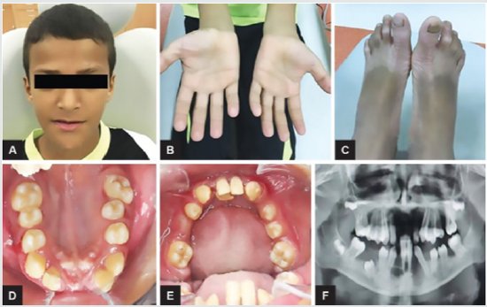
Figure 1:
Lupinepublishers-openaccess-pediatric-dentistry-journal

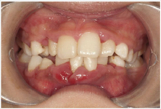
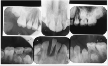
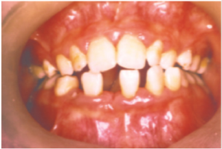
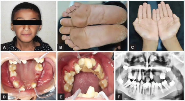
Periodontal disease is characterized by the imbalance between pathogens and host defenses leading to an inflammatory reaction around dental tissues. To diagnose aggressive periodontitis, it is necessary to investigate the health status of the subject and exclude the coexistence of systemic diseases. Some disorders can cause oral lesions clinically similar to those of aggressive periodontitis, but it is of fundamental importance to distinguish the various manifestations to make a correct diagnosis and intervene with the most appropriate treatment. Examples of significant conditions are AIDS, leukemia, diabetes or rare genetic disorders such as histiocytosis X and Papillon-Lefevre syndrome. The latter is a rare autosomal recessive disorder caused by mutations in the gene that codes for cathepsin C (dipeptidyl-peptidase I inhibitor). The syndrome is characterized by hyperkeratosis, destructive periodontitis that occurs from childhood, recurrent piogenic and systemic skin infections, susceptibility to bacterial infections and intra-cranial calcifications [21] (Figures 1&2). The prevalence is estimated to be between 1 / 250,000 and 1 / 1,000,000 subjects and is manifested in all ethnic groups. These dermatological features appear between the first year of life and 4 years and are accompanied by intraoral lesions that include gingival inflammation, mobility of the dental elements, even spontaneous bleeding and destruction of the periodontium. Patients with this syndrome show serious signs in the oral cavity until complete loss of deciduous bone, generating the normal appearance of the gum. However, with the eruption of permanent teeth, the form of aggressive periodontitis reappears. Any non-surgical but also surgical treatment is vain and almost always leads to partial or complete edentulism in the patient. The treatment is based on the intake of oral retinoids, which attenuate the palmoplantar keratoderma and slow down the lysis of the alveolar bone, and the skin lesions can also be treated with emollients in order to hydrate the affected area. Furthermore, good oral hygiene control, the use of mouthwashes and even antibiotics are recommended to slow the progression of periodontitis. Deciduous teeth or elements with excessive mobility must be extracted and eventually replaced by implants when the subject has completed growth.

Figure 2:
Lupinepublishers-openaccess-pediatric-dentistry-journal

The identification of the syndrome at an early age is something multidisciplinary that can improve the patients’ prognosis [22]. Leukemia is a malignant neoplastic disease of white blood cells and very often strikes in the pediatric age giving oral manifestations prior to systemic onset [23]. Acute lymphoblastic leukemia (ALL or ALL), specifically, develops when a cell destined to give rise to cells of the immune system turns into a tumor and starts to multiply in an uncontrolled way. Many studies show that acute lymphoblastic leukemia is the most frequent tumor in children, representing 75% of all newly diagnosed leukemias and 25% of all childhood malignancies [23]. The typical symptoms that characterize lymphoblastic leukemia are fatigue, dyspnea, fever, pallor and weight loss. Patients with this form of leukemia in the oral cavity have pale mucous membranes and an important gingival bleeding accompanied by lymphadenopathy in the head and neck region. It has been shown that sometimes the initial sign of the disease may correspond to a pericoronitis associated with a prolonged contraction of the masticatory muscles. Numerous studies have also reported a greater incidence of abnormalities in the oral cavity such as the presence of large, irregularly shaped ulcers, halitosis and a loose mucosa [24].
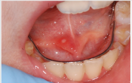
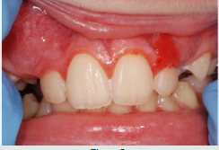
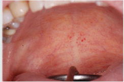
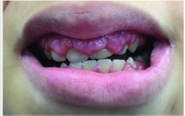
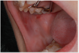
Acute myeloid leukemia (AML) is a disease that originates from the bone marrow. The disease is more common in adults over 60 years and infrequent before the age of 45. Patients with AML have symptoms related to complications related to anemia, neutropenia and thrombocytopenia, including weakness and easy fatigue, infections of varying severity, gingival bleeding, ecchymosis, epistaxis [25]. The oral examination can show pallor of the mucosa (Figure 3), ulcerations (Figure 4), spontaneous bleeding and bleeding (Figure 5) petechiae on gums, palate (Figure 6), tongue or lips, gingival hyperplasia (Figure 7) caused from leukemic infiltration. Usually the lesions of the oral cavity are the first manifestations of the disease, in particular, gingival swelling represents 5% of the early complications [26]. In leukemic patients, regardless of the form of the disease, oral manifestations occur as initial evidence or of its recurrence. Symptoms mainly include gum enlargement and bleeding, oral ulceration, petechiae, mucosal pallor and oral infections. These lesions may be the result of direct infiltration of leukemic cells or altered granulocyte function [26]. With regard to oral hygiene, patients must undergo periodic checkups and deplaquing or scaling sessions to reduce the level of inflammation of the mucous membranes, local antiseptics or antibiotics can be combined with active infections. Chemotherapy, often used in the treatment of leukemia, also has consequences on the subject that also affect the oral cavity. Often patients in therapy are predisposed to the appearance of ulcers, lesions, infections and are prone to have a partial xerostomia favoring plaque buildup.

Figure 3
Lupinepublishers-openaccess-pediatric-dentistry-journal

Figure 4:

Lupinepublishers-openaccess-pediatric-dentistry-journal

Figure 5:

Lupinepublishers-openaccess-pediatric-dentistry-journal

Figure 6:

Lupinepublishers-openaccess-pediatric-dentistry-journal

Figure 7:

Lupinepublishers-openaccess-pediatric-dentistry-journal

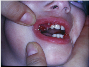
Scurvy is another disease that manifests itself with abundant gingival bleeding and which can be confused with aggressive periodontitis. The primary cause of scurvy is the insufficient intake of vitamin C mainly due to dietary imbalances. Childhood scurvy generally appears between the sixth and twelfth year of life. The child is easily irritated without appetite and fatigued. Sufferers of this disease develop anemia, weakness, fatigue, edema in some parts of the body, muscle pain in the lower limbs and ulceration of the gums (Figure 8) [27]. It occurs later as follicular hyperkeratosis and haemorrhage of the lower limbs, as well as bleeding in other areas such as the gingiva and joints [28]. If the disease is not treated it is potentially lethal due to important bleeding that can occur in the intracranial area or, due to the poor ability of the subject to heal due to open wound infections. At the oral level, the disease manifests itself in widespread hypertrophic areas of the violet-colored mucosa, a tendency for bleeding and the formation of hematomas. In small subjects, the symptoms that appear first are the pain and swelling of the joints accompanied by gingival hypertrophy [29].

Figure 8:
Lupinepublishers-openaccess-pediatric-dentistry-journal

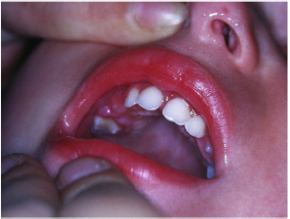
Patients suffering from this pathology are administered quantities of vitamin C orally or through injections and, in a short time , all symptoms disappear [28] (Figure 9). Diabetes mellitus is a disease that can present in the oral cavity as aggressive periodontitis and be confused with this. It includes a group of chronic metabolic disorders that turn out to be an altered glucose tolerance or an altered metabolism of lipids and carbohydrates [30]. It has been shown by numerous researches that in diabetic children with poor metabolic control, there is a greater tendency for gingivitis [30]. In fact, the high levels of glucose in the blood cause changes in microcirculation, promote bacterial proliferation and interact with the response of the host. Hyperglycemia caused by diabetes mellitus alters the immune system and the increased availability of glucose in the oral cavity environment increases the proliferation of periodontopathic bacteria and causes marked oral inflammation (Figure 10). In patients with diabetes, a microangiopathy occurs and this change in the periodontium reduces the functions of the polymorphonuclear cells, the chemotaxis, the adherence, the phagocytosis, the use of oxygen and the elimination of antigens, thus favoring the progression of periodontal disease. Hyperglycemia also reduces the solubility of collagen, reduces the production of fibroblasts and causes an increase in the levels of pro-inflammatory mediators responsible for the destruction of connective tissues. Changes to collagen metabolism result in accelerated degradation of both non-mineralized connective tissue and mineralized bone.

Figure 9:
Lupinepublishers-openaccess-pediatric-dentistry-journal

Figure 10:

Lupinepublishers-openaccess-pediatric-dentistry-journal

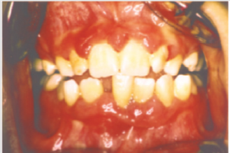
Even the saliva undergoes both qualitative and quantitative changes. Often in subjects with diabetes, the salivary flow is reduced leading to a further development of the bacterial species [31]. In diabetic patients, periodontal disease develops at a younger age than the healthy population and periodontal impairment usually occurs in adolescence but sometimes earlier in children with diabetes [32]. In the oral cavity, there is therefore an edematous and very inflamed gingiva, bleeding when the probe passes and bone resorption can occur especially in cases of poor metabolic control (Figure 11) [33], but in patients with a good diet with good glycemic supervision and good oral hygiene, do not show evident alteration in the mucosa (Figures 12&13). There is then a relationship between higher levels of plaque and a higher incidence of gingivitis in children with diabetes, moreover, the differences in oral microflora and the impact of metabolic control of diabetes on periodontal health have indicated a higher risk of periodontitis in children with type 1 diabetes [34]. In conclusion, when you are confronted with a child who has an oral situation of persistent inflammation, you need to perform more specific tests to understand if it is periodontitis as a manifestation of systemic pathology or aggressive periodontitis itself. The dentist or hygienist is usually the first to diagnose some diseases due to the involvement of the periodontium, and it is important to have multidisciplinary management to try to minimize the physical, psychological and social effects of the patient at an early age.

Figure 11:
Lupinepublishers-openaccess-pediatric-dentistry-journal

Figure 12:

Lupinepublishers-openaccess-pediatric-dentistry-journal

Figure 13:

Lupinepublishers-openaccess-pediatric-dentistry-journal

Conclusion

In conclusion, the review of the literature shows that periodontal disease does not only affect adults but, even if less frequently, it also involves children and adolescents. The forms of aggressive disease show a family aggregation, cause an important and rapid destruction even in the absence of local irritative factors and occur in systemically healthy subjects. It has been seen that particularly virulent bacteria trigger the process of bone destruction in the disease, the main micro-organism involved is Actinomycetemcomitans, which produces powerful leucotoxins that can also severely damage the subject’s immune system cells. Some clonal types of the bacterium are very pathogenic, and JP2 is always isolated from subjects suffering from aggressive periodontitis indicating that it is an important etiological agent. Regarding the epidemiological aspects, the statistics show very variable data also depending on the country, and so far the prevalence of aggressive periodontitis is not known exactly but it can be said that it occurs mainly in subjects of African descent and in individuals who are predisposed from the genetic point of view.

To date there is no specific criterion for defining a high-risk group for prepubertal pathology, and further research is needed to identify a robust set of genetic, microbiological risk markers and host factors that favor a diagnosis of the disease in association with young people and adolescents. The identification of aggressive periodontitis can be implemented through periodontal screening associated with radiographs, and the routine use of BPE can be helpful for early diagnosis. The treatment consists of the same methods applied also to adult patients, that is to say a causal therapy, a mechanic and a pharmacological one, in particular the studies have shown that antibiotics associated with professional hygiene in patients with aggressive periodontitis give very satisfactory results. If, despite careful treatment and hygiene, a child continues to have periodontal problems, it is necessary to investigate the general health with more specific examinations. A form of periodontal injury in a young person can also be a symptom of systemic diseases that are extraneous to the parent’s awareness and early diagnosis can become vitally important. Aggressive periodontitis, although infrequent, is not to be underestimated and it is important to take children to regular checkups and scaling sessions. Preventing and diagnosing problems early is the key to success.

Read More Lupine Publishers Pediatric Dentistry Journal Articles:
https://lupine-publishers-pediatric-dentistry.blogspot.com/


Friday, April 30, 2021

Lupine Publishers | Maryland Bridge : A Treatment Option in Children

 Lupine Publishers | Journal of Pediatric Dentistry


Abstract

The most common traumatic injury encountered in a pediatric dental set up is avulsion. Loss of permanent anterior teeth at a young age can have an impact on the psychological development of the child. Reimplantation if not possible, the anterior tooth can be restored with either removable or fixed prosthesis depending on the completion of the growth. To overcome the limitations of removable prosthesis, temporary fixed prosthesis can be an option in restoring the esthetics and functions when growth is not completed. Maryland bridge is an ideal option as a fixed temporary prosthesis in such patients.

Keywords: Avulsion; maryland bridge; re-implantation

Introduction

Over the last few decades dentistry has taken its turn towards more conservative and preventive treatment modalities. This has been possible not only because of the improved and advanced materials and techniques but also because of the understanding to preserve the natural tooth as far as possible. Reasons for tooth loss could be congenital absence, caries or trauma. Of these, traumatic injuries resulting in avulsion of the tooth is one of the common causes for tooth loss in the anterior region of the oral cavity. Trauma to the anterior teeth is common in childhood, one study reported that out of 2,100 children (aged 8-14 years) surveyed for teeth fractured due to trauma, 60.74% were aged between 11 and 14 with 13.8% cases involved incisors [1]. This case report presents a case of replacement of missing anterior teeth because of avulsion with direct fiber reinforced resin composite.

Case Report

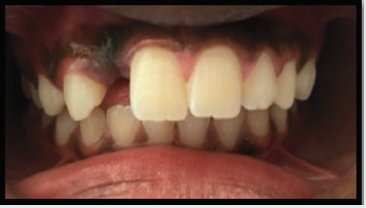
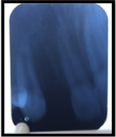
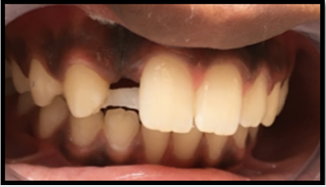
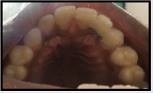
A 13-year-old boy reported to the Department of Pedodontics and Preventive dentistry with a chief complaint of avulsed tooth in the maxillary front region of the jaw for three weeks. Patient had a fall while riding bicycle. The patient was healthy without any significant medical and dental history. On extra-oral examination no abnormalities were found. On intra-oral examination soft tissues were normal avulsed socket was all healed. Patient had no bruises or scars on his body after the fall. Maxillary right lateral incisor had avulsed was seen with respect to maxillary right lateral incisor (Figures 1&2). Patient had reported to our department after three weeks with his natural avulsed tooth . The avulsed tooth was not stored in any media by the patient and was not in a good condition to be used as pontic as it had become dehydrated and had lost its color from the normal one. As the patient growth was not completed implant was not an option. Patient was very concerned about his esthetics and wanted immediate replacement. An option of conservative and conventional fiber reinforced composite resin bonded pontic was selected for the prosthetic rehabilitation. A composite buildup of maxillary right lateral incisor was done on a diagnostic cast. On the diagnostic cast the length of the FRC fiber (everstick, GC company) was pre-measured from the half the maxillary right canine to the half of the maxillary right central incisor. The fiber was pre-measured in such a way that it followed the contour of the ridge.

Figure 1: Intraoral view showing missing maxillary right lateral incisor.

Lupinepublishers-openaccess-pediatric-dentistry-journal

Figure 2: Radiographic view.

Lupinepublishers-openaccess-pediatric-dentistry-journal

Figure 3 Ever stick fiber placed on palatal Grooves.

Lupinepublishers-openaccess-pediatric-dentistry-journal

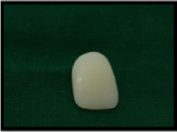
Figure 4: Composite build-up pontic.

Lupinepublishers-openaccess-pediatric-dentistry-journal

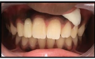
The palatal surface of the maxillary right canine and the right central incisor was roughened for better adaptability of the fiber for good retention. The surfaces were roughened using coarse flame shaped bur. Then the roughened surfaces of the abutment teeth was etched and bonded and then the fiber was placed starting from the canine to the central incisor and it was well adapted on the surface with a plastic carrying instrument. Then the fiber was bonded using composite in such a way that the fiber was at the center of the bonded tooth and was not in occlusion (Figure 3). Then a groove was made palatally on the composite build up pontic so that the fiber inserts well in the pontic for better retention (Figure 4). The pontic was bonded with composite keeping the arch form and function in harmony (Figures 5&6). The roughened surfaces were smoothened and polished with finishing and polishing kit (Enhance Composite Finishing and Polishing System Dentsply Caulk Dentsply International Inc, Milford, DE). The patient was followed for 6 months and had no complaints with the prosthesis.

Figure 5: Fiber reinforced bridge replacing missing tooth.

Lupinepublishers-openaccess-pediatric-dentistry-journal

Figure 6: Occlusal view with replaced missing tooth.

Lupinepublishers-openaccess-pediatric-dentistry-journal

Discussion

The patients with lost anterior teeth require immediate attention for the restoration of the aesthetics and function. The various treatment options available for restoring missing anterior tooth after avulsion are reimplantation, removable partial denture or fixed prosthesis depending on the case. In the present case the patient reported with the avulsed tooth after 3 weeks so reimplantation of the tooth was not a treatment option. Removable partial dentures are the most easily fabricated and cheapest options available, but they are often unacceptable to the patient because they are bulky, uncomfortable and not very esthetically pleasing [2]. Replacement of missing teeth with conventional fixed partial denture can involve tooth preparation of abutment teeth which can cause hypersensitivity, pulpal injury [3]. Fiber reinforced bridge was opted in this case, in order to provide a single visit, cost effective and a minimally invasive fixed solution to the patient. Clinical studies have shown a substantial clinical performance of the FRC FPDs, with an overall survival rate of 75% after about 5 years, which are higher than that of the FPDs with metal frameworks [4]. Hence high pulp horns of abutment teeth, expected transition in the position of the gingiva and age of the patient were factors that precluded the use of conventional fixed prostheses in this case. The resin material used in this study was GC Ever stick as a bridge for replacement of missing tooth. Accurate bonding between the fibers and composite is the key factor for a successful treatment. The resin fiber used has a unique, interpenetrating polymer network structure (IPN) which leads to superior bonding enabling reliable surface retained applications and perfect handling properties. The use of FRC bridges also allows utilization of a patient’s natural crown as a pontic if the tooth or tooth crown is still intact [5]. In our case the lost tooth was fractured and was not in good condition to be used as a pontic, so the composite build up tooth was used (Figure 4). A study was done involving 358 patients which concluded that the patient’s acceptance was better with resin bonded fixed partial dentures [6]. Maryland bridge is a minimal invasive procedure with less harm to the abutment tooth, minimum chair side time and less cost. It is well accepted by young patients and can be used as a treatment option for avulsion cases.

Read More Lupine Publishers Pediatric Dentistry Journal Articles:
https://lupine-publishers-pediatric-dentistry.blogspot.com/

Friday, April 23, 2021

Lupine Publishers | Assessment of Dental Neglect and It’s Relation to Oral Health Among School Children Aged 4-12 Years in Sri Ganganagar City, Rajasthan, India

 Lupine Publishers | Journal of Pediatric Dentistry


Abstract

Background: Dental neglect indicates that parents or guardians fail to give the necessary oral health requirements such that the child can take pleasure in function and free from disease, where reasonable means are accessible to parents or caretaker.

Aim: The aim of the study was to evaluate the dental negligence of parents towards their children aged 4-12 years in Sri Ganganagar city, Rajasthan and to correlate the oral health status of the children.

Materials and Methods: A cross‑sectional study was done involving 1000 school going children and their parents. Questionnaire were distributed among the parents of the children which includes the details of parents and their children . WHO form was used to assess the oral health status of the children i.e. the dental caries and bleeding on probing as per the World Health Organization criteria,2013 index. Statistical analysis was done by using SPSS software version 20.0.

Results: A significant higher dental negligence score was found among those parents who resided in the suburban area and whose educational qualification was higher secondary only and those who had visited the dentist only after the development of symptoms in their children.

Conclusion: Among the parents whose educational qualification was higher secondary only, those who resided in the suburban area and those who went to see dentist only after the development of symptoms in their children have shown poor health status.

Keywords: Dental neglect; dental caries; oral hygiene; parents

Introduction

Dental neglect indicates that how known knowledge of oral health care are not fulfilled which fails to do the proper maintenance of oral cavity. For proper oral health care people need to be aware of the dental problem consequences. In spite of giving so much dental professionals and audio-visual dental care measures, only a small number of people take proper dental home care and they do not go for periodic dental checkup. Oral health is considered as the central importance for our general health and well-being. A healthy oral cavity allows an individual to communicate, have food and participate in social activities without having any disease, discomfort or difficulty. Having oral disease can be expensive in terms of money and also life style can be changed [1]. The World Health Organization has stated that neglect should be differentiated from conditions like poverty, where it shows dental neglect is present only where there are available resources. It was found that there is a great deal of severity of dental neglect worldwide [2].

Materials and Methods

Ethical approval for the study were taken from the concerned authority of the institution. This ensured that participants involved in the study were randomly selected from the schools of Sri Ganganagar city. This study was conducted from January 2019 to August 2019. The age of the children were 4 to 12 years, and the questionnaire were given to the parents of the children to be answered. An informed consent was obtained from the parents earlier. The parents were asked to answer the questionnaire which had details of both parents and children. Consent form from parents were taken for the survey to be done. Oral examination of children was conducted by using disposable mouth mirror, straight probe and CPTIN probe in a natural day light. Oral status was recorded according to the WHO form, 2013. The collected data were tabulated and subjected to statistical analysis using SPSS software version 20.0, (SPSS Inc., Chicago Ill., USA) and levels of statistical significance were set at P < 0.05.

Results

Graphs 1&2: Shows the address and parental education level.

Lupinepublishers-openaccess-pediatric-dentistry-journal

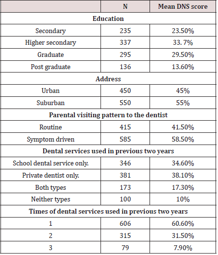
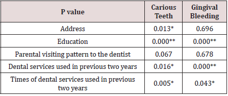
A total of 1000 pair of parents and children participated in the study, of which female and male parents were of 631 and 369, respectively. Table 1 illustrate the comparison of key background variables and dental service used in previous two years and Table 2 shows the p value for the oral status with respect to key background variables (Graphs 1&2). It is showed that with respect to dental neglect scores, a significant higher dental neglect score was reported among the people who resided in the suburban location, parents whose educational qualification was higher secondary only, parents whose visit to the dentist were only after the development of symptom, parents those who used only school dental services and those who used dental service only once in previous two years.

Table 1: Comparison of key background variables with respect to given demographic status.

Lupinepublishers-openaccess-pediatric-dentistry-journal

Table 2: .

Lupinepublishers-openaccess-pediatric-dentistry-journal

*P value is statistically significant, **P value is statistically highly significant.

Discussion

Oral health occupies a very significant role for the wellbeing of individuals, and parents’ behavior and way of thinking influence the oral health of their children [3]. Prevention is always better option than cure. People should be very thoughtful and particular to maintain oral health for the prevention of oral disease. Dental professionals and audio-visual media provide the essential dental care measures [4]. But the fact is that only few people take adequate regular home dental care and do not go for periodic/yearly dental check to the dentist to keep their oral cavity healthy [5]. It has been seen that dental neglect is mostly related to the illiteracy amongst low socio-economic class and the prevalence of oral diseases are highest amongst them [6]. Child neglect is a very important subject in terms of prevalence and severity–it is the most common cause for a child to be made subject to a child protection plan in the UK - and there is indubitable evidence and facts that it is harmful and damaging to children [7-10]. There is very scarce literature regarding the estimate of child dental neglect worldwide [11]. Hence, in this study, in addition to recording the caries status, the gingival status i.e. bleeding on probing was also evaluated which interprets the degree of failure to seek dental treatment. Out of 1000 children maximum number of the participants were 7 years with 596 male and 404 females. Majority of the children were in 1st standard and from private school. The caretakers who responded to the questionnaire were mostly mothers of the children. Hence, it helped us to know the complete home and professional dental care the child receives as the child is mostly with the mother. The results of the present study showed that dental neglect was found more in parents who have done only higher secondary education which showed lesser levels of positive dental attitudes which is in contrary to the result of Gurunathan D et al. [12] where it was stated that parents who have done secondary education showed lesser levels of positive dental attitudes which is similar to studies done by Freeman et al. and Williams et al. [13,14]. In the present study, a significant difference was observed in the dental neglect scores among parents residing in the suburban areas in comparison with parents of urban areas. This is essentially due to not much awareness of oral health, availability, and usage of dental services [15]. The dental neglect among children is higher among parents’ whose last dental visit was once or twice in previous 2 years, those who used only school dental services and were symptom driven which is similar to the findings in South Australia and Chennai [16]. The dental neglect is reflected in the poor oral health of these children with significantly higher caries prevalence and untreated carious lesions. This suggests that the knowledge of parents regarding oral health and utility of dental services is limited as the frequency of dental visits suggests the oral health awareness among parents [17].

Conclusion

Inadequate dental care by parents can be observed as dental neglect in children and therefore is a sign of child neglect. Health care workers involved in the care of children are thus in a unique and a very important position to understand early symptoms of child abuse and neglect. We believe and accept that the occurrence of dental caries is an important sign to this diagnosis. From this study it is concluded that, the dental neglect among school going children aged 4-12 years regarding oral hygiene is still far from satisfactory in certain respects in our population, but it is also seen that oral hygiene status can be improved after sessions of oral health education to the children and most importantly the parents.

Read More Lupine Publishers Pediatric Dentistry Journal Articles:
https://lupine-publishers-pediatric-dentistry.blogspot.com/
Saturday, April 17, 2021

Lupine Publishers | Newer Insights in Early Childhood Dental Caries

 Lupine Publishers | Journal of Pediatric Dentistry


Abstract

Early dental caries is a common devastating disease of early childhood but can be prevented easily by simple methods. It is more common in developing countries and poor socioeconomic status but can be found in all settings. Different bacteria interacting with host salivary molecules, play a role in the development and aggravation of these lesions. Newer techniques for diagnosis, bacterial isolation, as well as treatment are coming up. Clinicians and researchers should be abreast with new developments to ensure proper diagnosis and treatment of these cases.

Keywords: Caries; sugar; cariogenic; streptococcus mutans

Introduction

 Dental caries is a clinical challenge, especially in young children [1]. In fact, it is the commonest chronic infectious disease of childhood, and caused due to interaction or oral bacteria like Streptococcus mutans with sugary foods and saliva [1].  Of all dental caries, Early Childhood caries or ECC can be defined as “the presence of one or more decayed (non-cavitated or cavitated lesions), missing teeth (due to caries), or filled tooth surfaces in any primary tooth in a child 72 of months age or younger [1]. ECC is also called baby bottle-fed tooth decay, early childhood dental decay, comforter caries, nursing caries, maxillary anterior caries and rampant caries [1]. In children younger than 3 years of age, in addition, any sign of smooth-surface caries indicates severe early childhood caries (S-ECC) [1]. Early dental caries is frequently encountered in children of less than 71 months of age. Poor oral hygiene or insufficient dental plaque removal can lead to the rapid progression of ECC [2]. As regards bacterial etiology of dental caries, some strains, such as Streptococcus sanguinis, are reportedly associated with healthy teeth, while others, such as S. mutans, other oral Streptococcus spp., Veillonella spp., Actinomyces spp., Bifidobacterium spp., and Lactobacillus fermentum, were associated with caries [3]. There is also enough evidence that Gram positive bacteria like Actinomyces gerencseriae and other Actinomyces spp. play an important role in caries initiation [3]. In a study by Munson et al, in addition to S. Mutans and Lactobacillus spp., Rothia denticariosa and Propionibacterium acnes have also been found in caries lesions [4].

Epidemiology

ECC affects infants and preschool children worldwide, and its prevalence, though variable, can be up to 85% in disadvantaged groups [5]. Prevalence of ECC also varies widely, depending several variables like race, culture, and ethnicity, socioeconomic status, lifestyle, diet and oral hygiene practices and also according to the regional factors from country to country and from area to area [6]. A review of the literature suggests that in most developed countries the prevalence rate of ECC is between 1 and 12% [6]. In less developed countries and among the disadvantaged or poorer groups in developed countries, the prevalence of ECC has been shown to be as high as 70% [6]. However, it is not restricted to children with low socioeconomic status [2]. Recent data, for example, from Australia show a prevalence of ECC of more than 50% in 6-year-old children with caries on deciduous teeth [7]. Milsom et al. found that children with an already existing caries lesion have a 5–6 times higher incidence of developing new caries lesions compared to previously caries-free children [8]. Sleeping problems and insufficient sleep has also been identified as risk factor for ECC, because sleeping problems lead to more frequent use of night-time bottle use containing sugar-sweetened beverages [9]. There is a relationship with gender, since according to studies, the highest prevalence of ECC is found in the 3- 4-year-old age group; also, boys are significantly more affected than girls, aged between 8 months and 7 years [10].

Risk factors

Many risk factors have been identified like

a) Most of the studies have shown significant correlation between ECC and bottle-feeding and sleeping of the baby with a bottle [1].
b) There is substantial evidence that prolonged and nocturnal breastfeeding is associated with an increased risk of ECC, particularly after the age of 12 months [1].
These aggravate caries due to less saliva production at night and less bacterial clearing.
c) Fermentable carbohydrates are a major factor in the development of dental caries. The small size of these sugar molecules allows salivary amylase enzyme to split the molecules into components that can then be easily metabolized by the bacteria in the plaque [11]. This process leads to bacteria producing acidic end products with subsequent demineralization of teeth.
d) Enamel hypoplasia due to premature birth, low birth weight or malnutrition is also a very important risk factor for caries development [12].

Clinical features

In the initial phases, ECC appears as a dull, white demineralized enamel that quickly progresses to obvious decay along the gingival margin [13]. Primary maxillary incisors are usually afflicted earlier than the four maxillary anterior teeth which are often involved simultaneously [1]. Carious lesions can be found on either the labial or lingual surfaces of the teeth and, sometimes on both [1]. The decayed hard tissue is clinically apparent as a yellow or brown cavitated area [1].

Diagnosis

Diagnosis is clinical. Culture of the bacteria can be carried out in Mitis-Salivations agar, from where colonies can further be identified [14]. However, S. mutans may be slightly inhibited in this medium and S. mitis may need longer incubation [14].

Etiology and Pathogenesis

It has been depicted by Corby et al that some bacteria are associated with healthy or caries –free teeth, like Streptococcus parasanguinis, Abiotrophia defectiva, Streptococcus mitis, Streptococcus oralis, and S. sanguinis [15]. The same group also showed that Actinomyces species, S. mutans, and Lactobacillus spp. were consistently associated with disease [15]. S. mutans and S. sobrinus have been recognized over the years as the main culprits behind development of Early childhood Dental caries [16]. They damage the dental enamel in presence of fermentable carbohydrates like Glucose, sucrose and fructose. In fact, S. mutant is present in about 30% of the plaques in carious teeth compared to 0.1% in healthy teeth [17]. Nowadays, it is well studied that not only bacteria, but also fungi, such as Candida albicans and the interactions between several different microbes, can enhance the progression of caries [18] . Bacteria and other microbes degrade sugars and lead to acid production which causes demineralisation of teeth and caries development [1]. Enamel of deciduous teeth is more vulnerable to acid-mediated damage than permanent teeth because it is thinner and built quickly in about 24 months by ameloblasts than that of permanent teeth which takes about 16 years to be built [19].

Implications

If left untreated, ECC, also called rampant caries, can lead to rapid and complete destruction of the crown [5]. Hence there is need of rapid and accurate diagnosis of the condition and adoption of suitable preventive measures.

Newer developments

New research has unearthed new mechanisms of pathogenesis in ECC. Bacterial biofilms are rapidly produced and are made of exopolymer matrix or EPS. This EPS is formed more on exposure to sucrose and fructose, and lead to further adhesion and colonization by cariogenic bacteria on surface of damaged teeth. This increased formation of biofilm biomass or “visible plaque”, often found on the smooth surfaces of the children at risk of ECC shows the importance of EPS in the pathological process [20]. Research has also demonstrated a few biomarkers or host salivary molecules that affect ECC development. For example, host CSP-1 helps in adhesion of cariogenic bacteria to the enamel [20]. There are 2 types of Proline-rich glycoproteins in human saliva: acidic PRP and basic PRP. Basic PRP helps in ammonia production and neutralise sugar acids, whereas acidic PRPs bind strongly to teeth and enhance adhesion of the cariogenic bacteria [20]. These molecules can be biomarkers to help in assessing prognosis of ECC.

Newer bacterial and other agents

Atopobium vaginale has been found significantly in ECC in those cases that also grow Streptococcus mutans [3]. Bifidobacterium species have been associated with deep caries lesions [21]. In case of severe ECC, Porphyromonas catoniae has been found very commonly in the plaques [22]. Novel techniques like The Human Oral Microbe Identification Microarray (HOMIM) can be used for this type of assessment [22]

Newer options for treatment

Usually Children at low risk generally do not need any restorative therapy [1]. Children at moderate risk may require restoration of the progressing and cavitated lesions, while white spot and enamel proximal lesions should be treated by preventive techniques and then monitored for progression [1]. Children at high risk, however, may need earlier restorative interventions of enamel proximal lesions, and intervention of the progressing and cavitary lesions to minimize continual caries development [1]. Sometimes stainless-steel crown following pulpotomy and pulpectomy may be needed in cases of severe ECC [1].

Prevention

Prevention of ECC should focus on educating the parents about no nocturnal feeding and dietary modification [5]. Parents also should be advocated to maintain optimal dental health during pre- and postnatal periods [5]. Child health professionals, like physicians, assistants of physician, nursing practitioners, and nurses can play a very significant role in reducing the burden of this disease through monitoring, prompt diagnosis and health promotion activities [1]. Prevention of the progress of ECC can be carried out with the help of restorations, diet counselling, educating parents regarding decay-promoting feeding habits, maintenance of good oral hygiene, and the use of preventive agents like topical fluorides [5].

Discussion

ECC is a very common chronic disease of childhood and easily preventable by simple measures [5]. The associated pain from dental caries has got a negative impact on the child’s emotional status, sleep patterns, and ability to learn or perform usual daily activities. A wide range of risk factors are linked with ECC in children from underprivileged and low socioeconomic status [5]. Oral health has been recognized as an essential prerequisite for general health and quality of life. Therefore, both oral disease prevention and oral health promotion should be included as integral components of chronic disease prevention and general health promotion programmes [5].

Conclusion

Early childhood caries should be diagnosed and treated early. New research is showing new avenues of diagnosis and aetiopathogenesis study.

Read More Lupine Publishers Pediatric Dentistry Journal Articles:
https://lupine-publishers-pediatric-dentistry.blogspot.com/
Friday, April 9, 2021

Lupine Publishers | Cardiovascular Diseases in Children –Oral Findings and Dental Treatment Approaches

 Lupine Publishers | Journal of Pediatric Dentistry


Abstract

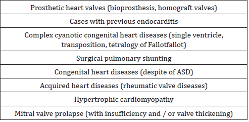
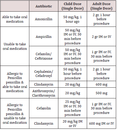
Oral and dental health is of vital importance in children with cardiovascular system disease. Congenital and acquired heart diseases make oral treatment complicate in children and increase the risk of infective endocarditis [1]. The dentists main aim in children with heart disease should be to prevent infective endocarditis, to maintain periodontal health and to ensure a well oral hygiene. In these children, medical history should be taken in detail before dental treatment. Consult with the pediatrician and inform about the condition of the disease and the drugs used recently. Consideration should be given, and antibiotic prophylaxis should be evaluated consultation to in the treatment planning of children with heart disease. In these children, dental treatments should be performed quickly and effectively, and family awareness should be given importance [1-3]. In this review, common oral findings related to cardiovascular system diseases will be discussed and the precautions and treatment approaches of dentist will be mentioned [4].

Keywords: Endocarditis, heart diseases, intraoral findings, pediatric dentistry

Review

Almost all of the heart diseases seen in children are congenital. The most common structural congenital heart diseases are; ventricular septal defect, patent ductus arteriosus, atrial septal defect, fallot tetralogy, pulmonary stenosis, aortic coarctation, aortic stenosis and transposition of the great arteries. Large defects are surgically closed in the first years of life, and some defects may require complex surgical treatment and eventually transplantation. Acquired heart diseases such as myocarditis and infected endocarditis are the cause of disability and death in children [5]. Oral and dental health may be insufficient in children with congenital heart disease. The reasons for this; chronic vomiting in children up to one-year, dry mouth due to drugs used, neglect of oral care due to cardiac problems as the primary priorities of families [1]. Poor oral hygiene in children with heart disease may pose a risk for bacteremia. Infective endocarditis can result with severe valvular dysfunction, dehiscence, congestive heart disease and various embolisms leading to death. In the long term, it may cause cardiac valve damage and prosthetic regeneration of the heart valve [1,6-8].

Oral findings

Oral findings in children with cardiovascular disease include cyanotic gingivitis, stomatitis, glossitis, mucositis, cleft lip and palate, clefts in the tongue, the fungiform and filiform papilla of the tongue becoming dark red [5,9]. In addition, enamel hypoplasia, high incidence of caries, delays in teeth exfoliation, periodontal diseases and intrinsic discolorations are observed [5,9,10]. In particular, discolorations in permanent teeth occur due to the ingestion of blood and blood products by drugs, increased caries activity and poor oral hygiene [5]. Poor oral hygiene, use of drugs, inadequate food and mineral consumption are effective risk factors for the onset of periodontal disease in children with congenital cardiovascular disease [11,12]. Pourmoghaddas et al. [12] reported that children with congenital heart disease had significantly higher periodontitis than healthy children and that periodontal disease caused endocarditis risk. Al Alousi et al. [13] reported that the rate of development of enamel defects was high in patients with ventricular septal defect and was observed due to malnutrition. The researchers stated that nutrient elements may affect the epithelial cell function and mineralization process and prepare the ground for defect formation in enamel hypoplasia [13]. In Fallot Tetralogy, wrinkled tongue is observed as an oral finding [1].

Infective endocarditis

Infective endocarditis is a rare disease that results in high morbidity and mortality, with an annual incidence of 0.05- 0.12/1000 in children [3,14,15]. Turbulent blood flow observed in heart diseases leads to endothelial injury, resulting in storage of platelets and fibrin on the endothelial surface [15]. As a result, non-bacterial thrombotic endocarditis occurs and bacteremia results in the adhesion of bacteria to this injured endocardium and proliferation in vegetation leading to infective endocarditis. Therefore, some heart diseases pave the way for the development of infective endocarditis [14,15]. Invasive dental procedures, gastrointestinal and genitourinary systems related operations may cause bacteremia and infective endocarditis [15].

Bacteremia in dental procedures

In children, oral flora changes and streptococci (alpha hemolytic streptococci), actinomyces and privately species increase after infancy and begins to resemble adult flora. Healthy children have streptococci, staphylococci, neissera and haemophilus species, while older children have bacteria such as capnocytophaga and aggregatibacter actinomycetemcomitans, which cause periodontal disease [15]. The host’s response to dental plaque due to poor oral hygiene is gingivitis. When this inflammation develops, bacterial colonies may become involved in the gum capillary circulation due to thinning and ulceration. Therefore, there is a risk of bacteremia even in minimal procedures such as daily tooth brushing The frequency of bacteremia after toothbrushing and flossing has been reported to be 20-68%, 20-40% after tooth cleaning with toothpicks, 7-50% after mouth rinsing, and 7-51% after chewing food [3]. In addition, 10-100% after tooth extraction, 36-88% after periodontal surgery, 8-80% after removal of dental plaque, 9-32% after plastic matrix placement, 20% after endodontic procedures have been reported to develop bacteremia [3]. Good oral hygiene and prevention from diseases (such as gingivitis, tooth decay) are important to minimize the risk of bacteremia [15]. The use of amoxicillin in the selection of antibiotics in dental procedures has been shown to have a statistically significant effect on reducing the incidence and duration of bacteremia.

While previous American Heart Association (AHA) guidelines recommend antibiotic prophylaxis for all dental interventions that may cause bleeding, this recommendation has been altered since there is no evidence that this is a predictive symptom of bacteremia when bleeding occurs during dental procedures [3]. In the latest guidelines published by AHA, it was decided to administer prophylactic antibiotics only to the highest-risk group before invasive dental procedures [3] (Table 1). Prophylactic antibiotic administration is recommended only if high-risk patients are scheduled for an operation to the gingival tissue or the periapical region of the tooth, or if there is a risk of mucosal perforation. Routine injections of anesthetic agents, x-rays, removable dental prostheses, orthoses, orthodontic material placement, prosthesis matching, primary tooth extraction, antibiotic prophylaxis are not recommended in uninfected tissue [3]. Antibiotic prophylaxis should be given 30-60 minutes before the procedure. However, it can be given within 2 hours after the procedure if it is not given before the procedure [3]. In patients using oral anticoagulants, intramuscular injection should be avoided, and oral antibiotics should be preferred. Clindamycin, clarithromycin, azithromycin should be given to patients who routinely use antibiotics as they may be resistant to penicillin and ampicillin [3]. These patients should receive dental treatment 10 days after antibiotic treatment to normalize oral flora. In patients receiving intravenous antibiotic therapy, the antibiotic dose should be adjusted 30-60 minutes before dental intervention [3,16] (Tables 1&2).

Table 1: High-risk patients in whom infective endocarditis prophylaxis is recommended [16].

Lupinepublishers-openaccess-pediatric-dentistry-journal

Table 2: Antibiotic prophylaxis recommended before dental procedures [16].

Lupinepublishers-openaccess-pediatric-dentistry-journal

Dentist approach

Patients with cardiovascular disease should be consulted with their doctors. Deciding on premedication, preoperative antibiotic requirements, anesthetic selection and surgical procedures should be decided in accordance with the recommendations of the physician of patient. In children diagnosed with cardiovascular disease, a preventive program including nutritional advice, fluoride therapy, fissure sealants and oral hygiene should be applied [1]. Arrhythmia due to endogenous catecholamines triggered by stress, fear and pain may develop during dental procedures, and patients who are prone to arrhythmia should be closely monitored [3]. Antibiotic prophylaxis should be performed in high-risk patients if a procedure is planned for the gingival tissue or the periapical region of the tooth, or if there is a risk of mucosal perforation [3]. Dental procedures in high and medium risk groups should not exceed the treatment of deep dentine caries. Direct pulp capping, pulpotomy and root canal treatments are not indicated because they increase the risk of bacterial endocarditis. In very deep decayed teeth, the treatment option should be extraction [1].

Conclusion

In children with cardiovascular disease, the risk of infectious endocarditis and bacteremia due to the risk of fatal outcome of the heart valve is very important in terms of oral and dental health in these patients. It is very important for dentists to consult with their physician before dental procedures in children with heart disease, and to take prophylaxis needs and stress tolerances of their patients by writing. These children and their families should be adequately informed about oral hygiene. In this way, the risk of bacteremia, even in oral cleaning procedures such as chewing and brushing, can prevent infective endocarditis [1].

Read More Lupine Publishers Pediatric Dentistry Journal Articles: https://lupine-publishers-pediatric-dentistry.blogspot.com/
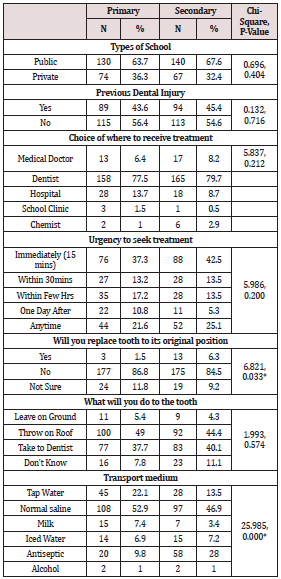

980 nm Diode Laser: A Good Choice for the Treatment of Pyogenic Granuloma

Abstract Pyogenic granuloma is a benign non/neo plastic mococutanous lesion . It is a reactional response to constant minor trauma and ca...