Friday, January 14, 2022

Lupine Publishers | Pregnant Women’s Knowledge about Children Oral Health

 Lupine Publishers | Journal of Pediatric Dentistry


Abstract

Bottle tooth decay is a common problem in dental medicine. Studies point to the relationship between mother’s knowledge and motivation to maintain oral hygiene and child’s oral health. The reason behind it is that mothers are usually the ones spending most of the time with the child during first years of life. The purpose of this study is to examine the level of pregnant women’s knowledge of the importance of primary teeth, caries and its causes, best time to begin tooth brushing and best time for first dental visit. In infant oral health lectures within the pregnancy class, 49 pregnant women were examined. The average age was 31, most of them were educated and employed. Before and after the lecture pregnant women got the same questionnaire and the results of the second questionnaire provides information on the importance of the lecture given within the course of pregnancy. Research results show that pregnant women consider primary teeth very important to childʹs development and general health. Most of them were aware that cleaning childʹs mouth starts even before the first primary tooth appearance and that primary teeth should be restored if necessary. Significant number of respondents knew the exact time of first tooth appearance. Knowledge of the recommended time for first dental visit and the caries risk factors was shown poor. The conclusion is that most pregnant women are unaware that breastfeeding, bottle feeding, using bedtime bottle and frequent feeding cause caries. Lecture on childʹs oral hygiene proved to be extremely useful because for most pregnant women that was the first time, they got that information.

Keywords: Pregnant women; bottle tooth decay; oral health knowledge; oral hygiene; prenatal care

Abbreviations:ECC: Early Childhood Caries; ADA: American Dental Association; ART: Atraumatic Restorative Treatment; EAPD: European Academy of Paediatric Dentistry; AAPD: American Academy of Paediatric Dentistry

Introduction

Early childhood caries (ECC) is a special type of tooth decay that occurs in children in the first three years of life [1]. The American Dental Association (ADA) defines ECC as the presence of one or more teeth affected by tooth decay, extracted teeth due to tooth decay and/or teeth provided with tooth filling in preschool children up to 71 months of age [2]. The reason why ECC is a separate clinical entity is the existence of a specific etiology of occurrence associated with breastfeeding and bottle feeding. Parents are often unaware of this, so they put their child to sleep with sweetened drinks, exposing the childʹs oral cavity to low pH throughout the night [3]. At night, the flow of saliva is greatly slowed down, which allows long-term retention of cariogenic solutions on the tooth surface, primarily upper milk incisors and canines [4]. Another reason why this type of tooth decay occurs is too frequent ad especially prolonged breastfeeding [5]. With the eruption of deciduous teeth, complex processes of demineralization and remineralization begin. Primary teeth are less resistant to tooth decay than permanent teeth due to poorer mineralization of enamel and consequently its tendency to wear out, thinner enamel and dentine and more voluminous pulp [1]. Research shows that the prevalence of bottle caries depends on the development of the country in which it is measured and on the criteria that are evaluated [6]. In developed countries that have good oral health programs, the prevalence of ECC is about 5%. Southeast European countries show a prevalence of about 20%, the Middle East 59%, and research results in North America vary from 11% to 72% [7]. Another study shows that the prevalence of bottle caries among preschool children in Zagreb is 30% (25% in girls and 48% in boys). The study was conducted on a sample of 145 children aged between 2 and 5 years. The results of the research showed that night feeding and feeding with sweetened drinks after first 24 months of life is the main risk factor for the development of ECC [8].

The clinical picture of ECC may vary depending on the frequency of feeding and oral hygiene. Depending on the number and location of teeth affected by caries, there is a mild type that involves isolated carious lesions on the incisors and / or molars of children aged 2 to 5 years. This is followed by a moderate type that affects the labial and palatal surfaces of the upper incisors with or without molar involvement and the absence of caries on the lower incisors. The severe type, also known as rampant caries, affects almost all teeth, including the lower incisors [9]. With the progression of caries, the first symptom appears, and that is pain. Pain when chewing can be the cause of a childʹs malnutrition and lack of nutrients. A link between ECC and childʹs slower physical development has been proven [10]. If left untreated, caries can progress to pulp and periapical disease, often resulting in tooth extraction. Premature extraction of deciduous teeth can have numerous consequences, from difficult chewing, impaired phonation, impaired aesthetics to orthodontic anomalies in the future. In addition, caries and early loss of deciduous teeth have an impact on the mental state of the child. The child experiences pain in the dental office early, which becomes a traumatic experience that often turns into anxiety. The approach to treatment depends on the severity of the clinical picture and the child’s cooperation. A good option for young and uncooperative children is the ART (atraumatic restorative treatment) program, which involves removing softened tooth tissue with chisels and excavators and filling with high-viscosity glass ionomer cement that releases fluoride and thus stops the spread of caries [1]. It is extremely important to start maintaining the oral hygiene of the child before his first baby teeth erupt. It is recommended to wrap a piece of gauze around the finger and pass it through the oral cavity after feeding. In this way, we not only remove food debris from the mouth, but also massage the alveolar ridge, which facilitates tooth eruption [11]. With the appearance of the first tooth in the oral cavity, most often the lower central incisor, brushing should begin. Cleaning the first teeth can also be done with gauze, and there are special toothbrushes for which it is indicated for which age they are suitable. The parent should consult a dentist which fluoride toothpaste is most suitable for his child’s age. The usual recommendation is from the European Academy of Pediatric Dentistry (EAPD). The concentration of fluoride in pastes suitable for children aged 6 months to 2 years is 500 ppm, for children aged 2 to 6 years 500-1000 ppm, and for children older than 6 years the amount of 1000-1450 ppm corresponds [12]. In addition to proper oral hygiene, proper nutrition of the child is also important. It is desirable to limit the frequency of feeding the child to 5-6 times a day. There should be a period of at least 3 hours between meals in which only water is allowed to be consumed. In this way, the saliva buffer mechanism neutralizes acids and mechanically flushes food debris from the oral cavity further into the digestive system [12]. The most common mistakes parents make, resulting in bottle caries, are putting the child to sleep with sugary drinks, holding the child to the chest after falling asleep, feeding at night, and allowing the child to walk with the bottle drinking its contents little by little [2]. Parents’ ignorance of the importance of proper and regular maintenance of a child’s oral hygiene is the cause of their lack of motivation to maintain it, which results in caries. The recommended time for the first visit to the dentist according to the AAPD (American Academy of Pediatric Dentistry) is from 6 months to a year or from the eruption of the first tooth to a year. The caries risk is assessed by talking to the child’s parent about his eating habits, oral hygiene, and the habits of the parents themselves. The aim of this research is to find out the level of knowledge about the oral health of the child of future parents, especially mothers who most often take care of the child in the first months of life. Lowereducated mothers and unemployed mothers are expected to have a lower level of knowledge about maintaining a child’s oral health.

Materials and Methods

In this research, the participants of the course for pregnant women held at the Clinic for Gynecology and Obstetrics, more precisely, the participants of the lecture “Care for the health and care of the child’s teeth in pregnancy and infancy” were examined. The research was conducted in the form of a questionnaire (Appendix 1), which was distributed to all participants at the beginning of the lecture, and which was completed by pregnant women before the beginning of the lecture. The same questionnaire, but without data on socio-economic status, was distributed and completed after the lecture. The questionnaire was designed at the Department of Pediatric Dentistry Study of Dental Medicine and consists of two groups of questions. The first group of questions focuses on the knowledge of pregnant women about maintaining oral hygiene of the child, caries, the importance of milk dentition and the recommended age of the child to start brushing teeth and the first visit to the dentist. The questions are in the form of adding, rounding YES-NO to the claims about oral hygiene, and in the form of rounding the statements that are closest to their opinion about caries, from “not at all” to “completely yes”. The second group of questions refers to the marital status, level of education, employment and occupation of the pregnant woman and the father of the child. Round off YES or NO to the question on employment and state the occupation, type of work and institutions where the pregnant woman and the child’s father work. Finally, data on family size and number of children in the family are filled out.

Results

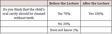
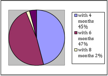
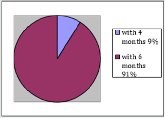
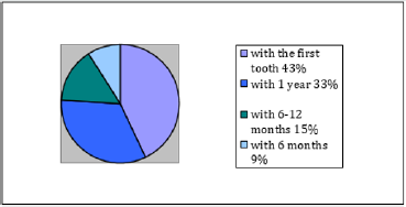
49 pregnant women with an average age of 31 participated in this study. The majority (61%) of pregnant women are married, have a university degree (67%) and are employed (87%). Fathers are mostly university educated (43%) or have completed vocational school (43%), and 98% are employed. The vast majority of respondents (92%) have this first child, and 71% claim that this is the first course for pregnant women attending and that they have never received information on maintaining oral hygiene (92%). The survey showed that most pregnant women know that oral hygiene must be maintained while the baby is still toothless. During the lecture, attention was once again drawn to the importance of cleaning the mouth of a child without teeth and the way in which this is done was described. All pregnant women answered in the affirmative after the lecture (Table 1). The next issue was the age of the baby in the months when the first baby teeth begin to erupt. The offered answers were 4, 6, 8, 10 and 12 months. Before the lecture, 47% of pregnant women answered for 6 months, and after the lecture, 91% answered for 6 months (Graphs 1 & 2). Pregnant women were aware even before the lecture that baby teeth were important for the baby. As many as 71% of respondents answered that deciduous teeth are extremely important, while 27% answered that they are mostly important. After the lecture, 96% of respondents claim that deciduous teeth are extremely important (Table 2). The next question was answered by overwriting, so there is a whole range of answers that needed to be sorted into correct or incorrect. The correct answer was considered to be all those who indicated that teeth should be brushed as soon as possible, when the first teeth erupt. Answers of the “2 months” and “3 months” types were also considered correct under the assumption that the respondents meant toothless oral hygiene which continued and dental hygiene when they erupted. Any responses that suggest brushing your teeth from 1 year onwards are considered guilty.

Table 1: Answers to the question on maintaining the hygiene of child’s oral cavity without teeth, before and after the lecture.
Lupinepublishers-openaccess-pediatric-dentistry-journal

Graph 1: Opinion of pregnant women on the time of eruption of deciduous teeth before the lecture.
Lupinepublishers-openaccess-pediatric-dentistry-journal

Table 2: Opinion of pregnant women on the importance of deciduous teeth for the child, before and after the lecture.

Lupinepublishers-openaccess-pediatric-dentistry-journal

Graph 2: Opinion of pregnant women on the time of eruption of deciduous teeth after the lecture./p>
Lupinepublishers-openaccess-pediatric-dentistry-journal

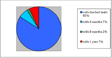
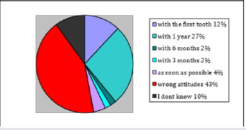
Before the lecture, 67% of respondents answered with one of the correct answers, and after the lecture 93% (Graphs 3 & 4). The next question was also answered by adding. Due to the large number of different incorrect answers, it is impossible to make a clear picture of the attitudes of pregnant women on this issue, so all the wrong attitudes will be classified into a single group. Some of the wrong attitudes were: “when we notice a defect”, “when all the teeth grow”, “when the first tooth falls out”, “at the age of 2” and many others. The correct attitude is considered to be all those who indicate the first visit to the dentist as early as 1 year of age. Before the lecture, 47% of pregnant women had the correct attitude about the time of the first examination, and after the lecture, all respondents (100%) had the correct attitude (Graphs 5 & 6).

Graph 3: Opinion of pregnant women about the time of the beginning of brushing the child’s teeth before the lecture.

Lupinepublishers-openaccess-pediatric-dentistry-journal

Graph 4: Opinion of pregnant women about the time of the beginning of brushing the child’s teeth after the lecture.
Lupinepublishers-openaccess-pediatric-dentistry-journal

Graph 5: Attitudes of pregnant women about the time of the first visit to the dentist before the lecture.
Lupinepublishers-openaccess-pediatric-dentistry-journal

Graph 6: Attitudes of pregnant women about the time of the first visit to the dentist after the lecture.
Lupinepublishers-openaccess-pediatric-dentistry-journal

Table 3: Attitudes of pregnant women about dental treatment of deciduous teeth.

Lupinepublishers-openaccess-pediatric-dentistry-journal

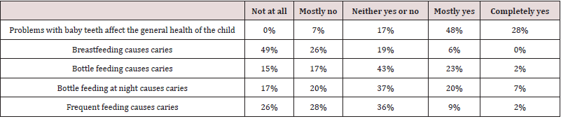
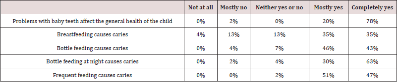
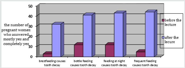
To the question „Are deciduous teeth being repaired“, 71% of respondents circled the answer YES before the lecture. After the lecture, 93% of respondents answered YES (Table 3). The next group of questions examines the attitudes of pregnant women about the causes of caries. With each statement, it was necessary to round off the position most similar to the position of the respondent, from “not at all” to “completely yes”. In this group of questions, pregnant women had the worst results (Table 4). Pregnant women’s knowledge of the causes of caries has greatly improved after the lecture (Table 5 and Graph 7).

Table 4:Attitudes of pregnant women about the impact of deciduous teeth on general health and factors for the development of caries before lectures.
Lupinepublishers-openaccess-pediatric-dentistry-journal

Table 5: Attitudes of pregnant women about the impact of deciduous teeth on general health and factors for the development of caries after lectures.
Lupinepublishers-openaccess-pediatric-dentistry-journal

Graph 7: Attitudes of pregnant women about risk factors that cause caries before and after the lectures.
Lupinepublishers-openaccess-pediatric-dentistry-journal

Discussion

Most of the respondents were not previously advised on maintaining the child’s oral hygiene. Nevertheless, this research has shown that pregnant women consider deciduous teeth important for the development and general condition of the baby. This figure coincides with the results of a study in Nigeria in which 79.2% of pregnant women stated that baby teeth are important for a child. However, pregnant women who participated in this study showed better knowledge regarding the repair of deciduous teeth (71% answered that they are being repaired) than pregnant women who participated in the study in Nigeria (43.6% did not know if deciduous teeth were being repaired) [13]. Most correctly answered the question about cleaning the oral cavity without teeth and the question about the time of eruption of the first teeth. Two descriptive questions about the time of the beginning of brushing and the first visit to the dentist have worse results, but half of the respondents answered these questions correctly. A similar study has been conducted in Germany among midwives. Midwives were asked questions about the advice they give to young mothers and pregnant women. Most claim to advise pregnant women about early childhood caries, but when asked about their first visit to the dentist, only 9% responded within the first year of life. Similar to the results of this research, 60% of midwives recommend starting brushing when the first tooth erupts [14]. A study conducted in India showed a poorer level of knowledge of pregnant women and young mothers about the time of the first visit to the dentist. Only 22.3% of pregnant women answered that the first visit to the dentist must be around the period of the eruption of the first teeth, while this research showed that about 47% of pregnant women have the correct attitude. The reason for this is because it is very likely the degree of illiteracy among women in India [15]. The area from which pregnant women showed the least knowledge is the development of caries. Specifically, they were unaware of the risky behaviors that lead to caries. 49% of respondents answered that breastfeeding does not cause caries at all, probably because they can hardly associate breastfeeding as a physiological action with negative consequences. A similar degree of ignorance of pregnant women is shown by questions about bottle-feeding, bottle-sleeping, and frequent feeding. Research has shown that pregnant women do not have sufficient knowledge of risky behaviors for caries, so they do not have the necessary knowledge to prevent caries in their child. The lecture on oral care for children as part of a course for pregnant women was the first encounter for most pregnant women with information on the causes of caries, harmful habits, tips for maintaining oral hygiene, the time to start brushing and the first visit to the dentist. Since the same but abbreviated survey was completed after the lecture, it is easy to notice an improvement in the knowledge of the mentioned topics, which supports the importance of timely education of young mothers on maintaining oral hygiene of the child.

Conclusions

This research showed that the participants in the course for pregnant women at the Clinic for Gynecology and Obstetrics in Rijeka do not have satisfactory knowledge about the oral health of the child, primarily about the risky behavior for early childhood caries. Most pregnant women were not aware that breastfeeding, bottle feeding and sleeping and frequent feeding cause tooth decay. More than half of pregnant women were of the opinion that the child should be taken to the dentist for the first visit at the age later than recommended (until the age of 1) or did not know the answer. For most pregnant women, the mentioned lecture was the first encounter with information on maintaining the oral health of the child. After the lecture, pregnant women had a much better knowledge of caries and all other issues, which indicates the importance of the mentioned lecture as part of the course for pregnant women.

Read More Lupine Publishers Pediatric Dentistry Journal Articles:
https://lupine-publishers-pediatric-dentistry.blogspot.com/

Saturday, January 8, 2022

Lupine Publishers | Children’s and Adolescents’ Pain, Discomfort, Fear, Cooperation, and Parental Presence During Dental Extractions

 Lupine Publishers | Journal of Pediatric Dentistry


Abstract

Introduction: Children’s reports of experiences from dental invasive procedures are scarce. The aim was to renew and actualize the understanding of children’s experiences of pain, discomfort, fear, and cooperation during the dental extraction procedures. A further aim was to study the frequency of parental presence during dental extractions.

Methods: The study was based on a sample of children and adolescents aged 3–19 years in the Region Västra Götaland (RVG) and Region Örebro County (ROC). It was a 5-year cohort study of an accelerated longitudinal design, named BITA (Barn I TAndvården = Children in dental care). Data on children’s pain, discomfort, fear, cooperation, and parental presence were assessed and compiled.

Results: 2363 out of 3134 children in the four age cohorts became eligible for inclusion. The cohorts included Cohort 1 (3-7 years old; n=695 children), Cohort 2 (7-11 years old; n=642 children), Cohort 3 (11-15 years old; n=574 children), and Cohort 4 (15-19 years old; n=452 children). There were 1215 girls (51%) and 1148 boys (49%). Extractions were assessed as painful by 62%. Discomfort was reported by 33%. Dental fear was reported by 47%. During painful extractions, no fear was reported by 38%, while in painless extractions, no fear was reported by 63%. Dental fear was most common in the age group 11-15 years. Full treatment acceptance was recorded in 73.6% during the extractions. In extractions reported to cause pain, 61% cooperated well. Most of the patients (77%) showed full treatment acceptance with parental presence in the dental room. The corresponding figure was 55% for the parent not being present.

Conclusion: Dental extractions continue to constitute one of the most complex and challenging treatment situations for the young patient. The oral injection was most frequently reported to cause pain, discomfort, and fear during dental extractions. Dentists should make efforts to prevent pain and discomfort, as well as utilize parental support for the child’s sense of security during dental extractions.

Keywords: Paediatric; pain management; pain assessment; clinical procedures

Abbreviations: RVG: Region Västra Götaland, ROC: Region Örebro County; BITA: Barn I T Andvården; DFA: Dental Fear and Anxiety; BMP: Behavior Management Problems

Introduction

Invasive dental treatments such as anesthesia and extractions have shown to stress children and adolescents the most [1,2]. In a study performed on 368 Swedish children and adolescents (8- 19 years), the prevailing reported painful dental procedures were injection, tooth extraction, drilling and performing a filling [1]. Negative treatment experiences may trigger a vicious cycle, i.e., leading to avoidance of dental appointments and hindering more positive experiences to take place [3-5]. Furthermore, avoiding dental appointments may affect the oral health [3-5] jeopardizing the well-being of young patients with long-term consequences [3-5]. Less traumatizing methods for invasive dental techniques during injection and extraction are available, although to date, studies report that the usage of pain preventive interventions such as topical anesthesia, local anesthesia, or analgesics are still substandard [6-8]. The routines applied by Swedish dentists have shown that ‘about 35% were more indifferent to their patient’s experiences of pain and psychological management’ [7]. Apart from pain experiences, invasive and/or noninvasive dental procedures may cause children discomfort. Although there is no universally prevailing definition, the phenomenon might be explained as psychological or bodily distress or annoyance, i.e., anything that disturbs the well-being [9]. Children’s experience of discomfort in dentistry has previously not been given much attention. Invasive procedures also test dentists’ technical skills with a need to be knowledgeable on children’s developmental levels, contemporary psychology, and pedagogical interactions. Furthermore, dentists may face difficulties in the interaction with the child/parent dyad [10].

Parental attitudes and behavior have been suggested to either facilitate or undermine the child’s cooperation and thus affect the treatment outcome [11-14]. It could be hypothesized that some dentists consider parents a hindrance, therefore not inviting them into the dental room. Seen from the young patients’ standpoint, such an approach may be controversial as parental absence may diminish the perception of security, predisposing to fear, pain, and discomfort experiences [14]. In Sweden, a prevailing tradition and the Dental Act have allowed parents to support their children in medical and dental settings [15]. The practice has thus become a norm in the pediatric dental community, despite alternative behavior management techniques [16]. At present, there is insufficient knowledge in the area of children’s and adolescents’ experience of invasive procedures, and dentists’ attitudes and praxis towards these. Furthermore, there is a lack of knowledge on how general dentists understand the parental interaction and how frequently they make use of parental help during extractions. The aim was to renew and actualize the understanding of children’s experiences of pain, discomfort, fear, and cooperation during dental extraction procedures. A further aim was to study the frequency of parental presence during dental extractions.

Patients and Methods

Patients

This study was based on a representative sample of children and adolescents aged 3–19 years in the Region Västra Götaland (RVG) and Region Örebro County (ROC). The project was a 5-year cohort study of an accelerated longitudinal design, named BITA (Barn I TAndvården = Children in dental care). It concerned different aspects of children in the dental situation when attending regular general dentistry. Four age cohorts with children aged 3, 7, 11 and 15 years, from seven Public Dental Service clinics in RVG and five in ROC, were invited to participate and then followed over a 5-year period in conjunction with regular dental visits. The clinics were selected to reflect the respective populations and to cover urban and rural areas. A total of 3134 children were considered for the study.

Procedure

Clinical registrations

The children’s ordinary dentists registered all performed dental treatments after each session. For this study, extraction treatments were selected, and assessments and self-ratings were made and registered.

Self-reported pain

Each child made a self-assessment of pain experienced after the extraction. The child was asked the question: Was anything painful? Yes or no? If the child answered yes, the pain intensity was measured using a visual analog scale (VAS) consisting of 11 points running from 0 = no pain, to 10 = worst pain possible. Parallel to the scale, six faces express different levels of pain/distress where young children point out the face matching their level of pain. Furthermore, the child was asked the question: What was painful?

Self-reported discomfort

The child was asked the question: Did anything cause discomfort? Yes or no? If the child answered yes, the discomfort was measured using a visual rating scale (VRS) consisting of 11 points running from 0 = no discomfort, to 10 = worst discomfort possible. Parallel to the scale, six faces express different levels of discomfort where young children point out the face matching their level of discomfort. Furthermore, the child was asked the question: What caused the discomfort?

Self-reported fear

Children assessed their fear at the dental treatment by answering the question, how did you feel today? The alternative answers were Not afraid at all = 0 or Afraid, on a scale graded 1-4.

Assessment of the child’s cooperation

The child´s cooperation was graded by the treating dental personnel according to the scale by Rud and Kisling, rated 3 to 0, where 3= full acceptance to treatment; 2= indifferent acceptance; 1= reluctant acceptance; and 0= non-acceptance.

Parental presence

Parental presence in the dental room was documented.

Ethics

The application for ethical review (No. 286-07) was approved.

Statistical methods

In addition to descriptive statistics in terms of frequency distributions, means, and standard deviations, differences between genders and between cohorts were analyzed using Chi-square tests. The statistical analyses were performed in IBM SPSS Statistics for Windows, Version 23.0 (IBM Corp., Armonk, NY, USA ) and p values below 0.05 represented statistical significance.

Results

3134 children were invited to participate in the study; 771 declined participation, whereby 2363 children in the four age cohorts became eligible for inclusion. These four age cohorts included Cohort 1 (born 2005; from 3-7 years old during the study period; n=695 children, 3906 visits), Cohort 2 (born 2001; 7-11 years old; n=642 children, 4588 visits), Cohort 3 (born 1997; 11-15 years old; n=574 children, 4085 visits), and Cohort 4 (born 1993; 15-19 years old; n=452 children, 3315 visits). The distribution between the genders was 1215 girls (51%) and 1148 boys (49%).

Pain and discomfort during extractions

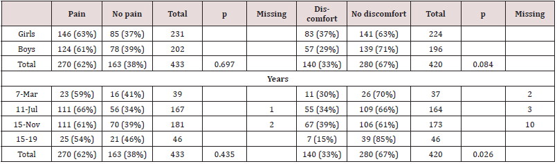
Extractions were assessed as painful by 62% (Table 1). The reasons were the extraction itself (16%), the injection (72%) or the combination of injection and extraction procedures (6%) (Table 2). Discomfort was reported in 33% of the extractions (Table 1). The reasons for discomfort pointed to the extraction per se (37%), followed by injection (26%) (Table 2). No statistically significant difference between the genders or cohorts was found regarding the experience of pain. There was a statistically significant difference between cohorts, with the lowest proportion of discomfort stated by the oldest group (30%, 34%, 39%, 15%; p=0.026) (Table 1). Primary (n=323) and permanent (n=102) tooth extractions were assessed as painful in equal proportions (63% and 61%), while discomfort was slightly less often reported for primary teeth, compared to permanent tooth extractions (32% and 37%). 11 tooth extractions lacked complete documentation. Topical anesthesia was applied in 35%, with no statistically significant difference between application and no application regarding the reported experience of pain and/or discomfort. Conscious sedation drugs were used in 6%.

Table 1: Extraction appointments (n=436 extractions) and the young patients’, reports of pain and discomfort in adjunction to the treatment, related to gender and age-cohorts.

Lupinepublishers-openaccess-pediatric-dentistry-journal

Chi-2 test, P probability value

Table 2: Children’s reported reasons for pain (n=270) and discomfort (n=140) in connection to the extraction procedures.

Lupinepublishers-openaccess-pediatric-dentistry-journal

Dental fear and pain during extractions

Children’s self-assessment of dental fear resulted in 47% reporting no dental fear in conjunction with the extractions. During painful extractions, no fear was reported by 38%, while in painless extractions, no fear was reported by 63% (Table 3). Dental fear was most common in the age group 11-15 years, followed by the age group 7-11 years (Table 3). No statistically significant difference between the genders or cohorts was found regarding the experience of pain. There was a statistically significant difference between cohorts, with the lowest proportion of discomfort stated by the oldest group (30%, 34%, 39%, 15%; p=0.026) (Table 1). Primary (n=323) and permanent (n=102) tooth extractions were assessed as painful in equal proportions (63% and 61%), while discomfort was slightly less often reported for primary teeth, compared to permanent tooth extractions (32% and 37%). 11 tooth extractions lacked complete documentation. Topical anesthesia was applied in 35%, with no statistically significant difference between application and no application regarding the reported experience of pain and/ or discomfort. Conscious sedation drugs were used in 6%.

Table 3:Young patients’ (age cohorts) self-rated dental fear and pain experiences. The subjective assessment of pain=Yes or No was given after the extraction procedure (missing data n=14). Dental fear, as rated on a scale 0 to 4, 0=no fear, fear =1-4, (missing data n=12).

Lupinepublishers-openaccess-pediatric-dentistry-journal

Dental fear and pain during extractions Children’s self-assessment of dental fear resulted in 47% reporting no dental fear in conjunction with the extractions. During painful extractions, no fear was reported by 38%, while in painless extractions, no fear was reported by 63% (Table 3). Dental fear was most common in the age group 11-15 years, followed by the age group 7-11 years (Table 3).

The child´s cooperation while experiencing pain

Full treatment acceptance was recorded in 73.6% during the extractions. In extractions reported to cause pain, 61% cooperated well (Table 4).

Table 4: Children’s self-rated pain experiences and the degree of treatment acceptance, as rated by the dental personnel, on a scale 3-0, by Rud and Kisling.

Lupinepublishers-openaccess-pediatric-dentistry-journal

The subjective assessment of pain=Yes or No was given by the child after the extraction (missing data n=16).

Treatment acceptance during parental presence

Most of the patients (77%) showed full treatment acceptance with parental presence in the dental room. The corresponding figure was 55% for the parent not being present. All 37 three-yearold had their parents present in the dental room. Among the 166 seven-year-olds, two were not accompanied by an adult. Among the 11-year-olds, 26 out of 179 had no parent present. Among the 15-year-olds, 25 out of 45 managed on their own.

Discussion

This longitudinal, 5-year study voiced children’s and adolescents’ self-reported pain, discomfort, and fear during dental extractions. The main results showed that more than 60% of the dental extraction appointments involved painful experiences. Furthermore, pain was equally reported among the different age groups. The extracted teeth were predominantly performed in young schoolchildren. This may indicate that more vulnerable children were at risk experiencing pain at an early stage of their dental care encounters. Within the extraction procedure, the injection was most frequently reported responsible for the negative experiences. During decades, pain has been connected with dental extractions and concerned children and adolescents [17]. It could be reasoned that the injection techniques have not developed sufficiently, despite alternative anesthesia delivery methods, Computer controlled local anesthesia, Jet injectors, Iontophoresis and Computer controlled intraosseous anesthesia [18,19]. An explanation might be that as these methods lie in the hands of numerous dentists, the injection performance may not be possible to calibrate. Other factors adding to the child’s pain experience may be the dentists handling the onset of the anesthetic, the amount of delivered anesthesia, or a complementary anesthesia differently. A noteworthy result was that topical anesthesia was applied in only 35% by the dentists, which is astonishingly low given the scientific evidence in support of the technique, which is in alignment with earlier result [7]. Furthermore, adding to the children’s uneasiness, discomfort was reported frequently during the extraction procedure. Thus, despite a complete anesthesia numbness, discomfort may have been experienced due to cracking sounds and/or the perceived pressure during the dental luxation. Discomfort has not been given much attention in the dental literature compared to general medical care [20]. The frequently reported discomfort also suggest that the dentists may not have practiced current theoretical knowledge sufficiently, which needs to be recognized [7,8].

No studies among dentists have confirmed the Knowledge Transition to have improved regarding dental invasive procedures in general or oral anesthesia specifically [21,22]. An explanation may be the complex interaction between the applied injection technique and the dentist’s way of psychological and pedagogical coaching [23-25]. Preferably, children should beforehand be prepared on anesthesia numbness sensation, luxation cracking sounds, increasing tissue-pressure during luxation, postoperative taste-sensation (iron, i.e., blood), and the subsequent inconvenience of not being able to enjoy a meal directly after the tooth extraction. Studies on Dental Fear and Anxiety (DFA) have showed that invasive dental procedures, such as extractions and injections, were by many children and adolescents associated with fear and anxiety [26,27]. In the current study, 47% of the extraction appointments were experienced without fear. From this perspective, the extractions were performed under satisfactory conditions. On the other hand, 53% of the children experienced fear, ranging from a bit nervous to terrified. Versloot et al. (2008) have reasoned that the level of dental anxiety may be of greater importance for the child’s reaction than the injection technique itself [27]. Given that young patients’ early pain encounters may not only trigger. Behavior Management Problems (BMP) and/or Dental Fear and Anxiety (DFA), but may also alter future pain responses, the results of this study are notable [28]. Contrary to the pain reports that were equally common in all age cohorts of the current study, DFA was more frequently reported in schoolchildren. This may define a vulnerable developmental stage, requiring parental presence and support. The relatively high proportion of extraction appointments performed with the parent does not present in the room calls for additional studies.
The current study presented that parental presence in the dental room was most frequent among children 3-12 years old, hereafter the presence significantly declined. Praxis has suggested that most parents are experts on their child’s behavior and reactions. Perets and Zadick (1998) found that most parents would assist, should the dentist not succeed to manage their child [29]. Vasiliki (2016) also stated that children responded more positively in the dental room with parental presence [11]. Pfefferle et al. (1982) reasoned that this may be due to parents knowing their child’s reactions and capabilities in challenging situations [30]. The current study demonstrated that most children cooperated fully during the dental extraction procedures despite experiencing discomfort or pain. A possible interpretation is that these children were not equipped to speak their needs, or they surrendered easily to authorities. Indirectly, the results also suggested that dentists practiced varied theoretical understanding, technical skills, and ethical attitudes during dental extraction procedures, not always in favor of the child. A limitation of this study may be argued that children below six years of age were not reliable to give accurate assessments of their experiences. However, there is a strong opinion among researchers that children from the age of 3-4 years are capable to communicate well and should be supported to express perceptions of discomfort and pain [31]. The strength of this study was that the results originated from a 5-year longitudinal perspective based on young patients’ experiences from the extraction appointments. These results, regarding pain, discomfort and DFA, have not been contradicted in the recent literature and are valuable for clinical discussions. In conclusion, dental extractions continue to constitute one of the utmost intricate and challenging treatment situations for young patients. The oral injection was most frequently reported to cause pain, discomfort, and fear during dental extractions. Dentists should make efforts to prevent pain and discomfort, as well as utilize parental support for the child’s sense of security during dental extractions.

Acknowledgements

This study was funded by the Public Dental Service, Region Västra Götaland and Region Örebro County, Sweden.

Conflict of Interest

The authors declare no conflicts of interest.

Read More Lupine Publishers Pediatric Dentistry Journal Articles:
https://lupine-publishers-pediatric-dentistry.blogspot.com/

Tuesday, December 21, 2021

Lupine Publishers | The Applied Importance of Pediatric Dentistry (An Overview)

 Lupine Publishers | Journal of Pediatric Dentistry


Abstract

Today, with the promotion of community awareness, fortunately, the importance of oral health and maintenance of primary teeth for parents is clear. Due to the important roles of primary teeth in the oral system, these teeth should be maintained until the eruption of the permanent teeth. For this reason, it seems the parent’s perspective has changed, and children’s dentistry is now a priority for most families. Pediatric dentists should be specialized in different fields of pediatric dentistry. They will be training in the fields of pediatric psychology, child anesthesia and sedation, and age-appropriate behavior management so that they can manage children’s treatments in the best way. This group of dentists is specialized in eliminating child concerns and anxiety too. They can distract children’s minds by talking to them from what they plan to do. This friendship makes the child’s fears go away and the treatments are accomplished positively so that the child would not feel any bad dental experiences at all. Pediatric dentists provide the parents with the necessary training information to protect their children’s teeth from the very beginning of their childhood.

Keywords: Pediatric dentists; pediatric psychology; child anesthesia; sedations; elimination of anxiety; child behavioral management

Introduction

Pediatric Dentistry is one of the branches of dentistry in which the dentist works with children and adolescents. In 1909, Minnie Evangeline Jordon established the first dental practice in the United States devoted only to pediatric patients [1]. This discipline was first recognized by the American Dental Association in the United States [2] and subsequently graded in other countries as a separate specialty. In general, for a general dentist to receive a child’s specialty, he must complete two to three years of special training. In this area, the dentist not only focuses on oral and dental health, but he will also be interlinked with children and the psychological behavior of the children. Children have their mental characteristics and do not tolerate oral examination and dental treatments; consequently, they may not cooperate well with the general dentist. In pediatric dentistry, the dental practitioner thoroughly performs examination and treatment with the use of applied techniques, so that the child will become fully cooperative. To ease the process of dental treatment, they will use the appropriate and fun tools for the child to accept the dental procedures. The dental office environment is also one of the factors that can be effective in improving the treatment in the child [3]. Entertaining the child before and during treatment with toys and cartoons is one of the things that play significant effects [3-5]. The pediatric dentist offers a range of therapeutic approaches that can help the improvement of the oral health status of the child. The pediatric dental specialization has given special attention to dentistry in children from birth to adolescence. Due to the importance of pediatric dentistry, a pediatric dentist must have the experience, expertise, and the ability to maintain the health of teeth, gums. In general, the oral conditions of children can be monitored by a pediatric dentist at different stages of the child’s development [4]. If oral health is neglected, the likelihood of developing dental caries and other illnesses that cause permanent complications for the teeth of the baby will be very high.

Pediatric Dentistry Features

In this article, we intend to introduce some of the most important features of pediatric dentistry.

Oral health education

Tooth decay, as well as non-compliance with health in children, reduce their school performances and create poor social relationships [6-9]. Children’s dentistry with providing a pleasant and educational environment for children and adolescents not only reducing their stress but also can greatly increase the awareness of children and prevent the occurrence of various types of oral and dental diseases [7].

Specialized children’s dental services

Primary teeth are very important in childhood and adolescence, which plays an important role in their correct chewing of foods. Not only does this contribute to the health of children, but it also helps children to speak the words correctly. All services such as primary teeth restoration, pulpotomy and pulpectomy procedures, SS crowns, fluoride therapy, and permanent teeth services in children are provided by children’s dentistry.

General anesthesia

One of the causes that prevent children from visiting the dentist is the fear and anxiety in referring to pediatric dentistry [5,10]. This fear causes the child to suffer from oral and dental illness due to a lack of regular referral. In a general dental office, due to a lack of knowledge of the psychology of the child, it is not possible to provide a warm and comfortable environment for children. Referring the child to the pediatric dentist, in addition to addressing the problems caused by the child’s lack of health, allows the child to address the dental and oral problems in a happy and intimate environment. Today, for children with anxiety and fear of referring to dentistry, general anesthesia is used during examination and dental procedures to increase the accuracy and quality of the work of the pediatric dentist.

Application of Hypnosis (artificial sleep)

Oral and dental hygiene is one of the things parents should consider from childhood. Since children are sensitive and fearful of the dental problem, many solutions have been proposed to counteract it. Sometimes children’s dental conditions are in a special situation where pediatricians turn to the use of hypnosis in children’s dentistry [11]. Dissatisfied children with a weak collaboration attitude could delay the treatment in most cases. This artificial sleep in dentistry is done by an anesthetist and is one of the newest dental care services. Although artificial sleep does not make the child feel unfamiliar with the office environment the advantage is the dental work is accomplished while they are sleeping.

Promoting the right habits

While a child may not need a dentist at the age of two, it is important to start training oral hygiene habits as soon as possible [12-14]. During dental examinations, the pedodontist can help the child learn how to use a toothbrush, or how to use dental floss to prevent dental caries and dental diseases. Considering that good habits can be formed from childhood and adolescence, so constant referral to the dentist can enhance that.

Features of Pedodontist

a) Pediatric dentists must be an expert in child psychology and know how to deal with children; they have to able to get the patient’s confidence in the shortest possible time [4].
b) Pediatric dentist should be able to interact with the parents; should not speak harshly and should not blame parents for the delayed treatment processes [4].
c) Because the child’s patience is very limited, the dentist should be on time, and the patient should not wait for a long time in the waiting room [4].
d) During the examination, the Pedodontist must show a high degree of accuracy and focus on the problem [4].
e) During the dental procedure, he has to avoid talking about things that are not suitable for a child’s age [4].
f) Treating adolescents like adults, should be another critical criterion that a Pedodontist must pay close attention to that.

Benefits of Pediatric Dentistry

Perhaps some parents think about what the difference between dentists is. All dentists are informed about dentistry, so they can refer to any dentist for treatment. But parents may not be fully aware of the benefits of pediatric dentistry. We review these advantages.

Maintaining oral and dental health

Perhaps one of the most important benefits of pediatric dentistry is that these professionals try to keep the child’s mouth clean and healthy by using the best training and educational tools, special features, and appropriate accessories. Constant and ongoing periodic examinations can help parents alleviate the child’s oral and dental illnesses, as well as help them to take care of regularly, and identify potential problems that may appear in the future.

Preventing early teeth loss

When parents think of the health of kids’ teeth, it is better to start their job right now! Pediatric dental care ensures that the child’s teeth will be healthy, free from decay and other dental diseases in the future. Dental caries is the most common chronic infectious disease of childhood [15]. Streptococcus Mutans, Lactobacilli, Actinomyces spp. and some other anaerobic bacteria are the most primary etiologic agents of dental caries [16]. Dental services and periodic examinations of children regularly can help prevent this complication and other types of other diseases of the mouth, teeth, and gums. Diagnosis of cavities in the teeth of a child is a critical issue insomuch as the lack of continuous care and periodic examinations can lead to more acute problems, such as tooth loss shortly.

Gaining awareness of the important issues of oral health of children

Oral health has to be started at the very beginning of the baby’s birth. Even if mothers are nursing or feeding with a bottle of milk, it is necessary to start a good oral hygiene program from the very beginning and as soon as possible. Before teething, mothers should keep the gums clean with soft and wet cloth. When teeth begin to erupt, they should also be cleaned. It is essential to use the right toolset for this daily work. For example, it is necessary to buy a special toothbrush for a baby that is more suitable for his little mouth. A Pedodontist keeps them healthy by removing microbial plaques and impacted food which may be stuck in between the teeth in a routine dental examination. This is an important part of pediatric dental care because it prevents the growth of bacteria that can lead to dental diseases such as tooth decay or inflammation of the gum tissue.

Children’s dentistry and dental care

The most important background for children’s dentistry is the prevention of dental decay and intensive care for their teeth. This care should be continued under the supervision of a pediatric dentist which starts at the age of one when the baby’s teeth begin to erupt and gradually would be replaced with permanent teeth until the age of ten to twelve. It is best to take the child to a pedodontist at the earliest age and repeat this procedure every six months. This prompts not only make the child become familiar with the dental environment but also it causes the child’s fears to diminish. More importantly, in both childhood and early youth, the kid’s oral health will be under the supervision of the pediatric dentist.

The Importance of Referring to the Pediatric Dentist

Oral health is an important part of public health that plays a significant role in the quality of life of people in society, especially children. Unfortunately, the frequency of teeth decay is often high due to the lack of attention paid by both parents and children. One of the most effective measures to improve the oral and dental health of children is conducting examinations by pediatric dentists [4,12]. To refer to a pedodontist office, parents should not wait for the pain and dental problems of the child because this delay can cause more severe problems. The primary teeth are very important and depend on the evolutionary stages necessary for the health of the children’s permanent teeth. Pedodontist can help parents to provide the necessary health care for children’s oral health and give them the necessary advice. A pediatric dentist has some special skills in oral health and dental care from childhood to adolescence. He has the expertise and experience to protect and maintain and treat the teeth, gums, and oral tissues of a child of all ages. Since the first teeth have erupted, regular consultation with a pedodontist is mandatory. This visit should be done every six months. These referrals help both parents get the right information and advice on how to protect the health of the child’s teeth. Furthermore, the child will get familiar with the office environment and its staff, and get some pleasant dental experience before having caries, pain, or dental abscesses. In this case, the child will be more cooperative in future treatment sessions.

Why are Periodic Dental Referrals Essential?

Nowadays, parents should be aware of the need for periodic examinations before further destruction of children’s teeth is taken into consideration. Regarding the principle of the existence of a problem, faster treatment will ensure longer teeth life. Moreover, it will increase the function of the teeth and will include a lower cost. Because if they close their eyes to the problems, those problems will get worse every day, and these will not ever stop. Problems can be accumulated and result in tooth loss, fabricating of space maintainers and apace retainers for the edentulous area, troublous and costly orthodontic treatments, implant-prone treatments, and so on. In other words, if parents fail to take the advantage of these referrals, they will be damaging to their children’s oral health which is hard to compensate and consequently, its effects remain until the end of their lives. Effects that are being handled on time definitely would be crucial in terms of cost, pain, and the length of the period of recovery for their parents and their children.

Conclusion

Pediatric dentistry is a branch of dentistry which concerns with dental and oral health in children. Since oral and tooth injuries start in childhood and adolescence ‘prevention and treatment of these injuries are very important in this period. For this reason, pediatric dentists not only can increase the level of knowledge of children and adolescents in oral health but also can prevent many abnormalities in the mouth and teeth. On the other hand, a pedodontist in addition to knowledge of dentistry has learned the principles of behavioral control of children. The dentist will provide attractive and pain-free behavioral therapy techniques for children. It is interesting to know that some dentists manage the treatment of children with cerebral palsy, mentally retarded, or even autism. The difference between children’s dentistry and other branches of dentistry is how to deal with children.

Read More Lupine Publishers Pediatric Dentistry Journal Articles:
https://lupine-publishers-pediatric-dentistry.blogspot.com/

Saturday, December 11, 2021

Lupine Publishers | The Challenges of Pediatric Dentistry in the 21st Century

 Lupine Publishers | Journal of Pediatric Dentistry

Editorial

According to the information available on the prevalence of early childhood caries in the United States and other industrialized societies and given the focus of this disease on poor children and minorities, pediatric dentistry has been focused on it. A pediatric dentist should be aware of the new challenges in keeping children free from dental caries in childhood. Today, we believe that on one hand the dental diseases, and on the other hand their prevention and control, are highly influenced by factors that are beyond the biological bacterial, sugars, and tooth factors in the Keyes interaction model. These factors include the impact of society, the system of life, and families that are not the primary biological factors. This means that the pediatric dentist should consider different factors that merely include the child, his family, and the child’s oral health. Preventive dentistry involves recognizing the effect of external factors and non-biological factors on the onset and progress of caries, especially in poor and heterogeneous societies. Nowadays, the most important considerations in childcare are as follows:

a) The importance of oral health for children as the best opportunity for preventing early childhood caries by providing preventive information to families and the role of the collaboration of general dentists and families as non-specialists in the field of dentistry.
b) The techniques of fissure sealants and composite resins, and their positions in children’s dentistry, as well as the considerations of the lifespan of these treatments in comparison with other treatments, and also concerns about dental toxicity.
c) Dentistry for disabled patients and other children with special care needs, which today have a longer lifespan thanks to medical advances, but usually dental treatments, require medication considerations, body health, and medical needs.
d) Early diagnosis and treatment of orthodontics, which is important in guiding the development of occlusion and reducing long-term future treatments. e) Complex treatments, such as sedation techniques, to control pain and anxiety of the patient, and the need for knowledge and mastery of the guidelines for the safe and practical use of these techniques.
f) The problem of developing fluorosis, due to the presence of fluoride in the environment and the need to change the therapies of fluoride therapy and their dose.
g) Obesity and other eating disorders, their effects on oral health, and the need for patient care in the dental office.
h) Extensive and complex radiographic techniques such as digital radiography and 3D imaging, providing better diagnostic information, as well as simultaneous concerns about radiation doses during childhood.
i) Substance abuse, such as painless tobacco, recreational drugs used in adolescents and pre-puberty, and misuse of prescription drugs that have become epidemic in many societies.
j) Digitization of health information, management, and ability to move them, as well as the role and the position of today’s electronic world.
k) Promote public health support activities, including the participation of dentists in health committees, school sports teams, health program counselors, and other out-of-office roles that require oral health care expertise.

Children’s dentistry is the key to health and prevention, and social macroeconomic planning at such an age can be used in a large community to reduce the teeth decays, periodontal diseases, the prevention of orthodontic treatment, the promotion of the national culture and health, and finally cost reduction of the family per capita. If prevention dentistry would be a national priority, then surely the dental insurance companies that usually do not cover the dental treatments, they will be persuaded to provide public health after a decade. This can happen only by eliminating the dangers of dental crisis which shows the importance of childhood preventive dentistry.

Read More Lupine Publishers Pediatric Dentistry Journal Articles:
https://lupine-publishers-pediatric-dentistry.blogspot.com/


Friday, December 3, 2021

Lupine Publishers | Impact in Oral Cavity due to the Use of Hydrogen Peroxyde in Dental Treatment

 Lupine Publishers | Journal of Pediatric Dentistry


Abstract

Background: Chemicals agents are commonly found in people activities. It has a big role, especially in the oral cavity. The adverse effects that have been reported in cellular, animal and human studies include: chemical burn, cervical root resorption associated with non-vital bleaching; increased tooth sensitivity associated with vital bleaching; alteration in the surface topography of enamel; reduction in bond strength of resin based materials and the possibility that H2O2 may have carcinogenic or tumor promoting capabilities Chemical agents are often used by people. But the chemical agents can cause the damage in human body such as oral cavity.

Objectives: To know the association between dental treatment and chemical burns in oral cavity. Problem statement: In dental treatment, chemical agents are often used by dentist due to its function. But chemical agents can cause the damage in oral mucosa.

Discussion: One of the chemicals that is often found can cause chemical burn in oral mucosa is hydrogen peroxide. Hydrogen peroxides or H2O2 is used to decrease plaque formation and to control pyorrhea (gum inflammation). The mechanism of antimicrobial action is due to the release of nascent oxygen which is detrimental to anaerobes. It acts on both Gram positive and Gram-negative organism. H2O2 solutions usually do not result in severe toxicity but may cause vomiting; mild irritation to mucosa; and burns in the mouth, throat, oesophagus, and stomach. Ingestion of higher concentrations (>10%) can result in more dangerous sequelae, such as burns to mucus membranes and gut mucosa.

Conclusion: The use of chemical agents in dental treatment can cause the chemical burns in oral cavity. So, the patients need to listen more towards the dentist about the instruction after treatment so the materials cannot cause the irritation in oral mucosa.

Keywords:Oral cavity, chemical burn, dental treatment, hydrogen peroxide

Introduction

Chemicals agents are commonly found in people activities. It has a big role, especially in the oral cavity. Chemical agents are often used by people. But the chemical agents can cause the damage in human body such as oral cavity. Mucosal damage in the oral cavity can be iatrogenic during dental procedures, misuse or improper application of medicine and non-pharmaceutical substances by the patients. Chemicals that are often used in the oral cavity when dental care such as bleaching, restorations for restoration to dentures [1]. The practice of dentistry is very closely related to the use of chemicals. Inappropriate or excessive use of chemicals can cause damage to the oral mucosa. Clinical presentation of the chemical injuries might differ according to the composition and concentration, the pH of the substance, the quantity applied, the manner and duration of tissue contact, and the extent of penetration into tissue. These oral mucosal changes can vary from diffuse erosive lesions ranging from simple mucosal sloughing to complete mucosal detachment with extensions into the submucosa [2]. Dental treatments which were provided by dentists cannot be separated from chemical agents that actually have an impact on the body, but the dentists use them as needed so that often it does not have an impact on the oral mucosa. One of the chemicals that is often found can cause chemical burn in oral mucosa is hydrogen peroxide [3].

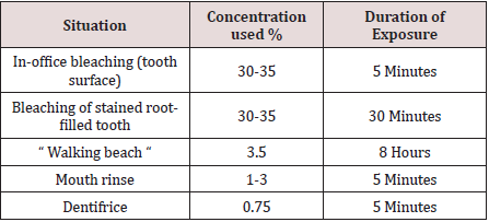
Hydrogen peroxide (H2O2) is a colorless liquid with a bitter taste and is highly soluble in water to give an acidic solution. H2O2 is an oxidizing agent with a wide number of industrial applications in for example, bleaching or deodorizing textiles, wood pulp, hair, fur and foods, in the treatment of water and sewage, as a seed disinfectant and neutralizing agent in wine distillation. Low concentrations of H2O2 have been found in rain and surface water, in human and plant tissues, in foods and beverages and in bacteria. Hydrogen peroxide is a reactive oxygen species, along with superoxide (O2-), hydroxyl (HO), peroxyl (ROO) and alkoxyl (RO). In human tissue, intrinsic sources of H2O2 are organelles (especially mitochondria), salivary cells, microorganisms, and the lungs. Hydrogen peroxide production can be followed by the liberation of highly reactive oxygen species in the body via enzymatic and spontaneous redox reactions that often involve interaction with transitional metals such as iron or copper [4]. Enzymes such as catalase, glutathione peroxidase and superoxide dismutase catalyze the decomposition of H2O2 into water and oxygen. Reactive oxygen radicals are a potential source of cell damage through causing DNA strand breaks, genotoxicity, and cytotoxicity, but these radicals tend neither to cross biological membranes nor travel large distances within a cell. Antioxidants provide a source of electrons that reduce hydroxyl radicals to water. However, when exogenous H2O2 levels overwhelm cellular protective mechanisms, H2O2 presents a health hazard. Individuals with catalase lack catalase activity, leading to high endogenous H2O2 levels causing necrosis and ulceration of soft and hard tissues [5]. Hydrogen peroxides or H2O2 is used to decrease plaque formation and to control pyorrhea (gum inflammation). The mechanism of antimicrobial action is due to the release of nascent oxygen which is detrimental to anaerobes. It acts on both Gram positive and Gram-negative organism. The other mechanism of antimicrobial property is the effect of H2O2 on debridement of bacterial cell walls [6]. It is widely used professional and self‑administered dental product. The most common applications of H2O2 include Mouth rinse (1%–3%) and bleaching agent (3%–5%). As patients' needs for aesthetic dental treatment shift from traditional treatment including caries and dentures to esthetic treatment, an increasing number of people are visiting the dental office with hopes for whiter teeth. Over the counter (OTC) bleaching products are sold as cosmetics and are freely available through stores, pharmacies, and the Internet. Although in-office bleaching is a particularly popular method for bleaching, the bleaching agent may sometimes come into contact with the patient's gingiva or oral mucosa during the in-office bleaching procedure, even if the gingiva is protected with a light-cured resin or rubber dam and bleaching is performed by an experienced dentist. This may result in temporary whitening and pain in the gingiva or oral mucosa, but the pain subsides within a few hours and the whitened spot eventually regains its original color. This has also been reported with at-home bleaching [7,8] (Table 1).

Table 1: Exposure to Hydrogen Peroxide.

Lupinepublishers-openaccess-pediatric-dentistry-journal

A study investigate the toxicity of in-office bleaching agents on gingiva, Hydrogen peroxide can be toxic in high concentrations and with exposures of prolonged duration, concentrated (30-35 per cent) hydrogen peroxide solutions have been used for in-office bleaching treatments with had adverse soft and hard tissue effects observed clinically, one of them might be chemical burn. Nevertheless, there have been occasions when accidental ingestion of hydrogen peroxide products in the home has led to hospitalization and adverse health outcomes, and this should be kept in mind when assessing the relative safety of hydrogen peroxide-containing products for home (unsupervised) use. Concerns have been expressed over the potential adverse effects of the use of H2O2 in dental treatment. The adverse effects that have been reported in cellular, animal and human studies include: chemical burn, cervical root resorption associated with non-vital bleaching; increased tooth sensitivity associated with vital bleaching; alteration in the surface topography of enamel; reduction in bond strength of resin based materials and the possibility that H2O2 may have carcinogenic or tumor promoting capabilities [9,10]. Aim of this article is to know the association between dental treatment and chemical burns in oral cavity due to chemical agents used especially impact H2O2.

Discussion

The response of mammalian cells to H2O2 is highly variable, dependent on factors such as catalase concentration and DNA repair activity. In one study, H2O2 induced a dose-dependent increase of hypoxanthine guanine phosphoribosyl transferase (HPRT) mutations in vitro in human T lymphocytes, most mutations being of the same kind as observed in T cells in vivo. Another study, using human lymphocytes, assessing genotoxicity showed the main event induced by extracellular application of H2O2 to be necrosis and not micronucleus formation [10,11].

Stomatitis

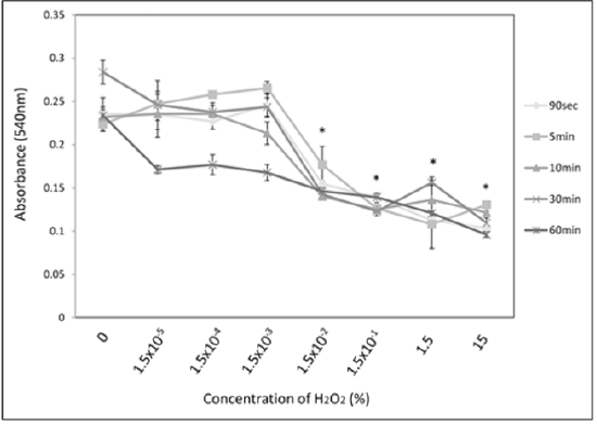
Oral ingestion of 3% H2O2 solutions usually do not result in severe toxicity but may cause vomiting; mild irritation to mucosa; and burns in the mouth, throat, oesophagus, and stomach. Ingestion of higher concentrations (>10%) can result in more dangerous sequelae, such as burns to mucus membranes and gut mucosa. It shows a dose‑dependent reaction where at high concentration eugenol causes adverse effect on fibroblast‑.and osteoblasts‑like cells. This leads to localized necrosis and compromised healing. In lower concentration, it causes localized hypersensitivity reactions to oral mucosa called “contact stomatitis” and on dermis causes “contact dermatitis,” possibly because it can react directly with proteins to form conjugates and reactive happens [5,11] (Figure 1). Study before found that almost no cytotoxicity from H2O2 was seen in the low-concentration stimulation group (1.5 × 10⁻³% or less) at any duration other than 60 minutes. However, 60-minute stimulation resulted in cytotoxicity at all concentrations. Stimulation at 15% H2O2, which is similar to the concentration used in clinical practice, showed marked cytotoxicity, lowering cell survival by half at 90 seconds (p<0.05), which is the shortest time period examined. Using the same stimulation samples as in the section above, microarray was used to analyze the gene clusters that were suppressed from the addition of vitamin E among the gene cluster induced by H2O2 stimulation. The results confirmed that H2O2 stimulation induced the expression of genes associated with the proinflammatory cytokine TNF-α cascade. Expression of these genes was suppressed with vitamin E addition. These results demonstrate that H2O2 stimulation response includes triggering the proinflammatory cytokine TNF-α cascade and that addition of vitamin E suppresses the response of this cascade [12,13].

Figure 1: Concentration of H2O2 and its absorbance.

Lupinepublishers-openaccess-pediatric-dentistry-journal

Gingival inflammation

Study in vitro examinations of signs such as whitening of the gingiva and pain that may result from tooth whitening. No studies to this effect in HGFs have been reported from at-home bleaching agents but mostly result from in-office bleaching agents and OTC products. Many studies have been carried out on tooth hypersensitivity from in-office or at-home bleaching agents. From in vitro studies, it has been concluded that whitening agents histologically penetrate the dentin and do not damage the pulp.  In current dental practice, pain incurred during the procedure is generally treated with medication or fluoride or by stopping the procedure. Several studies examining plaque control as an index have shown that whitening agents reduce plaque on the gingiva and reduce gingival inflammation.  Hydrogen peroxide and carbamide peroxide have been used for debridement during endodontic therapy, in mouth rinses to reduce plaque in individuals with gingivitis, and for treatment of periodontal diseases [13,14]. Gingival fibroblasts are known to be affected by H2O2.However, studies have shown that fibroblasts in other parts of the body are more strongly affected.  In the present study, highly concentrated H2O2 caused inflammation in gingival fibroblasts and had toxic effects, with marked changes in cell morphology. One study found that higher concentrations of H2O2 caused senescence-like changes in cells. It has long been known from in vivo animal experiments that H2O2 causes acute inflammation and even edema in skin. Simon and other  have shown H2O2 to cause necrosis in human fibroblasts. Cells are protected by various types of enzymes and other substances and manage the immune system that responds to inflammation. Inferring from the above studies, it is possible that H2O2 penetrates the cell membrane to cause damage, so that enzymes cannot do their job [15,16].

Chemical burn

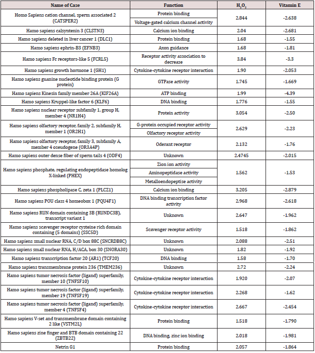
Recent studies have reported gingival irritation and chemical burn after at-home bleaching. Kirsten and others reported that patients experienced gingival irritation from at-home bleaching both immediately after the procedure and up to 45 days following treatment. Another study reported that hypersensitivity and gingival irritation disappeared within two days after in-office bleaching. Previous investigations have shown that 15% of patients reported gingival irritation after in-office bleaching, but it was possible to safely control contact of the bleaching gel with the gingival margin by using light-cured gingival dams [17,18]. Among the human genes listed in Table 2, TNFSF10 belongs to the tumor necrosis factor (TNF)–α ligand superfamily. TNFRSF4 and TNFRSF19 belong to the TNF-α receptor superfamily (TNFRSF). Other authors have examined the gingiva and proinflammatory cytokines. Two types of in-office were examined for bleaching agent and one type of at-home bleaching agent. Found that interleukin (IL)–1β expression increased with in-office bleaching but that there was no change in the expression of IL-10. In inflamed tissue, macrophages and other cells of the innate immune system synthesize TNF-α, a proinflammatory cytokine, to fight off infection and treat tissue damage. This TNF-α then binds to cell surface receptors and induces the production of other cytokines, triggering and maintaining inflammation. It is possible that H2O2 came into contact with the gingiva triggered a cellular response through the inflammatory cascade via TNF-α resulting in chemical burn [19,20].

Table 2: Correlation between H2O2 and its impact to human gene.

Lupinepublishers-openaccess-pediatric-dentistry-journal

Carcinoma potential

Another study also reported that using hydrogen peroxide and alcohol on a daily/weekly basis encourages the promotion of malignant neoplasm in the oral mucosa. Alcohol, for instance, potentiates in 50 times the harm caused by tobacco. Mouth washing with hydrogen peroxide, using products made with alcohol or drinking alcohol everyday may lead to the risk of oral chemical carcinogenesis. Also known as urea peroxide, sodium perborate, carbamide peroxide or other less common names, the hydrogen peroxide may have deleterious effects on the enamel, dentin, cementum, pulp and gingiva [6]. These names vary according to the presentation and formulation of the product. However, if both formulation and presentation of the product are controlled, and if the product is properly applied by a professional who takes the appropriate compensatory measures, its use is safe. In the oral mucosa, hydrogen peroxide potentiates the effect of many other carcinogenic agents found in patient's mouth. These carcinogenic agents may originate from food, cosmetics, hygiene products, pesticides, herbicides, tobacco, alcohol, virus, among others. Such potentiation happens due to the fact that these products are promoting agents of oral chemical carcinogenesis [21]. In mice low doses of hydrogen peroxide (0.1% and 0.4%) administered in drinking water caused adenomas or adenocarcinomas in the duodenum. These findings have been questioned and it has been proposed that the most likely cause of the lesions observed were the decreased water consumption of the mice and resultant abrasion of the luminal lining on ingestion of pelleted dry rodent food. Another animal study suggested that a known tobacco carcinogen 9, 10-dimethyl-1, 2-benzanthracene (DMBA) could be augmented in producing carcinogenesis in the buccal epithelium of hamsters by 3% H2O2.These results have been disputed due to the small number of animals studied. Further studies in hampsters given up to 70mg/kg of H2O2 by oral intubation for up to six months or up to 3% H2O2 in combination with DMBA applied five times daily for 16-20 weeks on the buccal epithelium did not show any sign of carcinogenesis. Further studies on skin have concluded that H2O2 is inactive as a tumour promoter or carcinogen [22,23].

Impact in root resorption and tooth sensitivity

An adverse effect that has been reported following internal tooth bleaching is cervical root resorption (an inflammatory-mediated external resorption of the root). Summarizes the available data to support a correlation between internal tooth bleaching and cervical root resorption. In these cases, it is very difficult to distinguish if the root resorption noted was due to the effect of the bleach or the trauma [21,24]. A high concentration of hydrogen peroxide in combination with heating seems to promote cervical root resorption. The underlying mechanism for this effect is unclear, but it has been suggested that the bleaching agent reaches the periodontal tissues through the dentinal tubules and initiates an inflammatory reaction.  In vitro studies using extracted teeth showed that  hydrogen peroxide placed in the pulp chamber penetrated the dentine, that heat increased the penetration and that the penetration is greater in teeth with cervical cemental defects. Intracorneal bleaching with 30% hydrogen peroxide reduces the micro-hardness of dentine and enamel  and mechanically weakens the dentine [5,19].

Case reports and small clinical studies have confirmed that a 10% carbamide peroxide gel used in a bleaching tray at night, (the so-called nightguard vital bleaching technique), produces predictable tooth whitening as do H2O2 strips and 'power bleaching' using 35% H2O2 with or without light and/or heat activation. Tooth sensitivity is a common adverse effect of external tooth bleaching. Data from various studies of 10% carbamide peroxide indicate that from 15-65% of patients reported increased tooth sensitivity. Higher incidences of tooth sensitivity (from 67-78%) were reported after bleaching with H2O2 in combination with heat [22,25]. There are many carcinogenic products available for sale, for instance: alcoholic beverages, cigarettes, ground tobacco, medicines, insecticides and others. These products are sold directly to the consumer after warnings and awareness campaigns had been carried out. That is how democracy works; products are sold directly to the consumers, but with a wide range of awareness, classification and explanatory campaigns stating that these products are carcinogenic. Hydrogen peroxide reddens the mucosa and gingiva by wounding them with tissue dissolution and inflammation. Hydrogen peroxide burns and may lead to necrosis of gingival papillae. It completely cleans the teeth because it demineralizes the enamel and also removes dirt or pigments. The enamel becomes porous and food stains it even more, increasing the need for mouth washing. Enamel becomes thicker every day. Should there be any restoration, it will induce microleakage through its interface with the tooth, causing the enamel to come out easily while eating [22,26].

If burning the mucosa and demineralizing the enamel were the biggest problems, we could think about using hydrogen peroxide with moderation. However, the biggest problem is that hydrogen peroxide is a promoting agent. In other words: it potentiates the effect of inducers of oral, throat, esophagus, stomach, and intestine cancer. Pesticides, tobacco products, alcohol, HPV and other oncogenic viruses, sunray and chemicals found in industrialized food are potentiated by hydrogen peroxide. Many doctoral dissertations, master's thesis, research and books confirm what the literature has already proved by means of different methodologies. In vivo chemical carcinogenesis experiments prove the carcinogenic effect of hydrogen peroxide on the mucosa that receives tooth bleaching products, antiseptics, and toothpaste [20,27]. Dental bleaching is based on the ability of H2O2 to penetrate through tooth structure and produce free radicals to oxidize the colored organic molecules. There are many reports investigated the effect of dose and exposure time of H2O2 to the pulpal tissues as the bleaching agents penetrated through the tooth structure.  The safety of H2O2 tooth bleaching is still controversial: its genotoxicity and carcinogenicity are under active discussion. Diaz-Llera et al. showed that 0.34–1.35 μM of H2O2 induced hypoxanthine guanine phosphoribosyl transferase (HPRT) mutation both in vitro and in vivo. High-dose H2O2 was reported to be mildly carcinogenic for the duodenum of catalase-deficient mice. In another report, 1% H2O2 (~ 0.3 M) in drinking water induced forestomach tumors in rats. These reports showed that exposure to high-dose H2O2 for a sustained period induces oxidative stress that leads to DNA damage in mammalian cells [26,27].

Conclusions

The use of chemical agents in dental treatment can cause the chemical burns in oral cavity. So, the patients need to listen more towards the dentist about the instruction after treatment so the materials cannot cause the irritation in oral mucosa.

Read More Lupine Publishers Pediatric Dentistry Journal Articles:
https://lupine-publishers-pediatric-dentistry.blogspot.com/
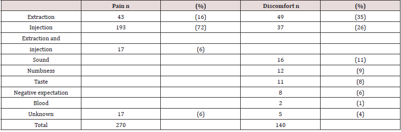
980 nm Diode Laser: A Good Choice for the Treatment of Pyogenic Granuloma

Abstract Pyogenic granuloma is a benign non/neo plastic mococutanous lesion . It is a reactional response to constant minor trauma and ca...