Friday, February 25, 2022

Lupine Publishers | Complete Unilateral Cleft Lip and Palate Repair: A Modification of the Millard Surgical Technique

 Lupine Publishers | Journal of Pediatric Dentistry


Abstract

Cleft lip is one of the most commonly encountered craniofacial deformities. The comprehensive treatment of cleft lip and palate deformities requires thoughtful consideration of the anatomic complexities of the deformity and the delicate balance between intervention and growth. As evidenced by the multiple techniques that have been developed for its repair, a functionally and aesthetically pleasing result is challenging to attain. In this study, we describe a modification of the surgical technique for the repair of complete unilateral cleft lip and palate, through the design of flag-shaped flaps without removing any tissue, as a modification to the rotation and advancement flaps or Millard technique.

Keywords: Cleft lip; cleft palate; rotation; advancement flap

Introduction

Since 390 B.C.E., the treatment of upper lip clefts has represented a challenge for surgeons [1]. The modern reconstruction of clefts requires an anatomical, functional, and tridimensional understanding of the cleft (and noncleft) lip, nose, and alveolar bone. Multiple repair techniques and modifications reflect the wide variety and constant evolution of the surgical principles for the repair of clefts [2]. Lip development occurs from the fourth to the seventh week of gestation [3,4]. Unilateral cleft lip occurs when the complete merging of the two maxillary processes and the medial nasal process on one side fails. Several anomalies can happen during the lip and palate development, finding the cleft lip and palate as the most frequent anomaly (and the cleft lip alone being less frequent) [5]. Cleft lip (with or without a cleft palate) is most frequently found in boys with a 6:3:1 ratio of left/ right/bilateral involvement, respectively [6]. This anomaly occurs in approximately 2 out of 1000 Asians, 1 out of 1000 Caucasians and 0.5 out of 1000 Afro-Americans. Cleft lip is usually isolated and associated with few syndromes like van der Woude, DiGeorge, Conotruncal anomaly, Velocardiofacial syndrome and Stickler [5]. The aim of this study is to describe a modification of the surgical technique for the nasal and complete unilateral cleft lip repair, with excellent aesthetical and functional results, without the need of removing any tissue

Technical Note

Preoperative consideration

The ideal timing of lip repair is between 3 and 6 months of age and it is recommended that the patient weigh at least 10 pounds and have a hemoglobin of at least 10 g/dL. Prior to surgery, patients wear presurgical orthopedics. These techniques are ideally initiated in the first week of life and are continued until the time of repair. The technique proposed by Dr. Tulio Chacín, has been used exclusively to repair defects in a complete unilateral cleft lip and palate (CLP). This technique consists of rotation and advancement flaps with the modification of a flag-shaped flap design for the reconstruction of the posterior side of the lip and vestibule, tractioning the displaced alveolar process and repositioning the displaced nasal structures, without removing any tissue. The goal of cleft lip repair is to recreate a continuous oral sphincter, attain adequate vertical lip height with symmetry of cupid’s bow, and generate symmetry of the nostrils and nasal sill with a minimally visible scar. This study was approved by the Hospital Coromoto de Maracaibo IRB and all participants signed an informed consent agreement.

Surgical technique

Surgical repair is performed under general anesthesia with an oral tube placed at the midline lower lip so that it does not distort the upper lip. The neck is slightly ex- tended with a small shoulder roll and the OR table is tilted slightly in reverse Trendelenburg position. An alcohol pad is used to dry the vermillion boarder and enhance its identification. Key landmarks (as described below) are marked with methylene blue using a 30-gauge needle. This is performed prior to injection of the local anesthetic.

Landmarks

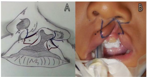
a) Point 1: Lateral peak of the lip (noncleft side).
b) Point 2: Center of the philtrum (located on the mucocutaneous line).
c) Point 3: Lateral peak of the lip (cleft side) (equal to the distance between 1-2).
d) Point 4: Lateral wall of the columellar base (noncleft side).
e) Point 5: Lateral wall of the columellar base (cleft side).
f) Point 6: Medial wall of the nasal ala (noncleft side).
g) Point 7: Medial wall of the nasal ala (cleft side). It’s drawn from point 9 with a medial direction, equal to the distance between 3-4.
h) Point 8: It is placed where the horizontal mucocutaneous line becomes vertical and the mucous border starts to get thin (the distance between 7-8 must be the same as the distance between 3-5).
i) Point 9: Lateral wall of the nasal ala (cleft side).
j) Flap A: Lip mucosa of the noncleft side: used to reconstruct the posterior wall of the lip, and it is fixated on the displaced alveolar zone.
k) Flap B: Lip mucosa of the cleft side: used to reconstruct the posterior wall of the lip, and it is fixated on the non-affected alveolar ridge.
l) Point C: Used to release a portion of the incorrectly positioned alar cartilage.
A – B = Flag-shaped flaps.
C = Nasal structural reposition (Figure 1).

Figure 1: Anatomic landmarks and flag-shaped flaps design.
Lupinepublishers-openaccess-pediatric-dentistry-journal

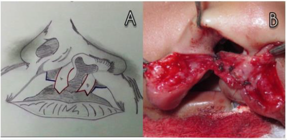
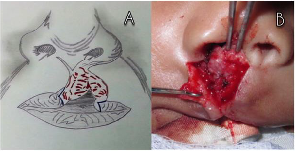
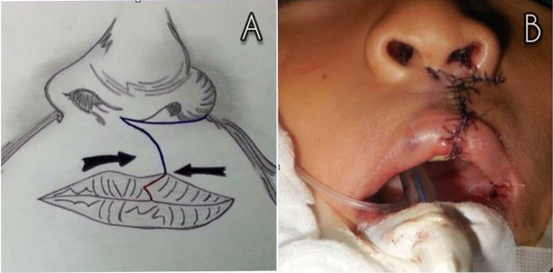
Taking into consideration these reference points and the principles of the rotation and advancement technique, the flagshaped flaps proposed by Dr. Tulio Chacín are designed. These flaps consist of an A flap on the lip vermillion of the noncleft side, having a triangular design with a distal end and a proximal base. A B flap on the contralateral vermillion, with an M form on its distal end and having a proximal base. Furthermore, a marginal incision is performed on the nasal vestibule of the cleft side in order to release the alar cartilage and improve the rotation of the nasal component. Once the design of the technique has been made, the nasal-labial reconstruction is conducted. Starting with the cephalic transposition of the flag shaped flaps, which are fixated on the nasal floor and the alveolar bone respectively, intertwining them (flap A with flap B), in order to create the posterior part of the lip and the oral vestibule as well as giving support to the nasal component (Figure 2). After that, the muscular and cutaneous component are settled (Figure 3). Additionally, on the remaining mucous plane, the coincidence of the triangular design on the lip vermillion is detailed, in order to break the wound and avoid its retraction, which could create a whistler deformity (Figure 4).

Figure 2: Release of the flag-shaped flaps and cephalic reposition of the flag-shaped flaps.
Lupinepublishers-openaccess-pediatric-dentistry-journal

Figure 3: Muscle component reconstruction.
Lupinepublishers-openaccess-pediatric-dentistry-journal

Figure 4: Skin component and vermilion border reconstruction.
Lupinepublishers-openaccess-pediatric-dentistry-journal

Discussion

The first record about unilateral cleft lip repair was written in 390 A.D. in the Tang dynasty in China. The edges of the cleft lip were cut and sutured, and the child could not speak for a period of 100 days [7]. Ambroise Pare repaired unilateral cleft lips by merging/ joining both sides of the cleft, with a large needle and thread [8]. Rose and Thompson reported a modification, consisting of straightline closures, for the unilateral cleft lip repair. All of these “straight line” techniques closed the cleft. However, vertical retractions and notches in the lip were frequently found after [9,10]. Several efforts have been made in order to improve the aesthetic and functional results obtained by the “straight line closure” technique. Within these, several types of geometrical repairs with full-thickness lip flaps are included. These techniques were designed with the purpose of breaking the scars on the lip and to prevent vertical retractions and notches [11]. LeMesurier reported a quadrangular full thickness (with a lateral base) flap on the cleft side, which intercepts the scar in the mucocutaneous union [12]. Tennison, Marcks et al, introduced a triangular flap, where a Z-plasty was designed on the inferior section of the lip [13,14]. Later, Randall uses the same design as Tennison, but reducing the size of the triangular flap [15]. Millard, in 1957, reports the rotation and advancement flaps technique for the repair of the unilateral cleft lip. This technique consists of 2 full-thickness flaps and its design is made in order to place the scar on the philtral ridge, and it is the most used technique to repair unilateral cleft lips nowadays [16].
The primary objective of the treatment of unilateral upper lip clefts is to restore the anatomy and function of the lip. Other objectives are to close the nasal floor, correct the asymmetry of the nasal tip and to approximate the alveolar cleft. The rotation and advancement technique involves two full-thickness flaps, which are approached in order to repair the cleft and avoid the notching of the lip. This design allows the rearrangement of the orbicularis oris muscles. The geometrical flaps techniques require a more accurate design, which allows creating flaps that are more precise in shape and extent. This is ideal for less experienced surgeons. However, with this technique the surgeon will have less flexibility during the surgical procedure. The main advantage of the rotation and advancement flaps is the flexibility of the technique, allowing continuous modifications during its design, incisions and the repair. Another advantage is that the incision is designed in order to place the scar on the new philtral ridge. Most of the geometrical flaps violate the philtral sub-unity. Within other advantages of the rotation and advancement technique we can find: the minimum waste of tissue and the maximum repair of the muscular component. One disadvantage of this technique is that less experienced surgeons could have difficulties with the flap design. Also, this technique requires an extensive dissection, having the tendency to create a small nostril on the cleft side. Because of this, the surgeon must try to make the cleft side nostril slightly wider than the one from the noncleft side, given the fact that it is easier to repair a wide nostril than a narrow one [17].
In conclusion, the technique proposed by Dr. Tulio Chacín for the nasal and complete unilateral cleft lip repair represents a valuable tool for plastic/maxillofacial surgeons when repairing this severe anomaly. Modifications in this technique include: the reconstruction of the posterior wall of the lip, not removing any tissue, the traction of the displaced alveolar process, the reposition of the displaced nasal structures (giving them support), and the breaking of the wound on the lip vermillion in order to avoid retractions. The aesthetical and functional sequels of the techniques used previously (such as: short and depressed lips, the retraction of scars, an inadequate reposition of the affected nasal ala and the collapse of the cleft alveolar process) create the need for modifications, and some of them are obsolete and not even used nowadays. We consider that the nasal and complete unilateral cleft lip repair, using Dr. Tulio Chacín’s technique, may be one procedure to choice during the correction of this complex anomaly.

Funding

No funding was received for this study

Conflicts of Interest

The authors declare that they have no conflicts of interest.

Acknowledgment

Special thanks to Michelle De Bacco for making the illustrations of the surgical technique.

Read More Lupine Publishers Pediatric Dentistry Journal Articles:
https://lupine-publishers-pediatric-dentistry.blogspot.com/

Friday, February 18, 2022

Lupine Publishers | Presurgical Infant Orthopaedics - Journey So Far

 Lupine Publishers | Journal of Pediatric Dentistry


Abstract

The successful management of patients with cleft lip and palate deformity requires a multidisciplinary approach. Historically, cleft lip and palate care starts with treatment modality of presurgical infant orthopaedics (PSIO). However, the necessity of presurgical orthopaedics in managing the resulting orofacial deformity is the discussion to ponder upon due to the variety of methodologies available and results produced by these devices. The objectives of this paper were to review the journey of PSIO appliances so far, basic principles of PSIO treatment, the various types of techniques and the protocol followed, and to critically appraise the advantages and disadvantages of these techniques. In conclusion, we believe that PSO treatment, with its objective to approximate the segments of the cleft maxilla may reduce the intersegment space in readiness for the surgical closure of cleft sites.

Keywords: Cleft lip and palate; presurgical infant orthopaedics; PSIO

Abbreviations: PSIO: Presurgical Infant Orthopaedics; CLP: Cleft Lip and Palate; NAM: Nasoalveolar Molding; DMA: Dentomaxillary Advancement Appliance; UCLP: Unilateral Cleft Lip and Cleft Palate

Introduction

Cleft lip and palate (CLP) are the most common congenital malformation caused due to variation in development of facial structure during gestation [1]. The incidence of patients with CLP is about 1.7 in 1000 live births globally [2]. The incidence is highest in Afghan population as 4.9 and lowest in Negroid population as 0.4 per 1000 live births [3,4]. The presence of cleft involving lip, palate and alveolus results in disfigurement and distorted growth and development. There is wide presentation of facial features among patients depending upon the severity of the cleft. A wide nostril base, separation in the upper lip of the cleft side is the characteristic feature of unilateral cleft defect. There is lateral and inferior displacement of affected lower lateral nasal cartilage which results distortions in the anatomic form of nose, tripod tilt in skeletal structure, a depressed dome, increased alar rim and deformities in apex of nostril. Shift in the base of the nose, deviation of septum to non-cleft side is also seen in patients with CLP. The separated premaxilla may overhang from the maxilla with variation in size [5,6]. Supervision and management of patients with CLP is a process that begins in infancy and continues in adulthood. Early treatment in the form of Presurgical Infant Orthopaedics (PSIO) is required to reduce the cleft width and to help maxillary arch development, thereby improving occlusion, feeding, speech, hearing, and language development and aesthetics [6,7]. PSIO has been defined as “use of forces to reposition tissues secondarily displaced due to a cleft deformity” [8]. Active and passive orthopaedic appliances have been developed for correction of CLP defect by using compressive & tensional forces or passively guiding growth. The aim of PSIO is to decrease the width of the cleft gap, to achieve a favorable alignment in the cleft segments within the initial few months of infancy prior to cheiloplasty, and to allow surgical repair with minimal tension [9]. In addition, there is improvement and ease in feeding, increased fluid intake, subsequently weight gain, improvement in functioning of tongue, reduced risk of aspiration and reduction in severity of dental & skeletal deviations.

Various methods and treatment protocol have been developed over ages and suggest PSIO prior to the primary surgery in patients with CLP for better surgical aesthetic results and prevent the social stigma. The aim of this review is to sum up the history, evolution, efficacy, advantages, disadvantages, complications, recent advances of different PSIO appliances, and critically analyze the evidence as well as the current status of PSIO.

Historical Perspective

PSIO has been a part of treatment strategy used in the management of patients with CLP for centuries. As early as in 1556, detailed explanation of the indications, surgical technique, and post-operative care of the cleft lip has been documented. A technique involving lip repair with cleft lip pins has been described by Amboise Pare in 1575. However, it was in the year 1689, Hoffmann demonstrated the use of facial binding to narrow the cleft and thereby prevent postsurgical dehiscence. The technique of retraction of the maxilla before surgical repair in patients with bilateral CLP was introduced in 1790 by Desault [10,11]. Adhesive tape binding usage in presurgical preparation was popularized by Hullihen [12]. Brophy in 1927 clinically demonstrated that silver wire passing cleft alveolus can be gradually tightened to approximate the alveolus before lip repair [13]. The modern school of presurgical orthopaedic to mould the alveolar segments using a series of plate system with active forces was introduced by 1950 McNeil [14] later popularized by Burston. Cupid’s bow and the philtrum symmetrical correction as Millard’s rotation advancement closure technique was introduced by Millard in1960 [15]. A pinretained active appliance which could simultaneously help in retraction of premaxilla and expansion of the posterior segments was introduced by Georgiade and Latham in 1975 [16]. The use of a passive orthopaedic plate for slow alignment of the cleft segments was described by Hotz in 1987 [17]. Matsuo’s (1988-91) series of research on molding of neonatal nasal cartilage and nostril with the help of silicone tubes was the gateway to invent newer modern methods [18-20]. The paradigm shift in the PSIO treatment was with the introduction of Nasoalveolar molding (NAM) by Grayson and Cutting in 1993, a novel technique in which presurgical molding of the alveolus, lip and nose is carried out in infants born with CLP [21].

Objectives of PSIO

Literature has highlighted the objectives of PSIO: to stimulate growth of patalal shelves, upper arch development, improvement in the projection of nasal tip leading to overall growth of the face. It also facilitates improvement in occlusion, feeding, speech, hearing, and language development. Eventually, PSIO aim at achieving a more uniform osseous base. The achievements of these objectives facilitate surgical closure and improve the final aesthetic result [22- 25].

Classification of PSIO appliances

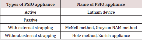
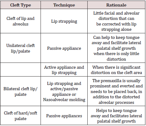
PSIO appliances can be classified into active and passive appliances based of force application (Table 1). Active appliances act by active forces being applied on the separated alveolar processes for growing them into desired anatomic position. The various appliances used for PSIO are discussed below and the technique of PSIO in different cleft types is summarized in Table 2.

Table 1: Classification of PSIO appliances.

Lupinepublishers-openaccess-pediatric-dentistry-journal

Table 2:PSIO techniques in different cleft types.

Lupinepublishers-openaccess-pediatric-dentistry-journal

Lip taping

Approximation of the alveolar segments within 5 mm of each other can be managed by using Lip taping. In this technique medical adhesive tape such as Steri-strips® is placed across the upper lip in the first week of life following which skin adherent dressing such as Tegaderm ®, are placed over the area of the cleft lip. Elastic forces will exert a retracting, backward pressure against the protruding premaxilla, improve their positions and allow definitive lip skin repairing. The clinical effectiveness of Lip taping is documented, however with only limited studies [26].

McNeil method

The pioneer work for alignment of the alveolar parts presurgical for patients with CLP was done by McNeil, who believed that a normal position of maxilla, alveolar & palatal cleft segment can be achieved by molding and approximating segments into correct preplanned position using a series of appliances. This method not only stimulated soft tissues to grow in cleft region but also modify the postnatal development of the maxilla. This method was further popularized by Burstone, an orthodontist. The advantage of this method is that a smaller number of appointment are required, hence encourage patients who may have to travel long distances for treatment [14,27].

Latham appliance

The Latham appliance also known as the Dentomaxillary Advancement Appliance (DMA) was developed to align the alveolar arch through rapid orthopedic correction and alignment of cleft segments was introduced by Dr Lantham and Georgiade [16,28]. Latham based his treatment concept on the facial growth hypothesis of Scott [29,30] with aim of the procedure ‘to carry the interrupted embryonic process to normal completion’ by maxillary alignment, stabilization of the alignment along with tunneling of the alveolar cleft with periosteum, and reconstruction of the nasal floor to support the alar base. This appliance is an active pin-retained appliance fixed surgically to the bone for patients with age around 2 to 5 months. The appliance works by simultaneously applying pressure to the cleft segments over a 4 to 6-week period to move the alveolar segments into proper position, which is followed by alveolo periosteoplasty and lip adhesion. According to Drs. Latham and Millard, these alignments allow the performance of gingiva periosteoplasty (GPP), providing stabilization of the maxillary segments and reconstruction of the nasal floor [31]. Greater values for cephalometric measurements in maxillary length, maxillary prominence and ANB angle has been found for patients treated with this appliance [32]. However, other authors have concluded that this appliance did not affect dental arch relationships in preadolescent children [33]. The problem associated with procedure is that, besides neonatal maxillary orthopedics, infant periosteoplasty is always performed, although it is more limited with less undermining of periosteum on the maxilla.

Hotz appliance

In Europe, the treatment principles of McNeil for neonatal maxillary orthopaedics were greatly modified by grinding away the acrylic in specific areas to bring out necessary alignment, known as Hotz appliance (Zurich approach). According to Hotz and Gnoinski, the primary aim of presurgical orthopedics is not to facilitate surgery or to stimulate growth, as postulated by McNeil, but to take advantage of intrinsic developmental potentials. In Zurich approach lip operation is performed at the age of 6 months while palate repair is postponed until 5 years of age [34,35]. The appliance is made of hard acrylic or a combination of hard and soft acrylic: it passively covers the alveolar segments and extends slightly into the area of the cleft and the buccal sulci. This appliance assists with both bottle-feeding and to allow some breast-feeding in infants with CLP. Harmonization in the vertical and transverse positions of the cleft segments has been found with Hotz plate therapy [36]. Long-term effects of the Hotz plate and early lip adhesion have been studied by several researchers and it has confirmed that arch width and length of the anterior part of the maxilla improves better than other treatment options [37]. Similarly, the two-stage palatoplasty in combination with application of the Hotz’ plate has good effects on the maxillary growth than one stage palatoplasty without Hotz plate [38].

Nasoalveolar molding

Earlier PSIO appliances were designed to correct the alveolar cleft only, despite the fact that the nasal deformity among these patients remains the greatest esthetic challenge. Grayson [21,39] described a new technique to presurgical mould the lip, alveolus and nose in infants born with CLP. The concept of naso alveolar molding (NAM) works on Matsuo’s principle; [18-20] that the nasal cartilage could be molded due to increased plasticity concurrent to increased levels of maternal estrogen if treatment is initiated within 6 weeks of life. The NAM appliance consists of an intraoral molding plate with nasal stents to mould the alveolar ridge and nasal cartilage concurrently. Beside other advantages of traditional plates, the main objectives of NAM appliances are improving nasal symmetry and lip aesthetics while elongating the columella and correcting nasal cartilage deformity. Hence, a less extensive surgery is required for the lip and nasal repair, and there is less tension on the reconstruction, greater nasal symmetry is be obtained after cleft lip repair using NAM therapy as well as better lip form, reduced oronasal fistulas and labial deformities, and a namely a 60% reduction in the need for secondary bone grafting [24].

Objectives of NAM in unilateral cleft lip and cleft palate (UCLP)

The main objective of NAM for UCLP is to reduce the severity of the original cleft deformity by reducing the width of the alveolar cleft segments and alignment of the base of the nose and lip segments [21]. Taping the lips together helps in correction of the inclined columella upright along the mid-sagittal plane. As the lower mid-face skeletal elements (alveolar ridge and lower maxilla) improve in relation to each other, the overlying soft tissue improves concurrently. The alar rim, which was initially stretched over a wide alveolar cleft deformity, shows some laxity that enables it to be elevated into a symmetrical and convex form. The nasal tip on the cleft side is overcorrected in its forward projection; this is achieved through the use of a nasal stent, an intra-oral acrylic plate, and surgical tapes [39-44].

Objectives of NAM in bilateral cleft lip and cleft palate cases

The main objective being the non-surgical elongation of the columella and also to center the pre-maxilla, along the mid-sagittal plane, retraction of the pre-maxilla in a slow and gentle process to achieve continuity with the posterior alveolar cleft segments. Reduction in the width of the nasal tip, improved nasal tip projection and increase in the nasal alar base width [21,39,43].

Benefits of NAM

Proper alignment of lip, nose and alveolus is achieved, thereby enabling surgeons for better surgical repair of the cleft deformity and hence reduce post-surgical breakdown [42,43]. Approximation of alveolar process before surgery also enables surgeons to perform gingivo-periosteoplasty successfully. NAM provides stable change in nasal shape with less scar tissue and better lip and nasal form. It also reduces the number of surgical revisions for excessive scar tissue, oro-nasal fistulas, nasal and labial deformities, due to proper columellar elongation and lengthening. With the alveolar segments in a better position and increased bony bridges across the clefts, the permanent teeth have a better chance of eruption in a good position with adequate periodontal support [39,40].

Complications of NAM

a) Locked-out segments: It may occurs due to the poor and un-volunteered molding process, wherein the greater segment moves more rapidly, without the change in position of the lesser segment, as a result, the lesser segment gets locked out behind the greater segment.
b) Nostril overexpansion (Mega-nostrils): This occurs when the nasal stent application is started before the size of the cleft gap is adequately reduced. The premature nasal stenting exerts excessive force against the nasal tissue leading to excessive alar expansion and resulting in mega-nostrils.
c) Tissue ulceration: It occurs due to application of pressure by the intra-oral acrylic appliance, which may be due to ill-fitting appliance. At times the area under the horizontal prolabium band may also get ulcerated, if the band is too tight.
d) Skin ulceration: It may result due to frequent application and removing of tape, resulting in irritated and ulcerated skin over the cheek region.
e) Dislodgement of the acrylic plate: It is the complication which may result in obstruction of the airway. This can only occur, if the arms of the appliance are taped too horizontally or with inadequate activation [39-44].

Prevention of the complications associated with NAM Therapy

a) NAM therapy must be closely monitored and volunteered at timely basis with adequate application of mechanics and robust principles of the therapy must be followed.
b) Tissue expanding direction and associated mechanics should be monitored vigorously and nasal stenting should commence only after the cleft gap is reduced by minimum 6 mm and softer denture liner must be covered over the nasal stent tip, so as to apply gentle forces. c) Tissue ulcerations can be prevented by coating of tissue lubricant over the appliance before insertion into the oral cavity.
d) Skin ulcerations over the cheek region can be prevented by using Duo-derm or Tega-derm, underneath the tape strapped.
e) Parents must be thoroughly educated to continue the use of NAM appliance for their child until the therapy lasts. Feeding instructions must also be given accordingly.
f) Motivating the parents to visit the dentist on scheduled appointments and in timely manner is of utmost importance for a successful NAM therapy [39-44].

Modifications

Recently NAM appliance has been modified by different authors in many ways. It includes modified muscle-activated maxillary orthopaedic appliance [45], incorporation of expansion screw [46], dynamic presurgical nasal remodeling intraoral appliance design [47], extra-oral nasal molding appliance [48], self-retentive appliance with orthodontic wire [49,50], use of TMA wire instead of SS wire for making nasal stent [51] and the latest technique of OrthoAligner “NAM” [52] & NAM custom aligners [53].

DynaCleft ® and Nasal Elevators

DynaCleft® is a premade nasal and alveolar molding device which can be used to successfully mold the upper lip, alveolus and nose prior to cleft lip repair. Traditional surgical adhesive tape (e.g. Silk tape, Steri-strips®) have been used in the past, unlike tape, DynaCleft® offers the benefit of being able to provide a constant approximation force with an elastic centre that allows it to conform to a baby’s mouth better because of its ability to expand and contract. Additionally, the controlled force provided to the prolabium and premaxilla could improve surgical results and decrease the necessity of early lip adhesion surgery. As the DynaCleft® device is pre-made; there is no need to create custom-made devices for the molding process. Studies have shown that with the use of DynaCleft® with a nasal elevator has produced results similar to that of NAM therapy. However, unlike the NAM appliance, it does not require adjustments with growth of the infant. Nasal elevators have been found to improve the shape of the nose and alae, thereby reduce the need for primary surgery to the nose in patients with UCLP. Due to its elastomeric core and stretch property, DynaCleft® allows the infant to feed and cry without limitation.

Conclusion

Many orthodontists working on patients with CLP have shown great enthusiasm for PSIO to improve surgical outcomes with minimal intervention. Although different forms of PSIO appliances are available, it seems that NAM therapy has been especially popular in all over the world. Undoubtedly, every orthodontist or surgeon aims to use the best treatment modality for their patients. Nevertheless, PSIO effects can be confounded by surgical type and timing of the primary repair, as is discussed in many studies. In such cases, one should be cautious when evaluating the particular outcomes for patients with CLP since it is difficult to differentiate the sole effect of an individual surgical or orthodontic intervention.

Read More Lupine Publishers Pediatric Dentistry Journal Articles:
https://lupine-publishers-pediatric-dentistry.blogspot.com/

Friday, February 11, 2022

Lupine Publishers | It’s Never Too Early to Cleanse the Oral Cavity of Our Babies

 Lupine Publishers | Journal of Pediatric Dentistry


Editorial

Dental caries continues to be the single most common chronic disease of infancy and one most widespread pathologies in the world. In Italy recent studies showed that 22% of 4-year old and 44% of 14-year-old suffer from dental caries. In industrialized nations this trend appears to be decreasing in 6-19-year old. Dental caries is increasing by 15.2%, according to recent epidemiological studies (2007), in children aged 2-5 years, in Milan, in the north-east of Italy, regrettably in the news, as the main site of the Covid-19 outbreak, last March 2020. Why are deciduous teeth more prone to caries? In part because although the enamel of deciduous teeth takes more time to develop, it demineralizes faster. Parent responsibility is also an important factor. Many parents don’t begin cleaning their children’s teeth until they reach 2-3 years of age and often it’s not performed properly. A diet rich in sugar, snacks, candy and chocolate also plays a key role. Another cariogenic feature is the prolonged use of the baby bottle filled with sugary or acidic liquids given to the child in the evening or during the night, when there’s a reduction in saliva flow.

For caries to form three factors must be present:

a) A tooth that easily demineralizes
b) Sugar in the diet
c) Specific bacteria

The first factor cannot be modified, while the other two are adjustable. Intervening on either one of these two aspects can prevent caries from forming. The easiest to correct, aimed at reducing the number of harmful bacteria, is oral hygiene. Bacteria begin developing in the infant’s oral cavity following birth. At birth the oral cavity is sterile. In the children’s saliva mainly bacterial strains of the respective mothers are found. The main source of infection is, therefore, maternal saliva. Primary prevention for the 2-5 age group can and must be initiated by the mother, followed by the father, the grandparents or other care givers. It’s never too early to begin cleaning the child’s or even the newborns oral mucosa. How? By using a clean or sterile gauze. It’s is a very simple but efficient means of removing bacteria from the oral mucosa. Place the index finger in the center of an open gauze then wrap it around the index finger of your dominant hand. To soften the gauze dampen it with a normal saline solution or plain tap water. The mouth is gently opened and the oral mucosa along with the gums are delicately rubbed. This simple procedure is effective in reducing the number of bacteria, while at the same time being non traumatic to the infant or small child.

Everyone agrees that prevention is more important than treatment, but only few reward acts of prevention.

An article entitled “Oral Diseases Affect Some 3.9 Billion People”, published in 2015 in the evidence-based literature search site (Pub Med) revealed how widespread oral disease is on a global level. Today our understanding of the ferocity and aggressiveness of bacteria and viruses that inhabit or transit via the oral cavity is greater than ever before. The need to implement procedures for their mechanical reduction is crucial and it must begin at infancy. In Italy, during the period of forced lock-down due to the Covid-19 pandemic, parents became teachers, playmates, and hairdressers for their children. They invented new forms of creative entertainment even in their children’s home care. “Digital brushing” might have been cleverly introduced. Children would mimic their parent’s gestures by rubbing their oral tissues and teeth with a gauze wrapped around their finger soaked in normal saline solution or plain water. For the child this was a game, but from an oral health standpoint this is extremely important not only in preventing disease, but in forming good, lifelong habits. A scrubbing action is indispensable to detach the adhesive biofilm from oral structures. Rinsing will not remove it. Rinsing only removes loose, floating debris and does not reach bacteria and viruses enclosed in the inter-microbial plaque matrix. Gentle scrubbing will produce adequate cleansing, even in areas of recession or dentinal sensitivity. It can also be used in situations where the bristles of the toothbrush could become irritating creating an “avoiding” reaction.
The forced lockdown of the Covid-19 pandemic brought on other important lessons in hygiene. Parents taught their children to wash their hands often and instructed them not to touch their eyes, nose and mouth with unwashed hands, simple but powerful weapons in helping to prevent disease. As clinicians the treatment of patients is our core mission, but prevention and education are just as important, following a patient for a lifetime. Quoting Elisabeth Stone: “Making the decision to have a child - it is momentous. It is to decide forever to have your heart go walking around outside your body”.

Read More Lupine Publishers Pediatric Dentistry Journal Articles:
https://lupine-publishers-pediatric-dentistry.blogspot.com/
Wednesday, February 9, 2022

Lupine Publishers | Effect of Thickness on the Fracture Resistance of Ceramic Partial Restorations: A Review

 Lupine Publishers | Journal of Pediatric Dentistry


Abstract

This publication describes the effect of thickness and type of material on the resistance fracture of ultra-thin ceramic restoration. The restorative phase of the treatment should not cause additional damage of the residual tooth structure. Ultrathin restorations (veneers, onlays , inlays) are considered as an alternative to traditional onlays and complete crowns. the technical aspects required for the success and the good prognosis of those new restorative design based on the control of tooth preparation with diagnostic wax-up, provisionalization, and the use of CAD-CAM technology.

Keywords: Ultrathin; restoration; ceramics; thickness; fracture

Introduction

The dental enamel is designed to withstand a lifetime. Her progressive reduction is biological consequence of advancing age. The loss of tissue may be due to the action of acidic foods, gastroesophageal reflux disease medications, and the reduction of salivary flow [1]. There are now many new protocols for a new concepts of ultrathin and non-preceramic restorations. Ceramic veneers and inlays, onlays and overlays are frequently presented as the major class of clinical conservative modalities [2]. CAD/CAM technology became popular during the last decade for the conception and fabrication of restorations. Different materials are supplied in the form of blocks that are milled to obtain the restorations [3]. Among ceramic CAD/CAM materials, lithium disilicate have recently expanded their indications to include ultra-thin restorations, with promising results. Recently, hybrid ceramic (vita Enamic) has been developed to allow faster milling of the ceramic block as ultrathin restorations with good mechanical behavior and good prognosis. Due to the importance of dental tissue preservation, it’s important to evaluate the fracture resistance of reduced thickness materials made with different restorative materials [4,5].

Effect of Thickness

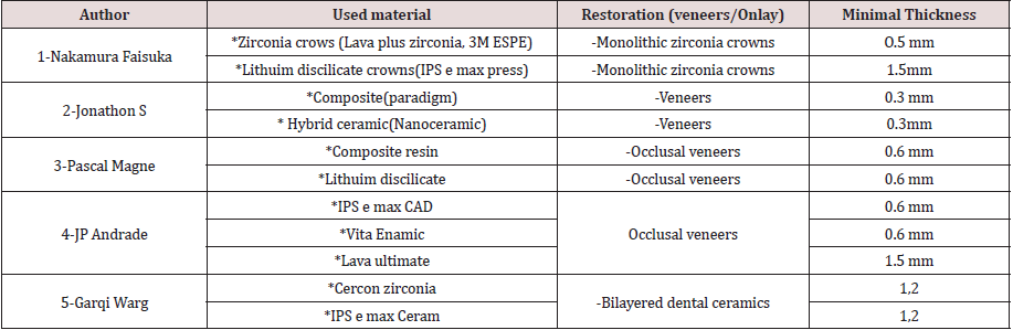
It’s very notable how well patients presenting with tooth fracture, moderate to severe loss of tooth structure when ultrathin restorations are proposed to them, both economic and biological costs are significantly lower compared to traditional and more invasive approaches. Minimal thickness can be used were in the Table 1 [1,2,6-8]. The possibility of making ultrathin (0, 3mm-0,6mm) ceramic restoration allows for a more conservative preparation with minimal wear to the tooth structure. It’s believed that these positive and promising results are due in part to the adhesive luting technique, dental substrate, and restorative material [13]. According to the study of Nordahi et al. comparing five thickness (0,3-0,5-0,7-1-1,5) for high-translucent(HTZ) and low –translucent (LTZ) zirconia restorations and glass ceramic (LDS) crowns. The lowest recorded load at fracture within & mm groups was 634 N, and 550N for the Y-TZP groups at thickness of 0,5mm.Compared to the forces measured during mastication (approximately 5 to 364N); the results suggest the possibility to reduce restorations thickness with good prognosis especially for Monolithic ceramic YTZP material [9]. On the other hand, the study of JP Andrade Showed that the fracture resistance was significantly higher at a thickness of 1,5 mm compared to a thickness of 0,6 mm for veneers made of lava ultimate and vita Enamic. Manufactures of lava Ultimate, vita 3namic and IPS e.max CAD, affirmed that restorations with a minimum thickness of 1, 5 mm on the occlusal surface of posterior teeth will support masticatory loads. Nevertheless, other studies showed that it is possible to treat severe erosive lesions or loss of wear on posterior teeth with ultrathin (0,5-1mm) ceramic and composite resin materials [1,5]. The study of JP Andrade evaluated 0,6 mm of thickness (veneers), which are considered ultrathin restorations. On the other hand, the study of Egbert et all, shows a fracture resistance of occlusal veneers with a 0,3 mm using Paradigm MZ 100, Vita Enamic and lava Ultimate; and found promising fracture resistances. Hence, it seems that the use of use of thickness smaller than 0,6 mm could be used with good prognosis [3].

Table 1.

Lupinepublishers-openaccess-pediatric-dentistry-journal

The Luting Material and Protocol

Ceramic indirect restoration looted by the adhesive luting technique provided better fracture resistance than conventional luting technique. Hence, the use of adhesive restorations has been recommended for reinforcing the remaining dental structure because It allows intimate contact between the dental substrate, luting agent, and ceramic material, therefore occlusal forces are dissipated through the root of tooth, periodontal ligament, and alveolar bone [3,8]. According to many studies, associating hydrofluoric acid with silane was the most effective surface treatment with which to potentiate the bond between the ceramic and the adhesive material [2,5,7]. The silane enhances the chemical bond between the silicon-containing materials and the resinous material used for luting.

The Type of Material [5,7,10]

The study of Katrin Heck et all showed that IPS e max CAD and lava ultimate should be preferred to IPS Empress CAD for the treatment of occlusal tooth loss with ultrathin restoration, whether this result is due to the viscoelastic proprieties of the composite material [11]. In another study , Johansson et al, compared fracture resistance of monolithic zirconia and monolithic lithium disilicate after cyclic loading and thermos-cycling. they reported higher strength for zirconia restorations with the same occlusal thickness (0,5mm and 1mm). According to Niklas Nordhal, Ceramic materials, such as glass ceramics and zirconia show a greater scatter in fracture strength compared to other material such as metal. This result calls for special factor approach when indicating reduced ceramic restoration [12].

Conclusions

Ultrathin restorations (inlay, onlays, veneers.) appear to be a promising restorative procedure in posterior and anterior teeth. The feasibility of their application depends on their fabrication options and fracture properties. Recent advances in technology and materials are offering new options for good treatment.

Read More Lupine Publishers Pediatric Dentistry Journal Articles:
https://lupine-publishers-pediatric-dentistry.blogspot.com/
Friday, January 14, 2022

Lupine Publishers | Pregnant Women’s Knowledge about Children Oral Health

 Lupine Publishers | Journal of Pediatric Dentistry


Abstract

Bottle tooth decay is a common problem in dental medicine. Studies point to the relationship between mother’s knowledge and motivation to maintain oral hygiene and child’s oral health. The reason behind it is that mothers are usually the ones spending most of the time with the child during first years of life. The purpose of this study is to examine the level of pregnant women’s knowledge of the importance of primary teeth, caries and its causes, best time to begin tooth brushing and best time for first dental visit. In infant oral health lectures within the pregnancy class, 49 pregnant women were examined. The average age was 31, most of them were educated and employed. Before and after the lecture pregnant women got the same questionnaire and the results of the second questionnaire provides information on the importance of the lecture given within the course of pregnancy. Research results show that pregnant women consider primary teeth very important to childʹs development and general health. Most of them were aware that cleaning childʹs mouth starts even before the first primary tooth appearance and that primary teeth should be restored if necessary. Significant number of respondents knew the exact time of first tooth appearance. Knowledge of the recommended time for first dental visit and the caries risk factors was shown poor. The conclusion is that most pregnant women are unaware that breastfeeding, bottle feeding, using bedtime bottle and frequent feeding cause caries. Lecture on childʹs oral hygiene proved to be extremely useful because for most pregnant women that was the first time, they got that information.

Keywords: Pregnant women; bottle tooth decay; oral health knowledge; oral hygiene; prenatal care

Abbreviations:ECC: Early Childhood Caries; ADA: American Dental Association; ART: Atraumatic Restorative Treatment; EAPD: European Academy of Paediatric Dentistry; AAPD: American Academy of Paediatric Dentistry

Introduction

Early childhood caries (ECC) is a special type of tooth decay that occurs in children in the first three years of life [1]. The American Dental Association (ADA) defines ECC as the presence of one or more teeth affected by tooth decay, extracted teeth due to tooth decay and/or teeth provided with tooth filling in preschool children up to 71 months of age [2]. The reason why ECC is a separate clinical entity is the existence of a specific etiology of occurrence associated with breastfeeding and bottle feeding. Parents are often unaware of this, so they put their child to sleep with sweetened drinks, exposing the childʹs oral cavity to low pH throughout the night [3]. At night, the flow of saliva is greatly slowed down, which allows long-term retention of cariogenic solutions on the tooth surface, primarily upper milk incisors and canines [4]. Another reason why this type of tooth decay occurs is too frequent ad especially prolonged breastfeeding [5]. With the eruption of deciduous teeth, complex processes of demineralization and remineralization begin. Primary teeth are less resistant to tooth decay than permanent teeth due to poorer mineralization of enamel and consequently its tendency to wear out, thinner enamel and dentine and more voluminous pulp [1]. Research shows that the prevalence of bottle caries depends on the development of the country in which it is measured and on the criteria that are evaluated [6]. In developed countries that have good oral health programs, the prevalence of ECC is about 5%. Southeast European countries show a prevalence of about 20%, the Middle East 59%, and research results in North America vary from 11% to 72% [7]. Another study shows that the prevalence of bottle caries among preschool children in Zagreb is 30% (25% in girls and 48% in boys). The study was conducted on a sample of 145 children aged between 2 and 5 years. The results of the research showed that night feeding and feeding with sweetened drinks after first 24 months of life is the main risk factor for the development of ECC [8].

The clinical picture of ECC may vary depending on the frequency of feeding and oral hygiene. Depending on the number and location of teeth affected by caries, there is a mild type that involves isolated carious lesions on the incisors and / or molars of children aged 2 to 5 years. This is followed by a moderate type that affects the labial and palatal surfaces of the upper incisors with or without molar involvement and the absence of caries on the lower incisors. The severe type, also known as rampant caries, affects almost all teeth, including the lower incisors [9]. With the progression of caries, the first symptom appears, and that is pain. Pain when chewing can be the cause of a childʹs malnutrition and lack of nutrients. A link between ECC and childʹs slower physical development has been proven [10]. If left untreated, caries can progress to pulp and periapical disease, often resulting in tooth extraction. Premature extraction of deciduous teeth can have numerous consequences, from difficult chewing, impaired phonation, impaired aesthetics to orthodontic anomalies in the future. In addition, caries and early loss of deciduous teeth have an impact on the mental state of the child. The child experiences pain in the dental office early, which becomes a traumatic experience that often turns into anxiety. The approach to treatment depends on the severity of the clinical picture and the child’s cooperation. A good option for young and uncooperative children is the ART (atraumatic restorative treatment) program, which involves removing softened tooth tissue with chisels and excavators and filling with high-viscosity glass ionomer cement that releases fluoride and thus stops the spread of caries [1]. It is extremely important to start maintaining the oral hygiene of the child before his first baby teeth erupt. It is recommended to wrap a piece of gauze around the finger and pass it through the oral cavity after feeding. In this way, we not only remove food debris from the mouth, but also massage the alveolar ridge, which facilitates tooth eruption [11]. With the appearance of the first tooth in the oral cavity, most often the lower central incisor, brushing should begin. Cleaning the first teeth can also be done with gauze, and there are special toothbrushes for which it is indicated for which age they are suitable. The parent should consult a dentist which fluoride toothpaste is most suitable for his child’s age. The usual recommendation is from the European Academy of Pediatric Dentistry (EAPD). The concentration of fluoride in pastes suitable for children aged 6 months to 2 years is 500 ppm, for children aged 2 to 6 years 500-1000 ppm, and for children older than 6 years the amount of 1000-1450 ppm corresponds [12]. In addition to proper oral hygiene, proper nutrition of the child is also important. It is desirable to limit the frequency of feeding the child to 5-6 times a day. There should be a period of at least 3 hours between meals in which only water is allowed to be consumed. In this way, the saliva buffer mechanism neutralizes acids and mechanically flushes food debris from the oral cavity further into the digestive system [12]. The most common mistakes parents make, resulting in bottle caries, are putting the child to sleep with sugary drinks, holding the child to the chest after falling asleep, feeding at night, and allowing the child to walk with the bottle drinking its contents little by little [2]. Parents’ ignorance of the importance of proper and regular maintenance of a child’s oral hygiene is the cause of their lack of motivation to maintain it, which results in caries. The recommended time for the first visit to the dentist according to the AAPD (American Academy of Pediatric Dentistry) is from 6 months to a year or from the eruption of the first tooth to a year. The caries risk is assessed by talking to the child’s parent about his eating habits, oral hygiene, and the habits of the parents themselves. The aim of this research is to find out the level of knowledge about the oral health of the child of future parents, especially mothers who most often take care of the child in the first months of life. Lowereducated mothers and unemployed mothers are expected to have a lower level of knowledge about maintaining a child’s oral health.

Materials and Methods

In this research, the participants of the course for pregnant women held at the Clinic for Gynecology and Obstetrics, more precisely, the participants of the lecture “Care for the health and care of the child’s teeth in pregnancy and infancy” were examined. The research was conducted in the form of a questionnaire (Appendix 1), which was distributed to all participants at the beginning of the lecture, and which was completed by pregnant women before the beginning of the lecture. The same questionnaire, but without data on socio-economic status, was distributed and completed after the lecture. The questionnaire was designed at the Department of Pediatric Dentistry Study of Dental Medicine and consists of two groups of questions. The first group of questions focuses on the knowledge of pregnant women about maintaining oral hygiene of the child, caries, the importance of milk dentition and the recommended age of the child to start brushing teeth and the first visit to the dentist. The questions are in the form of adding, rounding YES-NO to the claims about oral hygiene, and in the form of rounding the statements that are closest to their opinion about caries, from “not at all” to “completely yes”. The second group of questions refers to the marital status, level of education, employment and occupation of the pregnant woman and the father of the child. Round off YES or NO to the question on employment and state the occupation, type of work and institutions where the pregnant woman and the child’s father work. Finally, data on family size and number of children in the family are filled out.

Results

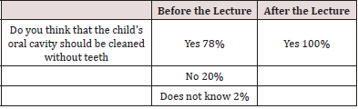
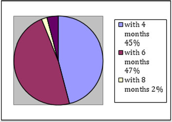
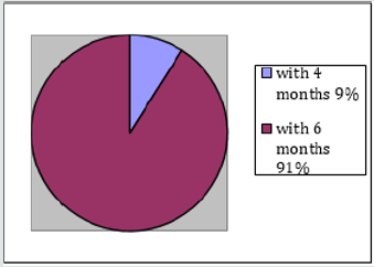
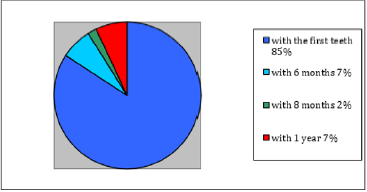
49 pregnant women with an average age of 31 participated in this study. The majority (61%) of pregnant women are married, have a university degree (67%) and are employed (87%). Fathers are mostly university educated (43%) or have completed vocational school (43%), and 98% are employed. The vast majority of respondents (92%) have this first child, and 71% claim that this is the first course for pregnant women attending and that they have never received information on maintaining oral hygiene (92%). The survey showed that most pregnant women know that oral hygiene must be maintained while the baby is still toothless. During the lecture, attention was once again drawn to the importance of cleaning the mouth of a child without teeth and the way in which this is done was described. All pregnant women answered in the affirmative after the lecture (Table 1). The next issue was the age of the baby in the months when the first baby teeth begin to erupt. The offered answers were 4, 6, 8, 10 and 12 months. Before the lecture, 47% of pregnant women answered for 6 months, and after the lecture, 91% answered for 6 months (Graphs 1 & 2). Pregnant women were aware even before the lecture that baby teeth were important for the baby. As many as 71% of respondents answered that deciduous teeth are extremely important, while 27% answered that they are mostly important. After the lecture, 96% of respondents claim that deciduous teeth are extremely important (Table 2). The next question was answered by overwriting, so there is a whole range of answers that needed to be sorted into correct or incorrect. The correct answer was considered to be all those who indicated that teeth should be brushed as soon as possible, when the first teeth erupt. Answers of the “2 months” and “3 months” types were also considered correct under the assumption that the respondents meant toothless oral hygiene which continued and dental hygiene when they erupted. Any responses that suggest brushing your teeth from 1 year onwards are considered guilty.

Table 1: Answers to the question on maintaining the hygiene of child’s oral cavity without teeth, before and after the lecture.

Lupinepublishers-openaccess-pediatric-dentistry-journal

Graph 1: Opinion of pregnant women on the time of eruption of deciduous teeth before the lecture.

Lupinepublishers-openaccess-pediatric-dentistry-journal

Table 2: Opinion of pregnant women on the importance of deciduous teeth for the child, before and after the lecture.

Lupinepublishers-openaccess-pediatric-dentistry-journal

Graph 2: Opinion of pregnant women on the time of eruption of deciduous teeth after the lecture./p>

Lupinepublishers-openaccess-pediatric-dentistry-journal

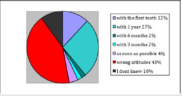
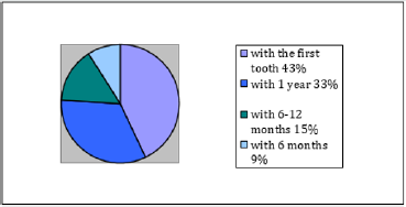
Before the lecture, 67% of respondents answered with one of the correct answers, and after the lecture 93% (Graphs 3 & 4). The next question was also answered by adding. Due to the large number of different incorrect answers, it is impossible to make a clear picture of the attitudes of pregnant women on this issue, so all the wrong attitudes will be classified into a single group. Some of the wrong attitudes were: “when we notice a defect”, “when all the teeth grow”, “when the first tooth falls out”, “at the age of 2” and many others. The correct attitude is considered to be all those who indicate the first visit to the dentist as early as 1 year of age. Before the lecture, 47% of pregnant women had the correct attitude about the time of the first examination, and after the lecture, all respondents (100%) had the correct attitude (Graphs 5 & 6).

Graph 3: Opinion of pregnant women about the time of the beginning of brushing the child’s teeth before the lecture.

Lupinepublishers-openaccess-pediatric-dentistry-journal

Graph 4: Opinion of pregnant women about the time of the beginning of brushing the child’s teeth after the lecture.

Lupinepublishers-openaccess-pediatric-dentistry-journal

Graph 5: Attitudes of pregnant women about the time of the first visit to the dentist before the lecture.

Lupinepublishers-openaccess-pediatric-dentistry-journal

Graph 6: Attitudes of pregnant women about the time of the first visit to the dentist after the lecture.

Lupinepublishers-openaccess-pediatric-dentistry-journal

Table 3: Attitudes of pregnant women about dental treatment of deciduous teeth.

Lupinepublishers-openaccess-pediatric-dentistry-journal

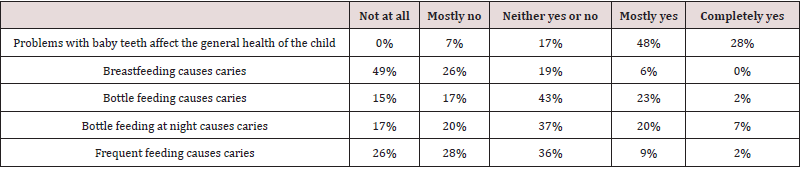
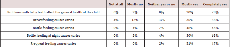
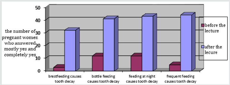
To the question „Are deciduous teeth being repaired“, 71% of respondents circled the answer YES before the lecture. After the lecture, 93% of respondents answered YES (Table 3). The next group of questions examines the attitudes of pregnant women about the causes of caries. With each statement, it was necessary to round off the position most similar to the position of the respondent, from “not at all” to “completely yes”. In this group of questions, pregnant women had the worst results (Table 4). Pregnant women’s knowledge of the causes of caries has greatly improved after the lecture (Table 5 and Graph 7).

Table 4:Attitudes of pregnant women about the impact of deciduous teeth on general health and factors for the development of caries before lectures.

Lupinepublishers-openaccess-pediatric-dentistry-journal

Table 5: Attitudes of pregnant women about the impact of deciduous teeth on general health and factors for the development of caries after lectures.

Lupinepublishers-openaccess-pediatric-dentistry-journal

Graph 7: Attitudes of pregnant women about risk factors that cause caries before and after the lectures.

Lupinepublishers-openaccess-pediatric-dentistry-journal

Discussion

Most of the respondents were not previously advised on maintaining the child’s oral hygiene. Nevertheless, this research has shown that pregnant women consider deciduous teeth important for the development and general condition of the baby. This figure coincides with the results of a study in Nigeria in which 79.2% of pregnant women stated that baby teeth are important for a child. However, pregnant women who participated in this study showed better knowledge regarding the repair of deciduous teeth (71% answered that they are being repaired) than pregnant women who participated in the study in Nigeria (43.6% did not know if deciduous teeth were being repaired) [13]. Most correctly answered the question about cleaning the oral cavity without teeth and the question about the time of eruption of the first teeth. Two descriptive questions about the time of the beginning of brushing and the first visit to the dentist have worse results, but half of the respondents answered these questions correctly. A similar study has been conducted in Germany among midwives. Midwives were asked questions about the advice they give to young mothers and pregnant women. Most claim to advise pregnant women about early childhood caries, but when asked about their first visit to the dentist, only 9% responded within the first year of life. Similar to the results of this research, 60% of midwives recommend starting brushing when the first tooth erupts [14]. A study conducted in India showed a poorer level of knowledge of pregnant women and young mothers about the time of the first visit to the dentist. Only 22.3% of pregnant women answered that the first visit to the dentist must be around the period of the eruption of the first teeth, while this research showed that about 47% of pregnant women have the correct attitude. The reason for this is because it is very likely the degree of illiteracy among women in India [15]. The area from which pregnant women showed the least knowledge is the development of caries. Specifically, they were unaware of the risky behaviors that lead to caries. 49% of respondents answered that breastfeeding does not cause caries at all, probably because they can hardly associate breastfeeding as a physiological action with negative consequences. A similar degree of ignorance of pregnant women is shown by questions about bottle-feeding, bottle-sleeping, and frequent feeding. Research has shown that pregnant women do not have sufficient knowledge of risky behaviors for caries, so they do not have the necessary knowledge to prevent caries in their child. The lecture on oral care for children as part of a course for pregnant women was the first encounter for most pregnant women with information on the causes of caries, harmful habits, tips for maintaining oral hygiene, the time to start brushing and the first visit to the dentist. Since the same but abbreviated survey was completed after the lecture, it is easy to notice an improvement in the knowledge of the mentioned topics, which supports the importance of timely education of young mothers on maintaining oral hygiene of the child.

Conclusions

This research showed that the participants in the course for pregnant women at the Clinic for Gynecology and Obstetrics in Rijeka do not have satisfactory knowledge about the oral health of the child, primarily about the risky behavior for early childhood caries. Most pregnant women were not aware that breastfeeding, bottle feeding and sleeping and frequent feeding cause tooth decay. More than half of pregnant women were of the opinion that the child should be taken to the dentist for the first visit at the age later than recommended (until the age of 1) or did not know the answer. For most pregnant women, the mentioned lecture was the first encounter with information on maintaining the oral health of the child. After the lecture, pregnant women had a much better knowledge of caries and all other issues, which indicates the importance of the mentioned lecture as part of the course for pregnant women.

Read More Lupine Publishers Pediatric Dentistry Journal Articles:
https://lupine-publishers-pediatric-dentistry.blogspot.com/

Saturday, January 8, 2022

Lupine Publishers | Children’s and Adolescents’ Pain, Discomfort, Fear, Cooperation, and Parental Presence During Dental Extractions

 Lupine Publishers | Journal of Pediatric Dentistry


Abstract

Introduction: Children’s reports of experiences from dental invasive procedures are scarce. The aim was to renew and actualize the understanding of children’s experiences of pain, discomfort, fear, and cooperation during the dental extraction procedures. A further aim was to study the frequency of parental presence during dental extractions.

Methods: The study was based on a sample of children and adolescents aged 3–19 years in the Region Västra Götaland (RVG) and Region Örebro County (ROC). It was a 5-year cohort study of an accelerated longitudinal design, named BITA (Barn I TAndvården = Children in dental care). Data on children’s pain, discomfort, fear, cooperation, and parental presence were assessed and compiled.

Results: 2363 out of 3134 children in the four age cohorts became eligible for inclusion. The cohorts included Cohort 1 (3-7 years old; n=695 children), Cohort 2 (7-11 years old; n=642 children), Cohort 3 (11-15 years old; n=574 children), and Cohort 4 (15-19 years old; n=452 children). There were 1215 girls (51%) and 1148 boys (49%). Extractions were assessed as painful by 62%. Discomfort was reported by 33%. Dental fear was reported by 47%. During painful extractions, no fear was reported by 38%, while in painless extractions, no fear was reported by 63%. Dental fear was most common in the age group 11-15 years. Full treatment acceptance was recorded in 73.6% during the extractions. In extractions reported to cause pain, 61% cooperated well. Most of the patients (77%) showed full treatment acceptance with parental presence in the dental room. The corresponding figure was 55% for the parent not being present.

Conclusion: Dental extractions continue to constitute one of the most complex and challenging treatment situations for the young patient. The oral injection was most frequently reported to cause pain, discomfort, and fear during dental extractions. Dentists should make efforts to prevent pain and discomfort, as well as utilize parental support for the child’s sense of security during dental extractions.

Keywords: Paediatric; pain management; pain assessment; clinical procedures

Abbreviations: RVG: Region Västra Götaland, ROC: Region Örebro County; BITA: Barn I T Andvården; DFA: Dental Fear and Anxiety; BMP: Behavior Management Problems

Introduction

Invasive dental treatments such as anesthesia and extractions have shown to stress children and adolescents the most [1,2]. In a study performed on 368 Swedish children and adolescents (8- 19 years), the prevailing reported painful dental procedures were injection, tooth extraction, drilling and performing a filling [1]. Negative treatment experiences may trigger a vicious cycle, i.e., leading to avoidance of dental appointments and hindering more positive experiences to take place [3-5]. Furthermore, avoiding dental appointments may affect the oral health [3-5] jeopardizing the well-being of young patients with long-term consequences [3-5]. Less traumatizing methods for invasive dental techniques during injection and extraction are available, although to date, studies report that the usage of pain preventive interventions such as topical anesthesia, local anesthesia, or analgesics are still substandard [6-8]. The routines applied by Swedish dentists have shown that ‘about 35% were more indifferent to their patient’s experiences of pain and psychological management’ [7]. Apart from pain experiences, invasive and/or noninvasive dental procedures may cause children discomfort. Although there is no universally prevailing definition, the phenomenon might be explained as psychological or bodily distress or annoyance, i.e., anything that disturbs the well-being [9]. Children’s experience of discomfort in dentistry has previously not been given much attention. Invasive procedures also test dentists’ technical skills with a need to be knowledgeable on children’s developmental levels, contemporary psychology, and pedagogical interactions. Furthermore, dentists may face difficulties in the interaction with the child/parent dyad [10].

Parental attitudes and behavior have been suggested to either facilitate or undermine the child’s cooperation and thus affect the treatment outcome [11-14]. It could be hypothesized that some dentists consider parents a hindrance, therefore not inviting them into the dental room. Seen from the young patients’ standpoint, such an approach may be controversial as parental absence may diminish the perception of security, predisposing to fear, pain, and discomfort experiences [14]. In Sweden, a prevailing tradition and the Dental Act have allowed parents to support their children in medical and dental settings [15]. The practice has thus become a norm in the pediatric dental community, despite alternative behavior management techniques [16]. At present, there is insufficient knowledge in the area of children’s and adolescents’ experience of invasive procedures, and dentists’ attitudes and praxis towards these. Furthermore, there is a lack of knowledge on how general dentists understand the parental interaction and how frequently they make use of parental help during extractions. The aim was to renew and actualize the understanding of children’s experiences of pain, discomfort, fear, and cooperation during dental extraction procedures. A further aim was to study the frequency of parental presence during dental extractions.

Patients and Methods

Patients

This study was based on a representative sample of children and adolescents aged 3–19 years in the Region Västra Götaland (RVG) and Region Örebro County (ROC). The project was a 5-year cohort study of an accelerated longitudinal design, named BITA (Barn I TAndvården = Children in dental care). It concerned different aspects of children in the dental situation when attending regular general dentistry. Four age cohorts with children aged 3, 7, 11 and 15 years, from seven Public Dental Service clinics in RVG and five in ROC, were invited to participate and then followed over a 5-year period in conjunction with regular dental visits. The clinics were selected to reflect the respective populations and to cover urban and rural areas. A total of 3134 children were considered for the study.

Procedure

Clinical registrations

The children’s ordinary dentists registered all performed dental treatments after each session. For this study, extraction treatments were selected, and assessments and self-ratings were made and registered.

Self-reported pain

Each child made a self-assessment of pain experienced after the extraction. The child was asked the question: Was anything painful? Yes or no? If the child answered yes, the pain intensity was measured using a visual analog scale (VAS) consisting of 11 points running from 0 = no pain, to 10 = worst pain possible. Parallel to the scale, six faces express different levels of pain/distress where young children point out the face matching their level of pain. Furthermore, the child was asked the question: What was painful?

Self-reported discomfort

The child was asked the question: Did anything cause discomfort? Yes or no? If the child answered yes, the discomfort was measured using a visual rating scale (VRS) consisting of 11 points running from 0 = no discomfort, to 10 = worst discomfort possible. Parallel to the scale, six faces express different levels of discomfort where young children point out the face matching their level of discomfort. Furthermore, the child was asked the question: What caused the discomfort?

Self-reported fear

Children assessed their fear at the dental treatment by answering the question, how did you feel today? The alternative answers were Not afraid at all = 0 or Afraid, on a scale graded 1-4.

Assessment of the child’s cooperation

The child´s cooperation was graded by the treating dental personnel according to the scale by Rud and Kisling, rated 3 to 0, where 3= full acceptance to treatment; 2= indifferent acceptance; 1= reluctant acceptance; and 0= non-acceptance.

Parental presence

Parental presence in the dental room was documented.

Ethics

The application for ethical review (No. 286-07) was approved.

Statistical methods

In addition to descriptive statistics in terms of frequency distributions, means, and standard deviations, differences between genders and between cohorts were analyzed using Chi-square tests. The statistical analyses were performed in IBM SPSS Statistics for Windows, Version 23.0 (IBM Corp., Armonk, NY, USA ) and p values below 0.05 represented statistical significance.

Results

3134 children were invited to participate in the study; 771 declined participation, whereby 2363 children in the four age cohorts became eligible for inclusion. These four age cohorts included Cohort 1 (born 2005; from 3-7 years old during the study period; n=695 children, 3906 visits), Cohort 2 (born 2001; 7-11 years old; n=642 children, 4588 visits), Cohort 3 (born 1997; 11-15 years old; n=574 children, 4085 visits), and Cohort 4 (born 1993; 15-19 years old; n=452 children, 3315 visits). The distribution between the genders was 1215 girls (51%) and 1148 boys (49%).

Pain and discomfort during extractions

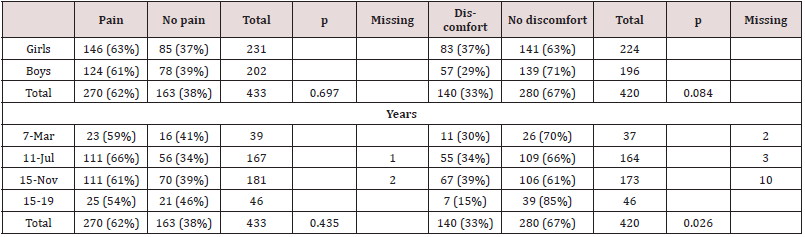
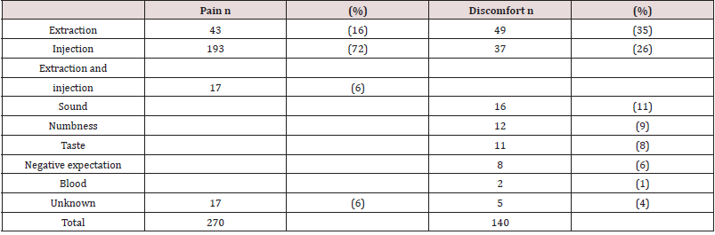
Extractions were assessed as painful by 62% (Table 1). The reasons were the extraction itself (16%), the injection (72%) or the combination of injection and extraction procedures (6%) (Table 2). Discomfort was reported in 33% of the extractions (Table 1). The reasons for discomfort pointed to the extraction per se (37%), followed by injection (26%) (Table 2). No statistically significant difference between the genders or cohorts was found regarding the experience of pain. There was a statistically significant difference between cohorts, with the lowest proportion of discomfort stated by the oldest group (30%, 34%, 39%, 15%; p=0.026) (Table 1). Primary (n=323) and permanent (n=102) tooth extractions were assessed as painful in equal proportions (63% and 61%), while discomfort was slightly less often reported for primary teeth, compared to permanent tooth extractions (32% and 37%). 11 tooth extractions lacked complete documentation. Topical anesthesia was applied in 35%, with no statistically significant difference between application and no application regarding the reported experience of pain and/or discomfort. Conscious sedation drugs were used in 6%.

Table 1: Extraction appointments (n=436 extractions) and the young patients’, reports of pain and discomfort in adjunction to the treatment, related to gender and age-cohorts.

Lupinepublishers-openaccess-pediatric-dentistry-journal

Chi-2 test, P probability value

Table 2: Children’s reported reasons for pain (n=270) and discomfort (n=140) in connection to the extraction procedures.

Lupinepublishers-openaccess-pediatric-dentistry-journal

Dental fear and pain during extractions

Children’s self-assessment of dental fear resulted in 47% reporting no dental fear in conjunction with the extractions. During painful extractions, no fear was reported by 38%, while in painless extractions, no fear was reported by 63% (Table 3). Dental fear was most common in the age group 11-15 years, followed by the age group 7-11 years (Table 3). No statistically significant difference between the genders or cohorts was found regarding the experience of pain. There was a statistically significant difference between cohorts, with the lowest proportion of discomfort stated by the oldest group (30%, 34%, 39%, 15%; p=0.026) (Table 1). Primary (n=323) and permanent (n=102) tooth extractions were assessed as painful in equal proportions (63% and 61%), while discomfort was slightly less often reported for primary teeth, compared to permanent tooth extractions (32% and 37%). 11 tooth extractions lacked complete documentation. Topical anesthesia was applied in 35%, with no statistically significant difference between application and no application regarding the reported experience of pain and/ or discomfort. Conscious sedation drugs were used in 6%.

Table 3:Young patients’ (age cohorts) self-rated dental fear and pain experiences. The subjective assessment of pain=Yes or No was given after the extraction procedure (missing data n=14). Dental fear, as rated on a scale 0 to 4, 0=no fear, fear =1-4, (missing data n=12).

Lupinepublishers-openaccess-pediatric-dentistry-journal

Dental fear and pain during extractions Children’s self-assessment of dental fear resulted in 47% reporting no dental fear in conjunction with the extractions. During painful extractions, no fear was reported by 38%, while in painless extractions, no fear was reported by 63% (Table 3). Dental fear was most common in the age group 11-15 years, followed by the age group 7-11 years (Table 3).

The child´s cooperation while experiencing pain

Full treatment acceptance was recorded in 73.6% during the extractions. In extractions reported to cause pain, 61% cooperated well (Table 4).

Table 4: Children’s self-rated pain experiences and the degree of treatment acceptance, as rated by the dental personnel, on a scale 3-0, by Rud and Kisling.

Lupinepublishers-openaccess-pediatric-dentistry-journal

The subjective assessment of pain=Yes or No was given by the child after the extraction (missing data n=16).

Treatment acceptance during parental presence

Most of the patients (77%) showed full treatment acceptance with parental presence in the dental room. The corresponding figure was 55% for the parent not being present. All 37 three-yearold had their parents present in the dental room. Among the 166 seven-year-olds, two were not accompanied by an adult. Among the 11-year-olds, 26 out of 179 had no parent present. Among the 15-year-olds, 25 out of 45 managed on their own.

Discussion

This longitudinal, 5-year study voiced children’s and adolescents’ self-reported pain, discomfort, and fear during dental extractions. The main results showed that more than 60% of the dental extraction appointments involved painful experiences. Furthermore, pain was equally reported among the different age groups. The extracted teeth were predominantly performed in young schoolchildren. This may indicate that more vulnerable children were at risk experiencing pain at an early stage of their dental care encounters. Within the extraction procedure, the injection was most frequently reported responsible for the negative experiences. During decades, pain has been connected with dental extractions and concerned children and adolescents [17]. It could be reasoned that the injection techniques have not developed sufficiently, despite alternative anesthesia delivery methods, Computer controlled local anesthesia, Jet injectors, Iontophoresis and Computer controlled intraosseous anesthesia [18,19]. An explanation might be that as these methods lie in the hands of numerous dentists, the injection performance may not be possible to calibrate. Other factors adding to the child’s pain experience may be the dentists handling the onset of the anesthetic, the amount of delivered anesthesia, or a complementary anesthesia differently. A noteworthy result was that topical anesthesia was applied in only 35% by the dentists, which is astonishingly low given the scientific evidence in support of the technique, which is in alignment with earlier result [7]. Furthermore, adding to the children’s uneasiness, discomfort was reported frequently during the extraction procedure. Thus, despite a complete anesthesia numbness, discomfort may have been experienced due to cracking sounds and/or the perceived pressure during the dental luxation. Discomfort has not been given much attention in the dental literature compared to general medical care [20]. The frequently reported discomfort also suggest that the dentists may not have practiced current theoretical knowledge sufficiently, which needs to be recognized [7,8].

No studies among dentists have confirmed the Knowledge Transition to have improved regarding dental invasive procedures in general or oral anesthesia specifically [21,22]. An explanation may be the complex interaction between the applied injection technique and the dentist’s way of psychological and pedagogical coaching [23-25]. Preferably, children should beforehand be prepared on anesthesia numbness sensation, luxation cracking sounds, increasing tissue-pressure during luxation, postoperative taste-sensation (iron, i.e., blood), and the subsequent inconvenience of not being able to enjoy a meal directly after the tooth extraction. Studies on Dental Fear and Anxiety (DFA) have showed that invasive dental procedures, such as extractions and injections, were by many children and adolescents associated with fear and anxiety [26,27]. In the current study, 47% of the extraction appointments were experienced without fear. From this perspective, the extractions were performed under satisfactory conditions. On the other hand, 53% of the children experienced fear, ranging from a bit nervous to terrified. Versloot et al. (2008) have reasoned that the level of dental anxiety may be of greater importance for the child’s reaction than the injection technique itself [27]. Given that young patients’ early pain encounters may not only trigger. Behavior Management Problems (BMP) and/or Dental Fear and Anxiety (DFA), but may also alter future pain responses, the results of this study are notable [28]. Contrary to the pain reports that were equally common in all age cohorts of the current study, DFA was more frequently reported in schoolchildren. This may define a vulnerable developmental stage, requiring parental presence and support. The relatively high proportion of extraction appointments performed with the parent does not present in the room calls for additional studies.
The current study presented that parental presence in the dental room was most frequent among children 3-12 years old, hereafter the presence significantly declined. Praxis has suggested that most parents are experts on their child’s behavior and reactions. Perets and Zadick (1998) found that most parents would assist, should the dentist not succeed to manage their child [29]. Vasiliki (2016) also stated that children responded more positively in the dental room with parental presence [11]. Pfefferle et al. (1982) reasoned that this may be due to parents knowing their child’s reactions and capabilities in challenging situations [30]. The current study demonstrated that most children cooperated fully during the dental extraction procedures despite experiencing discomfort or pain. A possible interpretation is that these children were not equipped to speak their needs, or they surrendered easily to authorities. Indirectly, the results also suggested that dentists practiced varied theoretical understanding, technical skills, and ethical attitudes during dental extraction procedures, not always in favor of the child. A limitation of this study may be argued that children below six years of age were not reliable to give accurate assessments of their experiences. However, there is a strong opinion among researchers that children from the age of 3-4 years are capable to communicate well and should be supported to express perceptions of discomfort and pain [31]. The strength of this study was that the results originated from a 5-year longitudinal perspective based on young patients’ experiences from the extraction appointments. These results, regarding pain, discomfort and DFA, have not been contradicted in the recent literature and are valuable for clinical discussions. In conclusion, dental extractions continue to constitute one of the utmost intricate and challenging treatment situations for young patients. The oral injection was most frequently reported to cause pain, discomfort, and fear during dental extractions. Dentists should make efforts to prevent pain and discomfort, as well as utilize parental support for the child’s sense of security during dental extractions.

Acknowledgements

This study was funded by the Public Dental Service, Region Västra Götaland and Region Örebro County, Sweden.

Conflict of Interest

The authors declare no conflicts of interest.

Read More Lupine Publishers Pediatric Dentistry Journal Articles:
https://lupine-publishers-pediatric-dentistry.blogspot.com/

980 nm Diode Laser: A Good Choice for the Treatment of Pyogenic Granuloma

Abstract Pyogenic granuloma is a benign non/neo plastic mococutanous lesion . It is a reactional response to constant minor trauma and ca...